Abstract
Blocking neurotransmission, botulinum neurotoxin is the most poisonous biological substance known to mankind. Despite its infamy as the scourge of the food industry, the neurotoxin is increasingly used as a pharmaceutical to treat an expanding range of muscle disorders. Whilst neurotoxin expression by the spore-forming bacterium Clostridium botulinum appears tightly regulated, to date only positive regulatory elements, such as the alternative sigma factor BotR, have been implicated in this control. The identification of negative regulators has proven to be elusive. Here, we show that the two-component signal transduction system CBO0787/CBO0786 negatively regulates botulinum neurotoxin expression. Single insertional inactivation of cbo0787 encoding a sensor histidine kinase, or of cbo0786 encoding a response regulator, resulted in significantly elevated neurotoxin gene expression levels and increased neurotoxin production. Recombinant CBO0786 regulator was shown to bind to the conserved −10 site of the core promoters of the ha and ntnh-botA operons, which encode the toxin structural and accessory proteins. Increasing concentration of CBO0786 inhibited BotR-directed transcription from the ha and ntnh-botA promoters, demonstrating direct transcriptional repression of the ha and ntnh-botA operons by CBO0786. Thus, we propose that CBO0786 represses neurotoxin gene expression by blocking BotR-directed transcription from the neurotoxin promoters. This is the first evidence of a negative regulator controlling botulinum neurotoxin production. Understanding the neurotoxin regulatory mechanisms is a major target of the food and pharmaceutical industries alike.
Author Summary
Botulinum neurotoxin produced by the spore-forming bacterium Clostridium botulinum is the most poisonous biological substance known to mankind. By blocking neurotransmission, the neurotoxin causes a flaccid paralysis called botulism which may to lead to death upon respiratory muscle collapse. Despite its infamy as the scourge of the food industry, the neurotoxin is attracting increasing interest as a pharmaceutical to treat an expanding range of muscle disorders. Whilst neurotoxin production by C. botulinum appears tightly regulated, to date only positive regulatory elements, thus enhancing the neurotoxin production, have been implicated in this control. The identification of negative regulators, responsible for down-tuning the neurotoxin synthesis, has proven to be elusive, but would offer novel approaches both for the production of safe foods and for the development of therapeutic neurotoxins. Here, we report a two-component signal transduction system that negatively regulates botulinum neurotoxin production. Understanding the neurotoxin regulatory mechanisms is a major target of the food and pharmaceutical industries alike.
Introduction
Botulinum neurotoxins are the most poisonous biological substances known to mankind. The neurotoxins are metalloproteases which block neurotransmission in cholinergic nerves [1], [2] in humans and animals to cause botulism, a potentially lethal flaccid paralysis. Botulinum neurotoxins are produced by vegetative cultures of the anaerobic spore-forming bacterium Clostridium botulinum which is widespread in the environment. The neurotoxins can enter the victim's body through intoxication with food or drink, or they can be produced from spores germinating and growing into active cultures in vivo, most likely in the gut of small babies with poorly developed gut microflora or in deep wounds. Despite their infamy, botulinum neurotoxins attract increasing interest as a pharmaceutical to treat an expanding range of muscular and other disorders [3], [4], such as torticollis, focal dystonia, inappropriate contraction of gastrointestinal sphincters, eye movement disorders, hyperhidrosis, migraine [5], genitourinary disorders [6], and even cancer [7]. Indications in cosmetic surgery are well known.
Seven antigenically distinct toxin types (A to G), and several subtypes therein, have been described [8]–[12]. Type A1 neurotoxins are the best characterized, a consequence both of their frequent involvement in human botulism worldwide and of their greater potency and, therefore, suitability for therapeutics [13]. Botulinum toxins are produced as a complex containing the neurotoxin itself and one or more non-toxic auxiliary proteins that protect the neurotoxin from environmental stress and assist in absorption [14]. Type A1 toxins are complexed with the non-toxic non-hemagglutinating (NTNH) protein and three hemagglutinins (HA17, HA33 and HA70) [15]–[18]. A typical A1-type gene cluster is transcribed in two operons, namely the ntnh-botA and ha operons [19] (Figure 1). Both operons have consensus −10 and −35 core promoter sequences, which are recognized by the alternative sigma factor BotR, directing RNA polymerase (RNAP) to transcribe the two operons [20]. The gene encoding BotR is located between the two operons within the neurotoxin gene cluster.
Figure 1. Schematic representation of the TCS CBO0787/CBO0786 and neurotoxin loci in C. botulinum ATCC 3502.

The neurotoxin operons are indicated with arrows. Predicted TCS domains are marked with gray color and the corresponding functions are listed under each gene. Insertional sites of ClosTron mutagenesis in cbo0786 encoding a response regulator and cbo0787 encoding a sensor histidine kinase are indicated with dashed lines.
Botulinum neurotoxin production is affected by the availability of certain nutrients [21]–[23] and is associated with transition from late-exponential to early-stationary phase cultures. A peak in the level of neurotoxin gene cluster expression in late-exponential to early-stationary phase cultures [19], [24] suggests that neurotoxin production is tightly regulated. To date only positive regulatory elements have been implicated in this control. These include the participation of BotR [25] and an Agr quorum sensing system [26]. The identification of negative regulators of botulinum neurotoxin production has until now proved to be elusive.
Two-component signal transduction systems (TCS) are conserved in bacteria and differentially specialized to control a range of cellular events in response to environmental stimuli. The histidine kinases sense cellular or environmental signals through the N-terminus of their sensor domains. This interaction leads to autophosphorylation at a histidine residue in their C-terminus and the subsequent activation of their cognate response regulator present in the cytosol by transmission of the phosphoryl group to an N-terminal aspartate residue of the response regulator and further to the C-terminal output domain. Response regulators possess DNA-binding activity, ultimately resulting in a specific response in the expression of their target genes. The individual roles of most TCSs in C. botulinum are not known, but their involvement in control of virulence in other pathogenic bacteria has been demonstrated [27]. Antisense mRNA inhibition of genes encoding three TCSs caused decreased neurotoxin production in C. botulinum type A strain Hall [28], suggesting these TCSs may play a role in positive control of neurotoxin synthesis. The model strain C. botulinum ATCC 3502 (Group I, type A) [29] encodes 29 putative TCSs and a set of orphan histidine kinases and response regulators [30]. One of the intact TCSs, CBO0787/CBO0786 (Figure 1), shares over 90% amino acid identity with other C. botulinum Group I strains and in many strains is located in the vicinity of the neurotoxin genes (3.6 to 24 kilobases [kb] up- or downstream of the toxin genes). Here we show that the TCS CBO0787/CBO0786 negatively regulates botulinum neurotoxin gene expression. Understanding the regulatory mechanisms that control the production of botulinum neurotoxin is a major target of the food and pharmaceutical industries.
Results
The cbo0787 and cbo0786 genes are down-regulated at transition into stationary growth phase
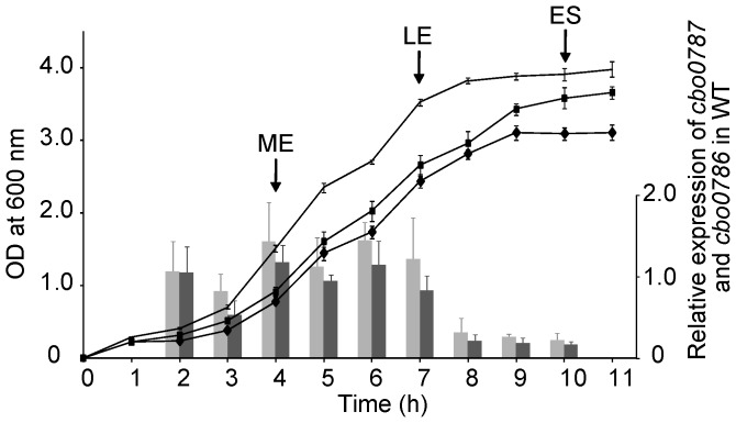
We used quantitative reverse transcription PCR (qRT-PCR) to measure the relative expression of cbo0787 and cbo0786 during the growth of C. botulinum Group I type A strain ATCC 3502, which is the most widely used model strain for genetic studies in C. botulinum [26], [31], [32]. The relative transcription levels of cbo0787 and cbo0786 followed an identical pattern, suggesting that the two genes are co-transcribed (Figure 2). In relation to growth, cbo0787 and cbo0786 were expressed at a relatively constant level throughout the logarithmic growth phase, and were down-regulated at the transition into stationary phase (Figure 2).
Figure 2. Expression of cbo0787 and cbo0786 in C. botulinum ATCC 3502 wild type (WT) and the growth curves of WT and the cbo0787 and cbo0786 mutants.
Quantitative RT-PCR analysis of relative cbo0787 (light grey bars) and cbo0786 (dark grey bars) transcript levels in WT. Target gene expression was normalized to 16S rrn and calibrated to 2-hour time point. Growth of WT (line without symbols), cbo0787 mutant (square) and cbo0786 mutant (diamond) in tryptose-peptone-glucose-yeast extract medium. Arrows indicate sampling at mid-exponential (ME), late exponential (LE) and early stationary (ES) growth phases. Error bars represent standard deviations of three replicate measurements.
Mutation of cbo0787 and cbo0786
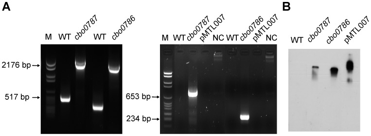
To address the role of CBO0787/CBO0786, we constructed single, insertional inactivation mutations in cbo0787 or cbo0786 using the ClosTron tool [33]. Single insertion of the group II intron from pMTL007 into the desired sites in cbo0787 or cbo0786 (Figure 1) was confirmed by PCR (Figure 3A) and Southern blotting (Figure 3B). Consecutive cultures showed the mutants to be erythromycin resistant and stable. No significant difference between the growth of the TCS mutants and the ATCC 3502 wild-type strain (WT) were observed (Figure 2), and the log cell counts per ml of WT and the cbo0787 and cbo0786 mutant cultures at early stationary phase (10 hours) were 8.9, 9.0 and 8.8, respectively.
Figure 3. Insertional inactivation of cbo0787 and cbo0786.
(A) PCR analysis of mutations. Ll.LtrB intron insertion was detected with primers flanking the insertional sites yielding a 2.3-kb PCR product in cbo0787 mutant and a 2.2-kb product in cbo0786 mutant (left), and with a gene-specific primer and the intron-binding primer EBS universal yielding a 785-bp product in the cbo0787 mutant and a 260-bp product in the cbo0786 mutant (right). (B) Southern blot analysis of HindIII digested genomic DNA from WT and cbo0787 and cbo0786 mutants with intron-specific probe.
The cbo0787 or cbo0786 mutants show induced expression of the neurotoxin cluster genes
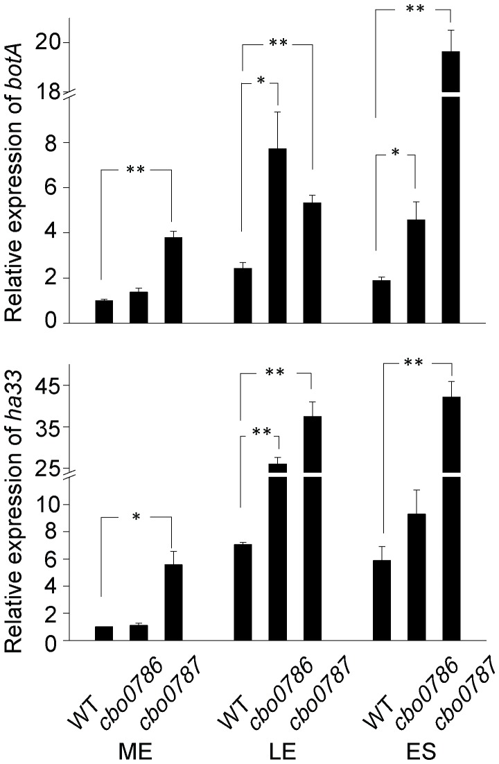
We used qRT-PCR to measure the relative expression of botA encoding botulinum neurotoxin type A and ha33 encoding one of the three haemagglutinins in WT and the two TCS mutants at mid-exponential, late exponential and early stationary growth phases. The two genes were selected to represent the two operons present in the neurotoxin gene cluster, and the three time points used have been shown to associate with induction and repression of the neurotoxin gene expression [19], [34], while later time points typically involve other cellular events, such as sporulation or lysis, and were thus not considered relevant. As expected, the WT levels of botA and ha33 expression peaked at late exponential or early stationary growth phases (Figure 4). Strikingly, the cbo0787 mutant had a maximum of 10.4-fold (P<0.01) and 8.0-fold (P<0.01) higher relative botA and ha33 expression levels, respectively, than the WT (Figure 4), the most prominent differences being observed at early stationary growth phase. This suggests that the histidine kinase CBO0787 is required to sense an as yet unidentified signal for the efficient ‘switch-off’ of the neurotoxin gene expression at transition into stationary growth phase.
Figure 4. Disruption of cbo0787 or cbo0786 increased the expression of neurotoxin genes.
Quantitative RT-PCR analysis of relative botA and ha33 transcript levels in WT and the cbo0786 and cbo0787 mutants. RNA was isolated from cells at mid-exponential (ME), late exponential (LE) and early stationary (ES) growth phases. Target gene expression was normalized to 16S rrn and calibrated to WT at ME. Error bars represent standard deviations of three replicate measurements. Statistical significance of differences between WT and each mutant is indicated with p-values (*, P<0.05; **, P<0.01).
The cbo0786 mutant also showed significantly increased botA and ha33 expression levels in relation to WT (Figure 4). A maximum of 2.4-fold (P<0.05) induction for botA at early stationary phase and 3.8-fold (P<0.01) induction for ha33 at late exponential growth phase were measured. These data suggest that CBO0786 negatively regulates neurotoxin gene expression, either directly or indirectly.
The cbo0787 or cbo0786 mutants show induced neurotoxin production
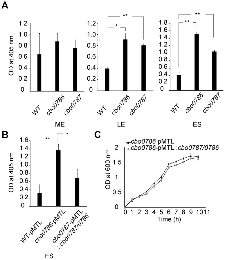
To confirm that our findings at the transcription level apply to protein level, we analyzed the relative amounts of botulinum neurotoxin in the WT and mutant culture supernatants collected at mid-exponential, late exponential and early stationary growth phases. Measurements were made using an enzyme-linked immunosorbent assay (ELISA). At mid-exponential growth phase, neurotoxin production was slightly but not significantly increased in the cbo0787 and cbo0786 mutant cultures compared to WT culture (Figure 5A). At late exponential and early stationary growth phases, neurotoxin production was significantly increased (2.1 to 3.7-fold higher OD405 readings, P<0.05) in both the cbo0787 and cbo0786 mutant cultures relative to WT culture (Figure 5A). To validate this result, we complemented the cbo0786 mutation by introducing pMTL::cbo0787/0786, containing the TCS genes and their putative native promoter, into the cbo0786 mutant and showed that neurotoxin levels in early-stationary phase cultures were restored to the vector-only control (WT-pMTL) level (P<0.05, Figure 5B). No difference was observed in growth between the cbo0786-pMTL control and the complemented strain (Figure 5C).
Figure 5. Neurotoxin ELISA to demonstrate elevated neurotoxin production by cbo0787 and cbo0787 mutants.
(A) ELISA analysis of botulinum neurotoxin in culture supernatants of WT and the cbo0786 and cbo0787 mutants at mid-exponential (ME), late exponential (LE) and early stationary (ES) growth phases. The samples were diluted 1∶10 at ME, 1∶20 at LE, and 1∶30 at ES. (B) ELISA analysis of botulinum neurotoxin in culture supernatants of the vector-only control strains WT-pMTL and cbo0786-pMTL, and the complemented strain cbo0786-pMTL::cbo0787/0786 at ES. All samples were diluted 1∶30. (C) Growth curve of cbo0786-pMTL and cbo0786-pMTL::cbo0787/cbo0786. Error bars represent standard deviations of three replicate measurements. Statistical significance of differences between WT and the two mutants is indicated with p-values (*, P<0.05; **, P<0.01).
The CBO0786 response regulator binds to neurotoxin promoters
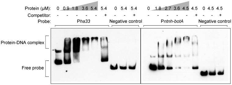
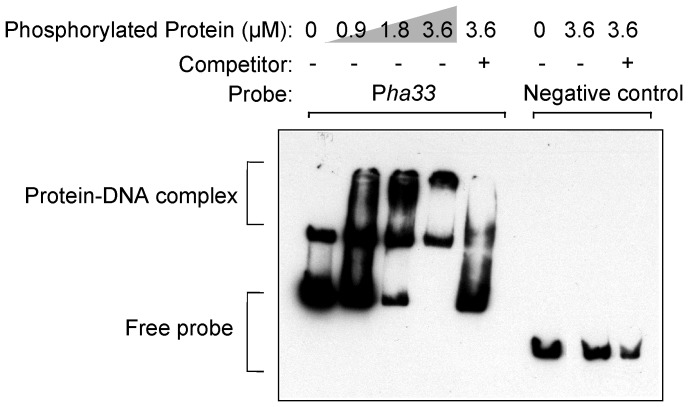
To test the hypothesis that the TCS response regulator CBO0786 is a transcriptional repressor of the neurotoxin gene cluster in ATCC 3502, we examined the binding of the recombinant CBO0786 protein to probes that encompassed the intergenic region between ha33 and botR containing the promoter of the ha operon [35] (Pha33 probe), or the intergenic region between botR and ntnh containing the promoter of the ntnh-botA operon [35] (Pntnh-botA probe), by electrophoretic mobility shift assay (EMSA). CBO0786 caused a shift in the mobility of both probes, although its binding affinity to the Pntnh-botA probe appeared somewhat lower than to the Pha33 probe (Figure 6). The specific nature of binding was further confirmed by disappearance of both protein-DNA complexes using competition with a 200-fold excess of unlabeled probe. Moreover, no electrophoretic shift was observed with the negative control probe (Figure 6). EMSA with recombinant CBO0786 phosphorylated by acetyl phosphate yielded a similar shift (Figure 7), indicating that phosphorylation is not essential for CBO0786 binding to DNA in vitro. These results suggest that CBO0786 recognizes and binds to the promoter regions of the ha and the ntnh-botA operons.
Figure 6. CBO0786 binds to botulinum neurotoxin promoters in vitro.
EMSA showing CBO0786 binding to Pha33 probe (left panel) and Pntnh-botA probe (right panel). Specificity was confirmed using 200-fold molar excess of non-labeled competitor DNA and a random gene fragment as negative control.
Figure 7. In vitro phosphorylated CBO0786 binds to Pha33 probe in vitro.
EMSA showing phosphorylated CBO0786 binding to Pha33 probe. Specificity was confirmed using 200-fold molar excess of non-labeled competitor DNA and a random gene fragment as negative control.
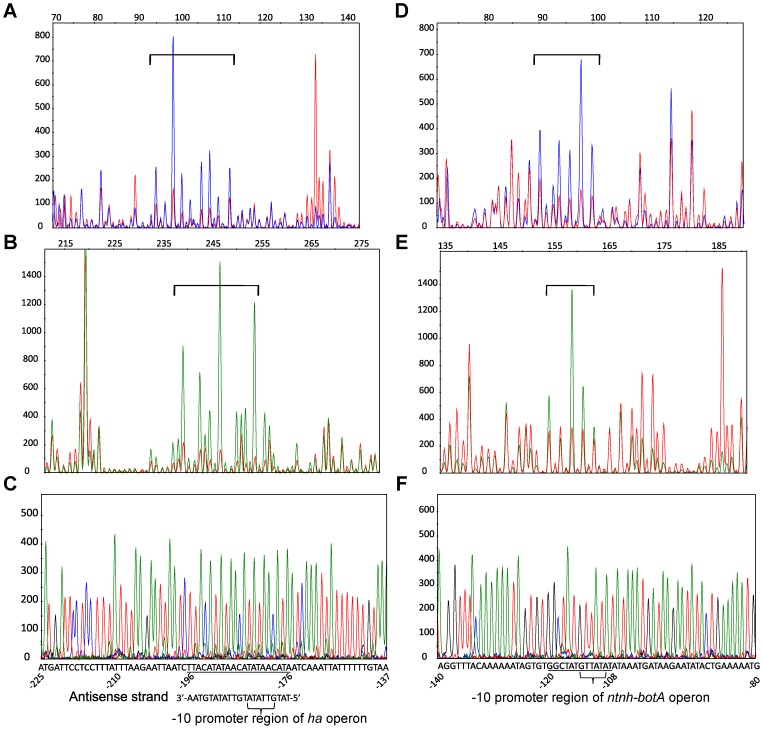
To understand the DNA-binding specificity of CBO0786, we identified its binding sites using DNase I footprinting and fluorescently end-labeled Pha33 and Pntnh-botA probes (Figure 8). With the Pha33 probe, the region protected by CBO0786 (−51 bp to −31 bp upstream of ha33 and −196 bp to −176 bp upstream of botR) was present in both strands (Figures 8A and 8B). Greater protection by CBO0786 was observed in the antisense strand containing the promoter of the ha operon [35], suggesting that the antisense sequence (TATGTTATATGTTATATGTAA, Figure 8C) is the major CBO0786 binding site. Interestingly, the core promoter −10 region (GTTATA) of the ha operon, recognized by the alternative sigma factor BotR [20], appeared as a direct repeat in the CBO0786 binding site, suggesting that CBO0786 represses transcription of the ha operon by binding to its core promoter.
Figure 8. CBO0786 binds to the core promoter −10 region of the ha and ntnh-botA operons.
DNase I footprinting analysis of 5′-6-FAM labeled sense strand (A, D) and 5′-HEX labeled antisense strand (B, E) of Pha33 probe (A, B) and Pntnh-botA probe (D, E). Comparison of DNase I digestion in the absence (blue peaks in A and D, green peaks in B and E) or with 10 µM of CBO0786 (red peaks). Protection regions are indicated by square brackets. Protection regions are underlined in sequencing electropherograms of Pha33 probe (C) and Pntnh-botA probe (F) in the sense strand. The consensus −10 regions of the ha and ntnh-botA promoters are indicated.
In the Pntnh-botA probe, a site of CBO0786 protection similar to that observed with the Pha33 probe was evident (Figures 8D and 8E). Both strands contained the same protection site (GGCTATGTTATAT) (−120 bp to −108 bp upstream of ntnh, Figure 8F). Accordingly, the −10 region (GTTATA) of the ntnh-botA operon core promoter was presented in the binding region, but in one copy only. In accordance with the EMSA analysis, the binding affinity of CBO0786 to the ntnh-botA promoter appeared lower than to the promoter of the ha operon.
The CBO0786 response regulator represses in vitro transcription from neurotoxin promoters
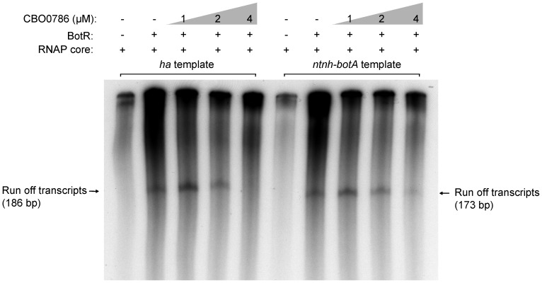
To demonstrate the direct effect of CBO0786 on transcription of the neurotoxin genes, we carried out in vitro run-off transcription assays using RNAP reconstituted with E. coli RNAP core enzyme and the purified sigma factor BotR (Figure 9). DNA fragments containing the promoter of the ha or ntnh-botA operons were cloned and used as transcription templates. As expected, transcripts produced from the promoters of ha or ntnh-botA operons were observed only in the presence of both RNAP core enzyme and BotR. Addition of increasing concentrations of recombinant CBO0786 caused gradual inhibition of both transcripts, suggesting CBO0786 directly represses the transcription from ha and ntnh-botA promoters. Transcription from the ha promoter was more efficiently repressed than that from the ntnh-botA promoter in the presence of 4 µM CBO0786 (Figure 9). Taken together, CBO0786 exhibits a higher binding affinity to the direct repeat of the −10 region (GTTATA) in the ha promoter than in the ntnh-botA promoter, and consequently shows a more effective inhibition of in vitro transcription from the ha promoter than from the ntnh-botA promoter. These results indicate that CBO0786 represses the transcription of neurotoxin gene cluster by binding to the consensus core promoter −10 region of ha and ntnh-botA operons.
Figure 9. CBO0786 represses transcription from ha and ntnh-botA promoters in vitro.
Run-off transcription from ha33 and ntnh-botA promoter-containing templates was repressed in the presence of increasing concentration of CBO0786. Arrow indicates the run-off transcripts.
To test whether CBO0786 represses the transcription of botR, we also performed in vitro run-off transcription assays with DNA template containing the botR promoter. No clear transcript from botR promoter was observed in the presence of RNAP core with BotR. Consistent with a previous study [20], the result demonstrates that botR is not auto-transcribed in vitro.
Discussion
We show that the TCS CBO0787/CBO0786 negatively regulates botulinum neurotoxin expression in C. botulinum Group I type A1 strain ATCC 3502. This was supported by enhanced toxin gene expression and increased toxin synthesis by cbo0787 and cbo0786 mutants, by specific binding of recombinant CBO0786 regulator to the neurotoxin promoters, and recombinant CBO0786 inhibiting in vitro transcription from the neurotoxin promoters. Identification of botulinum neurotoxin repressors has been an unattainable target of neurotoxin research for decades [13], [36], [37], and would open up novel strategies for controlling the public health risks caused by the toxin, but also for enhancing industrial processes for therapeutic neurotoxin preparations.
While the current work focused on the most well-characterized C. botulinum type A1 neurotoxin subtype, the role of CBO0787/CBO0786 homologs in strains of other subtypes will be an interesting line of future research. While the cbo0787/cbo0786 locus is found at an 11-kb distance from the neurotoxin gene cluster (cbo0801–cbo0806) in ATCC 3502, a TCS showing a strikingly high (>95%) amino acid identity to CBO0787/CBO0786 is similarly encoded by loci near the neurotoxin gene cluster in the genomes of type A2 strain Kyoto (3.6 kb distance), type A5 strain H04402 065 (12 kb), and type F strain Langeland (24 kb). This TCS, therefore, is an interesting candidate for a universal neurotoxin repressor in Group I C. botulinum.
Our biochemical data suggest that CBO0786 recognizes and binds to the consensus −10 region (GTTATA) of the ha and ntnh-botA operon promoters. The −10 region is also specifically recognized by the alternative sigma factor BotR [20], [38], suggesting that CBO0786 binding to the core promoter −10 region may prevent RNAP-BotR from binding and initiating the transcription. The in vitro run-off transcription assay demonstrated that CBO0786 directly inhibits RNAP-BotR-directed transcription from the ha and ntnh-botA promoters, supporting the hypothesis that CBO0786 inhibits the transcription of these operons.
BotR is specifically required for botulinum neurotoxin gene expression [25] but does not autoregulate its own expression [20]. While the −35 site (TTTACA) of the ha and ntnh-botA core promoters is also found upstream of botR, the −10 site of botR promoter is different (TTCGTA) [20]. Accordingly, we did not observe CBO0786 binding to botR promoter. Moreover, we did not find additional CBO0786 binding sites downstream of the botR promoter in vitro or in silico. Thus it is plausible that CBO0786 does not repress botR transcription.
While an early study detected both monocistronic botA and bicistronic ntnh-botA mRNA species and a putative transcription start site in the intergenic region between ntnh and botA [34], BotR was later shown to exclusively drive the bicistronic form of transcription [20]. Accordingly, we could not identify a CBO0786 binding site in the intergenic region between ntnh and botA, thus inhibition by CBO0786 of transcription from a putative botA-specific promoter is not likely.
BotR homologues in other pathogenic Clostridia, such as TetR controlling tetanus neurotoxin in Clostridium tetani [39], TcdR controlling toxins A and B in Clostridium difficile [40], and UviA controlling bacteriocins in Clostridium perfringens [41] all recognize the same −35 sequence as BotR [20]. Interestingly, TetR also recognizes the same −10 box (GTTATA) as BotR [36]. The closest homolog of CBO0787/CBO786 in C. tetani is CTC01420/CTC01421, with the response regulator CTC01421 showing 75% amino acid similarity to CBO0786. Thus CTC01420/CTC01421 may be an interesting candidate for a tetanus neurotoxin repressor.
Our qRT-PCR and ELISA analysis, albeit yielding significantly greater neurotoxin gene expression in both cbo0787 and cbo0786 mutants than in WT, suggest the mutant phenotypes to be somewhat less striking than reported for some well-characterized TCSs in other Gram-positive bacteria. For example, mutation of CsrS, reported to respond to environmental Mg2+ [42] and to repress virulence-related capsular polysaccharide production in group A Streptococcus, resulted in a 10-fold increase in cellular hyaluronic acid production in the presence of Mg2+ [42]. However, in medium lacking Mg2+, only a modest 1.5-fold induction in hyaluronic acid production was observed [42]. Thus the presence of the signal triggering a TCS kinase is a key to control the activity of the cognate response regulator and its subsequent effects on target gene expression. The signal triggering CBO0787 and the role of phosphotransfer between CBO0787 and CBO0786 remain to be elucidated. Although our EMSA analysis suggested a DNA-binding activity for CBO0786 both with and without phosphorylation in vitro, which is in line with reports on some other TCS response regulators showing similar DNA-binding properties regardless of phosphorylation state [43], in vivo phosphorylation of CBO0786 could lead to conformational changes that fine-tune its DNA binding affinity [44]–[46]. Identification of the signal triggering CBO0787 will be an important future task for optimization of the study conditions to detect maximal mutant phenotypes and thus to detect the maximal effect of CBO0786 on neurotoxin repression.
The sensor domain of CBO0787 is predicted to contain an extracellular loop consisting of 79 amino acid residues and is flanked by two transmembrane helices, indicating that the signal triggering it is most likely extracellular. Moreover, the increasing differences observed between the relative neurotoxin gene expression levels of the cbo0787 kinase mutant and WT towards stationary growth phase are consistent with cell density-dependent signals [37]. Neurotoxin overproduction associated with inability to sporulate by strain Hall A-hyper [13] suggests that regulation of these processes may be linked. Repression of toxin synthesis after logarithmic growth before initiation of sporulation may represent a survival mechanism when nutrient sources become limited. Moreover, uncontrolled synthesis of botulinum neurotoxin would waste energy since only nanogram quantities are sufficient for C. botulinum to kill mammals and thereby gain nutrients and establish anaerobiosis.
Previous studies have identified excess of arginine [21] and tryptophan [22] to repress botulinum neurotoxin formation. By contrast, glucose was shown to induce neurotoxin synthesis [21]. Interestingly, glucose has also been linked with regulation of toxin synthesis in C. difficile through carbon catabolite control [47], [48]. Further research will be required to clarify the mechanisms by which nitrogen sources or glucose control neurotoxin synthesis in C. botulinum; however, our preliminary data do not indicate that these compounds trigger CBO0787 (data not shown). Thus it is plausible that the regulation of neurotoxin synthesis is accomplished through a complex network and other regulators are involved in this control.
While the relative neurotoxin gene expression levels in the cbo0786 regulator mutant were significantly higher than those of the WT at logarithmic growth phase, this difference was smaller at early stationary growth phase. Moreover, the wild-type levels of cbo0786 and cbo0787 transcription, being stable throughout the logarithmic growth phase, collapsed at the transition into stationary phase. These observations further support the involvement of repressors other than CBO0786 in the ‘switching-off’ of the neurotoxin expression, ensuring efficient onset of stationary-phase cellular events.
Bearing in mind the emergence of repressor gene (tcdC) mutations in ‘hyper-virulent’ isolates of the notorious healthcare-associated pathogen C. difficile [49], [50], the emergence of C. botulinum strains with a mutated neurotoxin repressor would set challenges to public health and safety. Hence identification of neurotoxin regulators and their mutations is crucial.
In conclusion, we propose that the TCS CBO0787/CBO0786 negatively regulates botulinum neurotoxin gene transcription through the response regulator CBO0786 blocking the −10 core promoter sites of the ha and ntnh-botA operons, inhibiting transcription from the ha and ntnh-botA promoters. These data provide keys for controlling the production of botulinum neurotoxin, which is a major target of the food and pharmaceutical industries.
Materials and Methods
Strains, culture, plasmids, and oligonucoleotides
Bacterial strains and plasmids are described in Table S1. C. botulinum Group I type A1 strain ATCC 3502 [29] and derivative mutants were grown in anaerobic tryptone-peptone-glucose-yeast extract (TPGY) medium at 37°C under strictly anaerobic conditions. Cell counts were determined by plating serially diluted cultures on anaerobic TPGY agar plates. Escherichia coli conjugation donor CA434 [51] and E. coli TOP10 strain (Invitrogen) were grown in Luria-Bertani (LB) medium at 37°C. When appropriate, growth media were supplemented with 100 µg/ml ampicillin, 50 µg/ml kanamycin, 25 µg/ml chloramphenicol, 250 µg/ml cycloserine, 15 µg/ml thiamphenicol or 2.5 µg/ml erythromycin. All oligonucleotide primers are listed in Table S2.
Mutation of cbo0787 and cbo0786
Target genes were insertionally inactivated in C. botulinum ATCC3502 by using the ClosTron system as previously reported [33], in combination with the TargeTron gene knockout system kit (Sigma-Aldrich). Target sites in cbo0786 (between nucleotides 267–268) and cbo0787 (between nucleotides 603–604) were identified, and intron-retargeting PCR primers (Table S2) were designed by using the TargeTron algorithm (http://www.sigma-genosys.com/targetron/).
Plasmid retargeting was carried out as previously described [33] and the resulting plasmid pMTL007::cbo0786 or pMTL007::cbo0787 was transferred to C. botulinum ATCC3502 by conjugation from E. coli CA434 [51]. Successful transconjugants were screened on TPGY agar plates containing cycloserine (250 µg/ml) and thiamphenicol (15 µg/ml), and then resuspended in 1 ml of anaerobic TPGY containing 1 mM isopropyl-β-D-thiogalactopyranoside (IPTG) and thiamphenicol (7.5 µg/ml) and incubated at 37°C for 3 h. The bacteria were then harvested, resuspended in 1 ml of fresh TPGY and incubated for a further 3 h. The subsequent integrants were selected by plating bacteria on TPGY agar supplemented with erythromycin (2.5 µg/ml) and cycloserine (250 µg/ml) and incubated for 16 h at 37°C in anaerobic conditions to select clones harboring the spliced erythromycin retrotransposition activated marker (ErmRAM), which indicates intron integration.
To demonstrate the integration of the Ll.LtrB intron in the desired sites, PCR was performed using primers flanking the target sites in cbo0786 and cbo0787 (Table S2). PCR using ErmRAM primers demonstrated the spliced ErmRAM. In addition, to confirm that only a single intron insertion occurred in each mutant, genomic DNA from the ATCC3502 wild-type strain and mutants, and the pMTL007 plasmid DNA were digested overnight with HindIII and analysed by Southern blot probed with a DIG-labeled fragment derived from the Ll.LtrB intron sequence.
Complementation of mutation
For complementation, a 2441-bp fragment encompassing cbo0786, cbo0787, and the 5′ noncoding region including their putative promoter, was cloned into plasmid pMTL82151 [52] to make pMTL::cbo0787/0786. pMTL::cbo0787/0786 or pMTL82151 (empty-vector control) was transferred to C. botulinum ATCC3502 or cbo0786 mutant by conjugation from E. coli CA434. The complementation strain of cbo0786 mutant-pMTL::cbo0787/0786 and control strains of C. botulinum ATCC3502-pMTL and cbo0786 mutant-pMTL were obtained.
RNA isolation and qRT-PCR
Total RNA from C. botulinum ATCC 3502 and the two mutants was isolated using the RNeasy Mini Kit (Qiagen) as described [31]. Residual DNA was removed sequentially with RNase-free DNase set (Qiagen) and the DNA-free Kit (Ambion) according to the manufacturers' instructions. The RNA was dissolved in 50 µl of nuclease-free water (Sigma-Aldrich) and its concentration was determined using the NanoDrop ND1000 spectrophotometer (NanoDrop Technologies). The integrity of RNA was confirmed with the Agilent Technologies 2100 Bioanalyzer.
For qRT-PCR, samples were collected during mid-exponential, late exponential and early stationary growth phases (Figure 2). Duplicate cDNA samples were generated from 800 ng of RNA using the DyNAmo cDNA Synthesis Kit (Finnzymes). Quantitative real-time PCR reactions comprised of 1× DyNAmo Flash SYBR Green I Master Mix (Finnzymes), 0.5 mM of each primer (Table S2), and 4 µl of 102-fold (cbo0786, cbo0787, botA, ha33) or 105-fold (16S rrn) diluted cDNA template in a total volume of 20 ul. All PCRs were performed in duplicate for both cDNA replicates and three replicated experiments. Real-time PCR was performed using the Rotor-Gene 3000 real-time thermal cycler (Corbett Life Science). Cycling conditions included 7 minutes at 95°C, followed by 45 cycles of 95°C for 10 seconds and 60°C for 20 seconds, followed by 30 seconds at 60°C. PCR efficiencies were determined based on a standard curve made from serially diluted pooled cDNA for each primer pair. The calculated efficiencies were 0.97 for 16S rrn, 0.99 for botA, 0.92 for ha33, 1.04 for cbo0787 and 0.93 for cbo0786. Melting curve analysis was performed following the completion of the PCR to confirm specificity of the PCR amplification products. Target gene expression was normalized to the expression of 16S rrn based on the Pfaffl method [53]. All samples were calibrated against the wild type culture at mid-exponential growth phase.
Neurotoxin ELISA
Botulinum neurotoxin was quantified by using a commercial type A neurotoxin ELISA kit (Tetracore) in three independent WT and mutant culture supernatants collected at mid-exponential, late exponential, and early stationary growth phases [24], [26], [28]. The plates were read at 405 nm (Multiskan Ascent, Thermo Fisher). To keep the optical density readings in the dynamic range, the culture supernatants were diluted 1∶10 at mid-exponential, 1∶20 at late exponential, and 1∶30 at early stationary growth phase.
Expression and purification of recombinant CBO0786 and BotR
To construct the plasmids for the expression of N-terminal 6-histidine translation fusion to the response regulator CBO0786 or the alternative sigma factor BotR, PCR products were generated using the primers listed in Table S2. The PCR products of cbo0786 and botR were digested with appropriate restriction enzymes and cloned individually into plasmid pET28b (Novagen). The plasmids were then individually transformed into E. coli Rosetta 2(DE3) pLysS cells (Novagen).
CBO0786 expression was induced with 1 mM IPTG at 37°C for 5 h. Cells from a 500-ml culture were harvested, re-suspended in 10 ml of lysis/binding buffer (500 mM NaCl, 20 mM imidazole, 20 mM Tris-HCl, pH 7.9) and lysed by sonication. The lysate was centrifuged at 10 000 g for 15 min and filtered through a 0.45-µm filter. The lysate was loaded with 1 ml of Novagen His Bind affinity resin and allowed to bind for 30 min at 4°C. The resin was washed by 10 ml of lysis/binding buffer and 20 ml of wash buffer (500 mM NaCl, 60 mM imidazole, 20 mM Tris-HCl, pH 7.9). Bound protein was eluted by washing with 4 ml of elution buffer (500 mM NaCl, 500 mM imidazole, 20 mM Tris-HCl, pH 7.9).
BotR expression was induced with 1 mM IPTG overnight at 20°C and purified as described previously [20], with some modifications. Briefly, cells were lysed by sonication in lysis/binding buffer. The insoluble cell debris was separated by centrifugation and dissolved in denaturing lysis/binding buffer with 6 M guanidine hydrochloride. After centrifugation and filtration, the denatured solubilized proteins were collected and loaded onto column containing Ni-NTA affinity resin. The bound proteins were let to refold on column with a decreasing urea gradient (6 to 0 M) in lysis/binding buffer. Finally, the refolded proteins were obtained by washing with elution buffer.
Eluted fractions were examined by SDS-PAGE and fractions containing CBO0786 or BotR were pooled in the Novagen D-tube Dialyzer and dialysed against 1 l of dialysis buffer (300 mM NaCl, 20% glycerol, 50 mM Tris-HCl, pH 8.0) overnight at 4°C. Protein concentrations were determined by using the Bradford reagent (Bio-Rad) and BSA (Sigma-Aldrich) was used as a standard.
EMSA
A 354-bp fragment (Pha33 probe) covering the intergenic region between ha33 and botR (−287 bp to 67 bp of ha33) and a 262-bp fragment (Pntnh-botA probe) covering the intergenic region between botR and ntnh (−210 bp to 52 bp of ntnh) were amplified by PCR using 5′-end biotin labeled primers (Table S2). CBO0786 phosphorylation by acetyl phosphate was performed as described [54]. EMSA was performed with 1 nM of 5′-end biotin labeled, double-stranded oligonucleotide probes, 0 to 5.4 µM of recombinant CBO0786, 1 µg of poly(dI-dC), 2.5% glycerol and 5 mM MgCl2 in binding buffer (LightShift Chemiluminescent EMSA Kit, Pierce). For competition assays, a 200-fold molar excess of unlabeled double-stranded oligonucleotide was added. Binding was allowed to proceed for 30 min at room temperature. Band shifts were resolved on a 5% native polyacrylamide gel run in 0.5× TBE at 4°C for 1 h at 110 V.
DNase I footprinting
DNase I footprinting was performed in triplicate using a modification of [55]. The Pha33 and Pntnh-botA probes were amplified by PCR using 6-FAM-labeled forward primers and HEX-labeled reverse primers (Table S2). Binding reactions were performed as described for EMSA, with 20 nM of 5′-6-FAM-labeled probe and 10 µM protein in a final volume of 20 µl. After 20 min of incubation, DNA probes were partially digested by 0.002 to 0.2 Kunitz unit of DNase I (Sigma-Aldrich) for 5 min at room temperature. Reactions were stopped by addition of 22 µl of 0.5 M EDTA and heated at 70°C for 10 min. The digested DNA fragments were purified with the QIAquick PCR Purification Kit (Qiagen) and eluted in 25 µl of water. The purified fragments were separated in a capillary DNA analyzer (Applied Biosystems 3130×l DNA Analyzer) and the electropherograms were analyzed using the Peak Scanner software (Applied Biosystems). Protected regions were identified by sequencing of the fragments using the Thermo Sequenase Cycle Sequencing Kit (Affymetrix) and the same labeled primers as described above (Table S2).
In vitro run-off transcription
The upstream region of the ha operon spanning the −191 to +185 sites relative to the transcription start, and the upstream region of the ntnh-botA operon spanning the −108 to +172 sites relative to the transcription start, were cloned into pBluescript II KS- (Stratagene) and then linearized by SpeI or PstI to produce the run-off transcription templates. In vitro transcription assays were carried out in 10-µl reaction mixtures, in the absence or presence of CBO0786. E. coli RNAP core enzyme (Epicentre) was preincubated for 30 min at 37°C with six-fold molar excess of purified BotR. After incubation, 0.5 U RNAP, 0 to 4 µM of recombinant CBO0786, and 15 nM of linearized plasmid DNA were added in transcription buffer containing 40 mM Tris-HCl, pH 8.0, 10 mM MgCl2, 50 mM KCl, 0.1 mg/ml BSA, 5% glycerol, 4 U of RNasin (Promega), and incubated for 10 min at 37°C. Transcription was initiated by the addition of 200 µM each of ATP, GTP and CTP, 50 µM of non-radioactive UTP and 2.5 µCi [α-32P]-UTP (3000 Cimmol−1, Perkin-Elmer). After further incubation for 30 min at 37°C, the reaction was quenched by adding 10 µl of RNA loading buffer (95% formamide, 0.025% bromophenol blue, 0.025% xylene cyanol FF, and 5 mM EDTA), followed by heating for 10 min at 80°C. Samples were resolved by denaturing 6% PAGE and visualized by autoradiography.
Statistical analysis
For any pairwise comparisons between the WT and one of the two mutant strains, Student's t-test was used. For a multiple comparison among the cbo0786-pMTL::cbo0787/0786, cbo0786-pMTL, and WT-pMTL, one-way ANOVA with Tukey's post hoc test was used.
The NCBI ID numbers of the genes and genome studied
cbo0787 (ID number 5185042), cbo0786 (ID number 5185041), cbo0803, ha33 (ID number 5185058), cbo0805, ntnh (ID number 5185060), cbo0806, botA (ID number 5185061), Clostridium botulinum type A strain ATCC 3502 genome (Accession number NC_009495.1).
Supporting Information
Bacterial strains and plasmids.
(DOC)
Oligonucleotides.
(DOC)
Acknowledgments
We thank Hanna Korpunen and Esa Penttinen for technical assistance.
Funding Statement
The work was performed in the Centre of Excellence in Microbial Food Safety Research, and supported by the Academy of Finland (141140; www.aka.fi), Research Funds of the University of Helsinki (www.helsinki.fi), the European Community's Seventh Framework Programme (237942; http://cordis.europa.eu/), the ABS Graduate School (http://www.vetmed.helsinki.fi/abs/), and the Finnish Veterinary Foundation (http://www.sels.fi/index_eng.htm). The funders had no role in study design, data collection and analysis, decision to publish, or preparation of the manuscript.
References
- 1. Schiavo G, Benfenati F, Poulain B, Rossetto O, Polverino de Laureto P, et al. (1992) Tetanus and botulinum-B neurotoxins block neurotransmitter release by proteolytic cleavage of synaptobrevin. Nature 359: 832–835. [DOI] [PubMed] [Google Scholar]
- 2. Blasi J, Chapman ER, Link E, Binz T, Yamasaki S, et al. (1993) Botulinum neurotoxin A selectively cleaves the synaptic protein SNAP-25. Nature 365: 160–163. [DOI] [PubMed] [Google Scholar]
- 3. Johnson EA (1999) Clostridial toxins as therapeutic agents: Benefits of nature's most toxic proteins. Annu Rev Microbiol 53: 551–575. [DOI] [PubMed] [Google Scholar]
- 4. Lim EC, Seet RC (2010) Use of botulinum toxin in the neurology clinic. Nat Rev Neurol 6: 624–636. [DOI] [PubMed] [Google Scholar]
- 5. Diener HC, Holle D, Dodick D (2011) Treatment of chronic migraine. Curr Pain Headache Rep 15: 64–69. [DOI] [PubMed] [Google Scholar]
- 6. Chuang YC, Chancellor MB (2006) The application of botulinum toxin in the prostate. J Urol 176: 2375–2382. [DOI] [PubMed] [Google Scholar]
- 7. Ansiaux R, Gallez B (2007) Use of botulinum toxins in cancer therapy. Expert Opin Investig Drugs 16: 209–218. [DOI] [PubMed] [Google Scholar]
- 8. Smith TJ, Lou J, Geren IN, Forsyth CM, Tsai R, et al. (2005) Sequence variation within botulinum neurotoxin serotypes impacts antibody binding and neutralization. Infect Immun 73: 5450–5457. [DOI] [PMC free article] [PubMed] [Google Scholar]
- 9. Hill KK, Smith TJ, Helma CH, Ticknor LO, Foley BT, et al. (2007) Genetic diversity among botulinum neurotoxin-producing clostridial strains. J Bacteriol 189: 818–832. [DOI] [PMC free article] [PubMed] [Google Scholar]
- 10. Chen Y, Korkeala H, Aarnikunnas J, Lindström M (2007) Sequencing the botulinum neurotoxin gene and related genes in Clostridium botulinum type E strains reveals orfx3 and a novel type E neurotoxin subtype. J Bacteriol 189: 8643–8650. [DOI] [PMC free article] [PubMed] [Google Scholar]
- 11. Dover N, Barash JR, Arnon SS (2009) Novel Clostridium botulinum toxin gene arrangement with subtype A5 and partial subtype B3 botulinum neurotoxin genes. J Clin Microbiol 47: 2349–2350. [DOI] [PMC free article] [PubMed] [Google Scholar]
- 12. Macdonald TE, Helma CH, Shou Y, Valdez YE, Ticknor LO, et al. (2011) Analysis of Clostridium botulinum serotype E strains by using multilocus sequence typing, amplified fragment length polymorphism, variable-number tandem-repeat analysis, and botulinum neurotoxin gene sequencing. Appl Environ Microbiol 77: 8625–8634. [DOI] [PMC free article] [PubMed] [Google Scholar]
- 13. Schantz EJ, Johnson EA (1992) Properties and use of botulinum toxin and other microbial neurotoxins in medicine. Microbiol Rev 56: 80–99. [DOI] [PMC free article] [PubMed] [Google Scholar]
- 14. Matsumura T, Jin Y, Kabumoto Y, Takegahara Y, Oguma K, et al. (2008) The HA proteins of botulinum toxin disrupt intestinal epithelial intercellular junctions to increase toxin absorption. Cell Microbiol 10: 355–364. [DOI] [PubMed] [Google Scholar]
- 15. Lamanna C (1948) Hemagglutination by botulinal toxin. Proc Soc Exp Biol Med 69: 332–336. [DOI] [PubMed] [Google Scholar]
- 16. Sugii S, Sakaguchi G (1975) Molecular construction of Clostridium botulinum type A toxins. Infect Immun 12: 1262–1270. [DOI] [PMC free article] [PubMed] [Google Scholar]
- 17. Dineen SS, Bradshaw M, Johnson EA (2003) Neurotoxin gene clusters in Clostridium botulinum type A strains: sequence comparison and evolutionary implications. Curr Microbiol 46: 345–352. [DOI] [PubMed] [Google Scholar]
- 18. Gu S, Rumpel S, Zhou J, Strotmeier J, Bigalke H, et al. (2012) Botulinum neurotoxin is shielded by NTNHA in an interlocked complex. Science 335: 977–981. [DOI] [PMC free article] [PubMed] [Google Scholar]
- 19. Bradshaw M, Dineen SS, Maks ND, Johnson EA (2004) Regulation of neurotoxin complex expression in Clostridium botulinum strains 62A, Hall A-hyper, and NCTC 2916. Anaerobe 10: 321–333. [DOI] [PubMed] [Google Scholar]
- 20. Raffestin S, Dupuy B, Marvaud JC, Popoff MR (2005) BotR/A and TetR are alternative RNA sigma factors controlling the expression of the neurotoxin and associated protein genes in Clostridium botulinum type A and Clostridium tetani . Mol Microbiol 55: 235–249. [DOI] [PubMed] [Google Scholar]
- 21. Pattersoncurtis SI, Johnson EA (1989) Regulation of neurotoxin and protease formation in Clostridium botulinum Okra B and Hall A by arginine. Appl Environ Microbiol 55: 1544–1548. [DOI] [PMC free article] [PubMed] [Google Scholar]
- 22. Leyer GJ, Johnson EA (1990) Repression of toxin production by tryptophan in Clostridium botulinum type E. Arch Microbiol 154: 443–447. [DOI] [PubMed] [Google Scholar]
- 23. Bonventre PF, Kempe LL (1959) Physiology of toxin production by Clostridium botulinum types A and B. II. Effect of carbohydrate source on growth, autolysis, and toxin production. Appl Microbiol 7: 372–374. [DOI] [PMC free article] [PubMed] [Google Scholar]
- 24. Chen Y, Korkeala H, Lindén J, Lindström M (2008) Quantitative real-time reverse transcription PCR analysis reveals stable and prolonged neurotoxin complex gene activity in Clostridium botulinum type E at refrigeration temperature. Appl Environ Microbiol 74: 6132–6137. [DOI] [PMC free article] [PubMed] [Google Scholar]
- 25. Marvaud JC, Gibert M, Inoue K, Fujinaga Y, Oguma K, et al. (1998) botR/A is a positive regulator of botulinum neurotoxin and associated non-toxin protein genes in Clostridium botulinum A. Mol Microbiol 29: 1009–1018. [DOI] [PubMed] [Google Scholar]
- 26. Cooksley CM, Davis IJ, Winzer K, Chan WC, Peck MW, et al. (2010) Regulation of neurotoxin production and sporulation by a putative agrBD signaling system in proteolytic Clostridium botulinum . Appl Environ Microbiol 76: 4448–4460. [DOI] [PMC free article] [PubMed] [Google Scholar]
- 27. Beier D, Gross R (2006) Regulation of bacterial virulence by two-component systems. Curr Opin Microbiol 9: 143–152. [DOI] [PubMed] [Google Scholar]
- 28. Connan C, Brueggemann H, Mazuet C, Raffestin S, Cayet N, Popoff MR (2012) Two-component systems are involved in the regulation of Botulinum neurotoxin synthesis in Clostridium botulinum type A strain Hall. PLoS One 7: e41848 doi:10.1371/journal.pone.0041848. [DOI] [PMC free article] [PubMed] [Google Scholar]
- 29. Sebaihia M, Peck MW, Minton NP, Thomson NR, Holden MT, et al. (2007) Genome sequence of a proteolytic (Group I) Clostridium botulinum strain Hall A and comparative analysis of the clostridial genomes. Genome Res 17: 1082–1092. [DOI] [PMC free article] [PubMed] [Google Scholar]
- 30. Wörner K, Szurmant H, Chiang C, Hoch JA (2006) Phosphorylation and functional analysis of the sporulation initiation factor Spo0A from Clostridium botulinum . Mol Microbiol 59: 1000–1012. [DOI] [PubMed] [Google Scholar]
- 31. Selby K, Lindström M, Somervuo P, Heap JT, Minton NP, et al. (2011) Important role of Class I heat shock genes hrcA and dnaK in the heat shock response and the response to pH and NaCl stress of Group I Clostridium botulinum strain ATCC 3502. Appl Environ Microbiol 77: 2823–2830. [DOI] [PMC free article] [PubMed] [Google Scholar]
- 32. Söderholm H, Lindström M, Somervuo P, Heap J, Minton NP, et al. (2011) cspB encodes a major cold shock protein in Clostridium botulinum ATCC 3502. Int J Food Microbiol 146: 23–30. [DOI] [PubMed] [Google Scholar]
- 33. Heap JT, Pennington OJ, Cartman ST, Carter GP, Minton NP (2007) The ClosTron: a universal gene knock-out system for the genus Clostridium . J Microbiol Methods 70: 452–464. [DOI] [PubMed] [Google Scholar]
- 34. Couesnon A, Raffestin S, Popoff MR (2006) Expression of botulinum neurotoxins A and E, and associated non-toxin genes, during the transition phase and stability at high temperature: analysis by quantitative reverse transcription-PCR. Microbiol 152: 759–770. [DOI] [PubMed] [Google Scholar]
- 35. Henderson I, Whelan SM, Davis TO, Minton NP (1996) Genetic characterisation of the botulinum toxin complex of Clostridium botulinum strain NCTC 2916. FEMS Microbiol Lett 140: 151–158. [DOI] [PubMed] [Google Scholar]
- 36. Johnson EA, Bradshaw M (2001) Clostridium botulinum and its neurotoxins: a metabolic and cellular perspective. Toxicon 39: 1703–1722. [DOI] [PubMed] [Google Scholar]
- 37.Raffestin S, Couesnon A, Pereira Y, Mazuet C, Popoff MR (2009) Botulinum and tetanus neurotoxins: molecular biology, toxin gene regulation and mode of action. In Brüggemann H, Gottschalk G, editors. Clostridia: molecular biology in the post-genomic era. Norfolk: Caister Academic Press. pp. 1–28.
- 38. Dupuy B, Raffestin S, Matamouros S, Mani N, Popoff MR, Sonenshein AL (2006) Regulation of toxin and bacteriocin gene expression in Clostridium by interchangeable RNA polymerase sigma factors. Mol Microbiol 60: 1044–1057. [DOI] [PubMed] [Google Scholar]
- 39. Marvaud JC, Eisel U, Binz T, Niemann H, Popoff MR (1998) tetR is a positive regulator of the tetanus toxin gene in Clostridium tetani and is homologous to botR . Infect Immun 66: 5698–5702. [DOI] [PMC free article] [PubMed] [Google Scholar]
- 40. Mani N, Dupuy B (2001) Regulation of toxin synthesis in Clostridium difficile by an alternative RNA polymerase sigma factor. Proc Natl Acad Sci U S A 98: 5844–5849. [DOI] [PMC free article] [PubMed] [Google Scholar]
- 41. Garnier T, Cole ST (1988) Studies of UV-inducible promoters from Clostridium perfringens in vivo and in vitro . Mol Microbiol 2: 607–614. [DOI] [PubMed] [Google Scholar]
- 42. Gryllos I, Levin JC, Wessels MR (2003) The CsrR/CsrS two-component system of group A Streptococcus responds to environmental Mg2+ . Proc Natl Acad Sci U S A 100: 4227–4232. [DOI] [PMC free article] [PubMed] [Google Scholar]
- 43. Gao R, Stock AM (2009) Biological insights from structures of two-component proteins. Annu Rev Microbiol 63: 133–154. [DOI] [PMC free article] [PubMed] [Google Scholar]
- 44. He H, Zahrt TC (2005) Identification and characterization of a regulatory sequence recognized by Mycobacterium tuberculosis persistence regulator MprA. J Bacteriol 187: 202–212. [DOI] [PMC free article] [PubMed] [Google Scholar]
- 45. Mittal S, Kroos L (2009) A combination of unusual transcription factors binds cooperatively to control Myxococcus xanthus developmental gene expression. Proc Natl Acad Sci U S A 106: 1965–1970. [DOI] [PMC free article] [PubMed] [Google Scholar]
- 46. Sinha A, Gupta S, Bhutani S, Pathak A, Sarkar D (2008) PhoP-PhoP interaction at adjacent PhoP binding sites is influenced by protein phosphorylation. J Bacteriol 190: 1317–1328. [DOI] [PMC free article] [PubMed] [Google Scholar]
- 47. Antunes A, Martin-Verstraete I, Dupuy B (2011) CcpA-mediated repression of Clostridium difficile toxin gene expression. Mol Microbiol 79: 882–899. [DOI] [PubMed] [Google Scholar]
- 48. Antunes A, Camiade E, Monot M, Courtois E, Barbut F, et al. (2012) Global transcriptional control by glucose and carbon regulator CcpA in Clostridium difficile . Nucleic Acids Res 10.1093/nar/gks864. [DOI] [PMC free article] [PubMed] [Google Scholar]
- 49. Kunehne SA, Cartman ST, Heap JT, Kelly ML, Cockayne A, et al. (2010) The role of toxin A and toxin B in Clostridium difficile infection. Nature 467: 711–713. [DOI] [PubMed] [Google Scholar]
- 50. Carter GP, Douce GR, Govind R, Howarth PM, Mackin KE, et al. (2011) The anti-sigma factor TcdC modulates hypervirulence in an epidemic BI/NAP1/027 clinical isolate of Clostridium difficile . PLoS Pathog 7 e1002317: 10.1371/journal.ppat.1002317. [DOI] [PMC free article] [PubMed] [Google Scholar]
- 51. Purdy D, O'keeffe TAT, Elmore M, Herbert M, McLeod A, et al. (2002) Conjugative transfer of clostridial shuttle vectors from Escherichia coli to Clostridium difficile through circumvention of the restriction barrier. Mol Microbiol 46: 439–452. [DOI] [PubMed] [Google Scholar]
- 52. Heap JT, Pennington OJ, Cartman ST, Minton NP (2009) A modular system for Clostridium shuttle plasmids. J Microbiol Methods 78: 79–85. [DOI] [PubMed] [Google Scholar]
- 53. Pfaffl MW (2001) A new mathematical model for relative quantification in real-time RT-PCR. Nucleic Acids Res 29: e45 doi:10.1093/nar/29.9.e45. [DOI] [PMC free article] [PubMed] [Google Scholar]
- 54. Mccleary WR, Stock JB (1994) Acetyl phosphate and the activation of two-component response regulators. J Biol Chem 269: 31567–31572. [PubMed] [Google Scholar]
- 55. Zianni M, Tessanne K, Merighi M, Laguna R, Tabita FR (2006) Identification of the DNA bases of a DNase I footprint by the use of dye primer sequencing on an automated capillary DNA analysis instrument. J Biomol Tech 17: 103–113. [PMC free article] [PubMed] [Google Scholar]
Associated Data
This section collects any data citations, data availability statements, or supplementary materials included in this article.
Supplementary Materials
Bacterial strains and plasmids.
(DOC)
Oligonucleotides.
(DOC)


