Abstract
Eccrine angiomatous hamartoma (EAH) is a rare, benign cutaneous tumor. It is characterized by the proliferation of the eccrine gland elements that are closely associated with capillary proliferation. Patients usually present with a solitary nodule on the extremities that appeared at birth or during the prepubertal years. We report a rare case of EAH in a 13-year-old boy, with clinical features suggesting melanoma. Histologically, an EAH with changes resembling a verrucous hemangioma was noted.
Keywords: angiomatous hamartoma, eccrine glands, verrucous
Introduction
Eccrine angiomatous hamartoma (EAH) is a rare benign neoplasia characterized by the proliferation of the secretory portions of the eccrine glands associated with capillary angiomatosis and proliferation in other elements, such as in the adipose tissue, hair and epidermis. (1) Most cases occur at birth or in early childhood, with various forms of clinical presentation, ranging from simple angiomatous nodules to erythematous-purpuric plaques. Multiple lesions may occur, but the predominant picture is a solitary lesion.
Case report
A 13-year-old boy presented with a skin lesion on the right ankle since birth. There was mild pain, but no hyperhidrosis was noted. His medical and family histories were unremarkable. Physical examination revealed a 1.5 cm raised keratotic, dark- brown plaque overlying a 3 cm light brown to bluish, ill-defined plaque on the right ankle. The lesion was excised with clinical suspicion of melanoma.
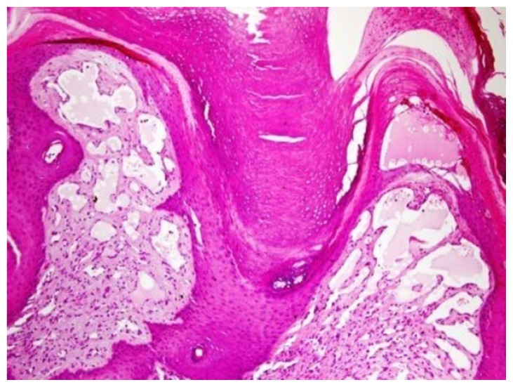
The histological examination of the excised biopsy showed a hyperkeratotic verrucous surface, with numerous dilated capillaries in the papillary dermis (Fig. 1)
Fig. (1).
Hyperkeratosis, papillomatosis, and blood vessel proliferation in the superficial dermis (H&E, ×200).
A vague nodular proliferation of eccrine glands was intermingled with numerous small vessels in the middle and deep dermis (Fig. 2-A).The epithelial cells of the eccrine glands were positive for pan-cytokeratin (Fig. 2-B), and the endothelial cells were positive for CD31 (Fig. 2-C). A diagnosis of eccrine angiomatous hamartoma with features resembling a verrucous hemangioma was made.
Fig. (2A).
The deep dermis and subcutaneous fat reveals eccrine structures proliferation intermingled with vascular channels (H&E, ×100).
Fig. (2B).
Pan-cytokeratin immunostaining highlights the eccrine glands and ducts (Pan-cytokeratin, PAP immunohistochemistry, ×100).
Fig. (2C).
CD31 immunostaining highlights the vascular structure (CD31, PAP immunohistochemistry, ×100).
Discussion
Eccrine angiomatous hamartoma (EAH) is an exceedingly rare benign tumor-like lesion prevalent in childhood that may produce pain and marked sweating. The term EAH was coined by Hymann and co-workers in 1968. (2)
EAH occurs with an equal incidence in both sexes, with approximately one third of the cases manifesting at birth or in early childhood. There are few reports of adult- onset cases up to age 71. Clinical manifestations may vary from nodules to plaques with an erythematous-bluish or brownish color. They may be located in any part of the body, although they are more frequently found in the palmoplantar regions. (3) Our case was located on the right ankle of a13 year-old boy.
EAH is commonly associated with hyperhidrosis and pain that is spontaneous or follows local pressure. Most likely, the pain occurs due to nerve fiber involvement, and hyperhidrosis occurs because of the stimulation of the eccrine components, which is caused by the elevated local temperature within the angioma. (3) The clinical differential diagnosis of EAH includes eccrine nevus, blue rubber bleb nevus, and other angiomatous lesions, such as tufted hemangioma, macular telangiectatic mastocytosis, nevus flammeus, glomus tumor, and smooth muscle hamartoma. Our case was excised to exclude melanoma.
The etiology of EAH has not been clearly defined. According to Zeller and Goldman it might be caused by the abnormal induction of heterotypic dependency during organogenesis. (4)
Histopathologically, EAH is an unencapsulated, dermal proliferation of mature appearing eccrine secretory and ductal structures that are intimately associated with thin- walled angiomatous channels, usually of a capillary nature but of variable size. The lesion typically has a lobular appearance, though rare ill-defined lesions have been described. Our case showed marked verrucous changes in the epidermis and numerous dilated capillaries in the papillary dermis. Pan-cytokeratine and CD 31 immunohistochemical stains have been performed to highlight the epithelial cells of the eccrine glands and the endothelial cells of the capillaries, respectively. To our knowledge, only few cases have been reported showing the coexistence of EAH and verrucous hemangioma-like features. (5, 6)
Conclusion
Eccrine angiomatous hamartoma is a rare condition characterized histologically by increased numbers of eccrine structures and numerous capillary channels. Patients characteristically have a solitary, congenital nodule/plaque that may be painful. It is important to recognize this condition because it is a benign lesion for which aggressive treatment is not indicated. We report a case of an eccrine angiomatous hamartoma with a rare association of verrucous hemangioma like-features.
References
- 1.Martinelli PT, Tschen JA. Eccrine Angimatous Hamartoma: A Case Report and Review of the Literature. Cutis. 2003;71:449–455. [PubMed] [Google Scholar]
- 2.Hyman AB, Harris H, Brownstein MH. Eccrine angiomatous hamartoma. NY State J Med. 1968;68:2803–6. [PubMed] [Google Scholar]
- 3.Laeng RH, Heilbrunner J, Itin PH. Late-onset eccrine angiomatous hamartoma: clinical, histological and imaging findings. Dermatology. 2001;203:70–4. doi: 10.1159/000051709. [DOI] [PubMed] [Google Scholar]
- 4.Zeller DJ, Goldman RL. Eccrine-pilar angiomatous hamartoma: report of a unique case. Dermatologica. 1971;143:100–104. [PubMed] [Google Scholar]
- 5.Cheong SH, Lim JY, Kim SY, Choi YW, Choi HY, Myung KB. A Case of Eccrine Angiomatous Hamartoma Associated with Verrucous Hemangioma. Ann Dermatol. 2009;21:304–307. doi: 10.5021/ad.2009.21.3.304. [DOI] [PMC free article] [PubMed] [Google Scholar]
- 6.Galan A, McNiff JM. Eccrine angiomatous hamartoma with features resembling verrucous hemangioma. J Cutan Pathol. 2007;34:68–70. doi: 10.1111/j.1600-0560.2007.00739.x. [DOI] [PubMed] [Google Scholar]


