Abstract
Efficacy of hippocampal fetal cell (HFC) grafting for restraining spontaneous recurrent motor seizures (SRMS) in chronic temporal lobe epilepsy (TLE) is unknown.We investigated both survival and anti-seizure effects of 5′-bromodeoxyuridine (BrdU) labeled embryonic day 19 (E19) HFC grafts pretreated with different neurotrophic factors and a caspase inhibitor. Grafts were placed bilaterally into the hippocampi of F344 rats exhibiting kainate (KA) induced chronic TLE, where the frequency of SRMS varied from 3.0 to 3.5 seizures/8-h duration. The first group received standard (untreated) HFC grafts, the second group received HFC grafts pretreated and transplanted with brain-derived neurotrophic factor (BDNF), neurotrophin-3 (NT-3) and caspase inhibitor Ac-YVAD-cmk (BNC-treated HFC grafts), the third group received HFC grafts pretreated and transplanted with fibroblast growth factor-2 (FGF-2) and caspase inhibitor Ac-YVAD-cmk (FC-treated HFC grafts), and the fourth group served as epilepsy-only controls. Epileptic rats receiving standard HFC grafts exhibited 119%increase in the frequency of SRMS at 2 months post-grafting consistent with 125% increase in seizure frequency observed in epilepsy-only controls during the same period. However, in epileptic rats receiving HFC grafts treated with BNC or FC, the frequency of SRMS was 33–39%less than their pre-transplant scores and 73–76% less than rats receiving standard HFC grafts or epilepsy-only rats. The yield of surviving neurons was equivalent to 30%of injected cells in standard HFC grafts, 57% in HFC grafts treated with BNC and 98% in HFC grafts treated with FC. Thus, standard HFC grafts survive poorly in the chronically epileptic hippocampus and fail to restrain the progression of chronic TLE. In contrast, HFCs treated and grafted with BNC or FC survive robustly in the chronically epileptic hippocampus, considerably reduce the frequency of SRMS and blunt the progression of chronic TLE.
Keywords: Brain-derived neurotrophic factor, 5′-Bromodeoxyuridine, Brain injury, Fibroblast growth factor-2, Caspase inhibitors, Dentate gyrus, Graft cell survival, Graft integration, Hippocampus, Neurotrophin-3, Temporal lobe epilepsy
Introduction
Epilepsy affects nearly 1–2% of the general population (Ang et al., 2006) and 40% of patients suffering from epilepsy have chronic temporal lobe epilepsy (TLE). The TLE is epitomized by the progressive expansion of spontaneous recurrent motor seizures (SRMS) that arise from the limbic system regions, especially the hippocampus (Engel, 1998; French et al., 1993). The TLE with hippocampal sclerosis, one of the most prevailing types of partial seizure disorders (Engel, 1998; Wieser and Hane, 2004), is generally linked to an initial precipitating event such as febrile convulsions, head trauma, status epilepticus (SE) or encephalitis (Harvey et al., 1997; Fisher et al., 1998; Cendes, 2004). The hippocampal sclerosis is characterized by pervasive neuronal loss in the dentate hilus and the CA1 and CA3 subfields, deviant mossy fiber sprouting into the dentate supragranular layer and deeply diminished dentate neurogenesis (Sutula et al., 1989; Dalby and Mody, 2001; Buckmaster et al., 2002; Hattiangady et al., 2004). Thirty-five percent of the people with TLE have chronic seizures that are resistant to antiepileptic drugs (Engel, 2001; Litt et al., 2001; McKeown and McNamara, 2001) and most TLE patients have learning and memory impairments and depression, likely due to the persistence of seizures (Devinsky, 2004; Detour et al., 2005). Thus, careful investigation of alternative therapies that have the potential for both reversing the epileptogenic circuitry and suppressing chronic epileptic seizures after the inception of TLE is needed.
Grafting of hippocampal fetal cells (HFCs) into the hippocampus may be effective for suppressing chronic seizures in TLE because these grafts have been found to be efficacious for healing hippocampal injury (Shetty and Turner, 1996; Turner and Shetty, 2003). Indeed, previous studies have shown that HFCs exhibit enhanced integration into the injured adult hippocampus when placed early after the injury (Shetty and Turner, 1995a, 2000; Zaman et al., 2000). Furthermore, these grafts facilitate restitution of the disrupted circuitry and restrain the abnormal synaptic reorganization that typically crops up in the hippocampus after injury (Shetty and Turner, 1997a,b; Shetty et al., 2000, 2005). These results are clearly encouraging for application of HFC grafting for conditions such as acute stroke or brain injury. However, in potential clinical application of grafting for chronic conditions such as the TLE with hippocampal sclerosis, grafting needs to be performed at protracted time-points after the initial precipitating injury, especially when the injured hippocampus is afflicted with neuron loss, extensive abnormal synaptic reorganization and potent seizures that are clinically refractory to treatment (Turner and Shetty, 2003). Although a recent study demonstrates that HFC grafts treated with appropriate neurotrophic factors survive well and undo some of the epileptogenic circuitry such as the mossy fiber sprouting in the chronically injured hippocampus (Hattiangady et al., 2006), the efficacy of HFC grafting for restraining SRMS in chronic TLE has not been tested hitherto. Rigorous testing of the effects of HFC grafts on chronic seizures in animal models of TLE is vital for potential clinical application of this approach in future because some earlier studies report that grafts themselves may generate seizures under certain conditions (Buzsaki et al., 1989, 1991).
Therefore, we quantified both survival and anti-seizure effects of embryonic day 19 (E19) HFC grafts pretreated with different neurotrophic factors and a caspase inhibitor following transplantation into the hippocampi of F344 rats displaying chronic TLE. We induced epilepsy via intraperitoneal injections of kainic acid (KA), which triggered continuous stages III–V seizures (i.e. SE) for over 3 h after the last KA injection (acute seizure phase) and reliable SRMS at 4 months post-SE (chronic epilepsy phase). For grafting studies, we chose rats exhibiting similar frequency of SRMS (3.0–3.5 seizures/8-h duration) at 4 months post-KA and divided into 4 groups. The first group received 5′-bromodeoxyuridine (BrdU) labeled HFC grafts (standard HFC grafts). The second group received BrdU-labeled HFC grafts pretreated and transplanted with brain-derived neurotrophic factor (BDNF), neurotrophin-3 (NT-3) and the caspase inhibitor Ac-YVAD-cmk (BNC-treated HFC grafts), the third group received BrdU-labeled HFC grafts pretreated and transplanted with fibroblast growth factor-2 (FGF-2) and the caspase inhibitor Ac-YVAD-cmk (FC-treated HFC grafts), and the fourth group served as epilepsy-only controls. We measured the frequency of SRMS for 2 months post-grafting through behavioral observation, after which the yields of surviving cells derived from different grafts were analyzed using BrdU immunostaining and the optical fractionator cell counting method. Furthermore, the yields of surviving neurons, CA3 pyramidal-like neurons and gamma-amino butyric acid-ergic (GABA-ergic) inhibitory interneurons from different grafts were measured using BrdU and neuron-specific nuclear antigen (NeuN), BrdU and non-phosphorylated neurofilament protein (NPNFP), and BrdU and GABA dual immunofluorescence methods and confocal microscopy. The selection of distinct graft augmentation strategies in groups 2 and 3 was based on our prior findings that HFC grafts treated with BNC or FC exhibit greatly enhanced survival than standard HFC grafts in the chronically injured hippocampus (Hattiangady et al., 2006).
Materials and methods
Animals and kainic acid induced acute seizures and status epilepticus
Young adult (5 months old) Fischer 344 (F344) rats acquired from Harlan Sprague–Dawley (Indianapolis, IN) were employed in these experiments. All animal experiments were carried out in accordance with the NIH Guide for the Care and Use of Laboratory Animals (NIH Publications No. 80-23), and all surgical protocols have been approved by the Duke University Institutional Animal Care and Use Committee and animal studies subcommittee of the Durham Veterans Affairs Medical Center. In addition, all efforts were made to lessen animal suffering and to use only the number of animals necessary to produce a reliable scientific data. The protocol for inducing SE and chronic epilepsy in F344 rats is adapted from the procedure developed by Hellier et al. (1998) for Sprague–Dawley rats, and seizures were scored as per the modified Racine’s scale (Racine, 1972; Ben-Ari, 1985; Hellier et al., 1998). For induction of acute seizures and SE, rats received graded intraperitoneal injections of KA (3.0 mg/kg b.w./h). As majority of animals (>90%) exhibited >10 stages IV–V seizures during the 60 min after the 3rd KA injection, the 4th KA injection was reduced to 1.5 mg/kg b.w. Thus, each animal received a total KA dose of 10.5 mg/kg b.w., which is consistent with our recent study (Rao et al., 2006a). The motor seizures were characterized by unilateral forelimb clonus with lordotic posture (stage III seizures), bilateral forelimb clonus and rearing (stage IV seizures) and bilateral forelimb clonus with rearing and falling (stage V seizures). All animals receiving a total KA dose of 10.5 mg/kg b.w. exhibited >10 stages IV–V seizures during the 3-h observation after the onset of the SE. The motor seizures subsided gradually thereafter and were not apparent at 5–6 h after the SE. Rats were given moistened rat chow and subcutaneous injections of lactated Ringer’s solution (10 ml/day) for 3–5 days after the SE.
Characterization of chronic epilepsy after KA-induced acute seizures and selection of animals for grafting
All animals that underwent KA-induced acute seizures and SE received standard animal care. From the beginning of 4th month after the SE, KA-treated rats were observed for the occurrence of SRMS for 1 month. During this period, we scored the numbers of SRMS every week for 8 h (total=32 h/month) and calculated the average frequency of seizures per 8-h duration. The scoring of SRMS was based on a modified Racine’s scale (Racine, 1972; Ben-Ari, 1985). Twenty rats having similar seizure frequency (ranging from 3.0 to 3.5 seizures/8-h duration) were selected for the current studies and were divided into 4 groups (n=5/group). The first group received BrdU-labeled HFC grafts (standard HFC grafts). The second group received BrdU-labeled HFC grafts pretreated and transplanted with brain-derived neurotrophic factor (BDNF), neurotrophin-3 (NT-3) and the caspase inhibitor Ac-YVAD-cmk (BNC-treated HFC grafts), the third group received BrdU-labeled HFC grafts pretreated and transplanted with fibroblast growth factor-2 (FGF-2) and the caspase inhibitor Ac-YVAD-cmk (FC-treated HFC grafts), and the fourth group served as epilepsy-only controls.
Labeling of HFCs with BrdU
To facilitate the labeling of donor hippocampal cells prior to grafting, we employed intraperitoneal injections of BrdU into timed pregnant rats (day of conceptio n=E1). Between embryonic days 15 and 19, each pregnant rat received a daily injection of BrdU (Sigma, St. Louis, MO) at a dose of 50 mg/kg body weight. Our previous studies have documented that this BrdU injection protocol to pregnant rats is efficient for permanently labeling a vast majority of HFCs with BrdU (Shetty et al., 1994, 2000; Zaman et al., 2000, 2001; Zaman and Shetty, 2002a, 2003a,b). Because HFCs destined to become CA1 and CA3 pyramidal neurons were already post-mitotic at the time of harvesting in this study (i.e. at E19; Altman and Bayer, 1990), BrdU is retained permanently in these cells, which facilitates their tracking in the host brain even at extended periods after grafting (Shetty and Turner, 1997a,b; Shetty et al., 2000; Zaman and Shetty, 2001).
Preparation of HFCs from E19 hippocampi, pretreatment of HFCs with neurotrophic factors and caspase inhibitor and assessment of BrdU labeling index
The dissection and trituration of hippocampi from E19 fetuses were performed as described in our earlier reports (Shetty and Turner, 1997a,b, 2000; Shetty et al., 2000; Zaman and Shetty, 2002a,b, 2003a,b). A suspension of E19 HFCs was first prepared using a culture medium comprising Dulbecco’s modified Eagle’s medium/F12, l-glutamine and B-27 nutrient mixture containing vitamins, essential fatty acids, hormones and antioxidants (Life Technologies, Grand Island, NY). This culture medium has been shown to support the long-term cultures of fetal hippocampal neurons and hippocampal stem cells (Brewer et al., 1993; Shetty et al., 1994; Shetty, 2004). Dissociated HFCs suspended in the B-27 containing culture medium were washed via centrifugation, resuspended and the viability of cells assessed using trypan blue exclusion test in a hemocytometer. The live and dead cells were counted and the percentage of live cells relative to the total number of cells was calculated. The viability ranged from 75 to 85%. For standard HFC grafting studies, the final volume of the cell suspension was adjusted to a cell density of 1.0×10⁵ viable cells/µl of the culture medium and stored on ice until grafting. These cells were transplanted into the hippocampi of chronically epileptic rats within 3 h after the preparation of cell suspension.
For studies on BNC-treated HFC grafts, a batch of cell suspension (3 ml containing ~5 million cells) was incubated with neurotrophic factors BDNF and NT-3 (each at 200 ng/ml; Peprotech, Rocky Hill, NJ) and a caspase inhibitor Ac-YVAD-cmk (500 µM; Calbiochem, La Jolla, CA). Whereas for studies on FC-treated HFC grafts, another batch of cell suspension (3 ml containing ~5 million cells) was incubated with the neurotrophic factor FGF-2 (200 ng/ml; Peprotech, Rocky Hill, NJ) and the caspase inhibitor Ac-YVAD-cmk (500 µM). Incubation continued for 3 h at 37 °C with gentle shaking in a water bath. The cell suspensions were then washed twice in a fresh medium (without trophic factors and caspase inhibitor), which comprised centrifugation and reconstitution of tissue pellets obtained after centrifugation with fresh medium. The final medium used for adjusting the density of cells contained either BNC or FC. The cells were counted and the viability of cells assessed using trypan blue exclusion test in a hemocytometer. The live and dead cells were counted and the percentage of live cells relative to the total number of cells was calculated. The viability ranged from 85 to 95%. The final volume was then adjusted to a cell density of 1.0×10⁵ viable cells/µl of the respective culture medium.
To ascertain BrdU labeling index at the time of grafting in different cell suspensions, samples from different cell suspensions were plated onto poly-lysine coated 35 mm culture dishes and incubated in the culture medium for an hour. The adhered cells were then fixed via treatment with 2% paraformaldehyde solution and the dishes were processed for rapid BrdU immunocytochemical staining (Shetty et al., 1994). This revealed that 92% of HFCs (mean±SEM=92.2±1.8, n=4) were immunopositive for BrdU at the time of grafting, which is consistent with the BrdU labeling index obtained in multiple previous studies from our laboratory (Shetty and Turner, 1995a; Zaman et al., 2000, 2001). Thus, based on the BrdU labeling index, ~55,260 cells (out of 60,000 live cells per transplant) express BrdU in each transplant at the time of transplantation.
HFC grafting into the hippocampi of chronically epileptic rats
Rats exhibiting chronic TLE (3.0–3.5 seizures/8-h duration) at 4 months after KA injections were chosen, anesthetized and fixed into a stereotaxic apparatus. Although the frequency of seizures reported for different animal models of epilepsy in published reports varies considerably, rats exhibiting 3.0–3.5 seizures/8-h duration can be classified as rats with significant chronic epilepsy. However, in the same KA model, we have encountered rats with highly robust chronic epilepsy (8.0–12 seizures/8-h duration; Rao et al., 2006a). As this study represents our initial assessment of the effects of HFC grafting on chronic seizures, we chose rats with significant chronic epilepsy (instead of rats with highly robust chronic epilepsy) for all groups. Grafting was performed as described in our earlier reports (Shetty et al., 1994, 1997a,b; Zaman and Shetty, 2002b, 2003a,b). Sixty thousand live cells in 0.6 µl of the cell suspension were injected into each of the four sites in the host hippocampus bilaterally using the following stereotaxic coordinates: (i) antero-posterior (AP)=3.0 mm from bregma, lateral (L)=1.8 mm from midline, and ventral (V)=3.5 mm from the surface of the brain; (ii) AP=3.6 mm, L=2.5 mm, V=3.5 mm; (iii) AP=4.2 mm, L=3.0 mm, V=3.5 mm; (iv) AP=4.8 mm, L=3.5 mm, V=4.0 mm. Thus, each hippocampus of chronically epileptic rats received 240,000 live HFCs.
Behavioral analyses of seizures following grafting of HFCs
Following grafting, rats in all four groups (rats receiving standard HFC grafts, rats receiving BNC-treated HFC grafts, rats receiving FC-treated HFC grafts and epilepsy-only rats) were observed for the occurrence of SRMS for 2 months (i.e. at 5th and 6th months after the SE). This comprised 8 h of observation every week during the day light period (total=32 h/month, and 64 h for the total duration of 2 months). The scoring was again based on a modified Racine’s scale (Racine, 1972; Ben-Ari, 1985).
Tissue processing, Nissl staining and selection of grafts for BrdU immunostaining and quantification
Two months after grafting (i.e. at 6 months after the SE), rats in all groups were perfused transcardially with 4% paraformaldehyde. Brains were cryoprotected in sucrose and sliced coronally (30-µm-thick sections) through the hippocampus using a cryocut and collected serially in phosphate buffer (PB). Every 10th section through the entire hippocampus was stained for Nissl and the sections were scanned to identify the presence and location of transplants in relation to different cell layers of the hippocampus. In all animals, grafts were located either predominantly or partially inside the hippocampus. When grafts were partially inside the hippocampus, a portion of the graft projected either into the corpus callosum above or into the lateral ventricle and thalamus below. For quantitative analyses of the yield of surviving grafted cells using BrdU immunostaining, we chose transplants that were predominantly intrahippocampal in all groups. Every 10th section through each chosen transplant was then processed for BrdU immunostaining using the avidin–biotin-complex (ABC) method, as detailed in our recent reports (Rao and Shetty, 2004; Rao et al., 2005).
Measurement of the extent of hippocampal injury inflicted by grafting and placement of grafts
Transplantation procedure and placement of grafts inside the hippocampus cause some damage to hippocampal cell layers. As typical surgical treatment for intractable epilepsy is resection of the hippocampus (which is somewhat equivalent to inducing significant damage to the hippocampus), we examined whether differential hippocampal injury inflicted by the grafting procedure and intrahippocampal placement of grafts influences the frequency of SRMS in animals receiving different types of HFC grafts. For this, we quantified the extent of injury inflicted by grafts to hippocampal cell layers in all three transplant groups using Neurolucida. We specifically measured the volume of damage in different hippocampal cell layers (i.e. dentate granule cell layer and CA1 and CA3 pyramidal cell layers) by marking segments of cell layers that are disrupted by transplants or needle track using serial (every 15th) Nissl stained sections. The volume of damage in individual hippocampal cell layers and the entire hippocampus (cumulative damage in all hippocampal cell layers) was then compared between the three transplant groups.
Quantification of graft cell survival using the optical fractionators method
For analyses of the absolute graft survival at 2 months post-grafting, five distinct transplants were analyzed in each of the three transplant groups. In each transplant, BrdU+ cells were counted in every 10th section through the entire antero-posterior extent of the transplant using the StereoInvestigator system (Microbrightfield Inc., Williston, VT). The StereoInvestigator system consisted of a color digital video camera (Optronics Inc., Muskogee, OK) interfaced with a Nikon E600 microscope. In each transplant, BrdU+ cells were counted from 14 to 54 randomly and systematically selected frames using the 100× oil immersion objective lens. Numbers and densities of frames were determined by entering the parameter grid size in the optical fractionator component of the StereoInvestigator system (Rao and Shetty, 2004). Measurement of the thickness of sections immediately following sectioning using the StereoInvestigator system incorporating XYZ stage controller equipped with Z-axis position control (LEP Electronic Products Ltd, Hawthorne, NY) has revealed that the variability between sections is minimal (i.e. ±1 µm). With BrdU immunostaining, sections showed significant shrinkage along the Z-axis with an average thickness reduced to 50% of the initial thickness. Hence, at the time of BrdU data collection, the original 30-µm-thick section was reduced to 15 µm.
For cell counting, in every section, the contour of transplant area was first delineated using tracing function of the Stereo-Investigator. The optical fractionator component was then activated, and the number and location of counting frames and the counting depth for that section were determined by entering parameters such as the grid size (150×150 µm), the thickness of top guard zone (4 µm) and the optical dissector height (i.e. 6 µm). A computer driven motorized stage then allowed the section to be analyzed at each of the counting frame locations. In every counting frame location, the top of the section was set, after which the plane of the focus was moved 4 µm deeper through the section (guard zone) to get rid of the problem of uneven section surface. This plane served as the first point of the counting process. All BrdU+ cells that came into focus in the next 6-µm section thickness were counted if they were entirely within the counting frame or touching the upper or right side of the counting frame. Based on the above parameters and cell counts, the StereoInvestigator program calculated the total number of BrdU+ cells per transplant by utilizing the optical fractionator formula (Rao and Shetty, 2004).
BrdU and neuron-specific nuclear antigen (NeuN) dual immunostaining
To calculate the percentages of neurons among surviving BrdU+ grafted cells in different groups, representative sections through transplants were processed for BrdU and NeuN dual immunofluorescence. The sections were first rinsed in Trisbuffered saline (TBS) and were subjected to BrdU pretreatment protocols (Rao and Shetty, 2004) and incubated overnight in a rat anti-BrdU solution (1:200; Serotech, Raleigh, NC). The sections were then washed in TBS, treated with anti-rat IgG conjugated to Alexa Fluor 594 for 60 min and rinsed in TBS. Following this, the sections were incubated overnight in mouse anti-NeuN (1:1000; Chemicon, Temecula, CA) solution, washed in TBS, treated with a biotinylated anti-mouse IgG (1:200; Vector labs Burlingame CA) solution for 60 min, rinsed in TBS and treated with streptavidin fluorescein (1:200; Molecular Probes, Eugene, OR) solution for 60 min. Sections were mounted on clean glass slides using a slow-fade mounting medium (Molecular Probes Eugene, OR). The above protocol facilitated visualization of BrdU as red fluorescence and NeuN as green fluorescence. The percentages of BrdU cells expressing NeuN were counted in every transplant from multiple Z-stacks taken at 1-µm intervals through the transplant area using a confocal laser scanning microscope (LSM-410, Carl Zeiss).
BrdU and non-phosphorylated neurofilament protein (NPNFP) dual immunostaining
Antibody to NPNFP recognizes hippocampal CA3 pyramidal neurons (Shetty and Turner, 1995b). To visualize CA3 pyramidal neurons among the surviving BrdU+ grafted cells, representative sections through transplants from different groups were processed for BrdU and NPNFP dual immunofluorescence (Rao et al., 2006b). In brief, after BrdU pretreatment protocols (Rao and Shetty, 2004) sections were incubated overnight in a cocktail solution containing rat anti-BrdU (1:200; Serotec, Raleigh, NC) and mouse anti-NPNFP (1:1000; Sternberger Monoclonals, Baltimore, MD) (Zaman and Shetty, 2002a, 2003a). The sections were washed in TBS, incubated for 60 min in a solution containing mixture of secondary antibodies (anti-rat IgG conjugated to Alex flour 594 and biotinylated anti-mouse IgG), washed in TBS and treated with streptavidin fluorescein solution for 60 min. The sections were washed in TBS and mounted on clean slides using a slow fade mounting medium (Molecular Probes). The above protocol facilitated visualization of NPNFP immunoreactivity as green fluorescence and BrdU as red fluorescence.
BrdU and GABA dual immunofluorescence
To identify the presence of GABA-ergic interneurons among the surviving BrdU-immunopositive grafted cells, representative sections were processed for BrdU and GABA dual immunofluorescence. For this, after BrdU pretreatment protocols, sections were incubated overnight in a solution containing rat anti-BrdU (1:200; Serotec, Raleigh, NC), washed in TBS, treated with goat anti-rat Alexa Fluor 594 for 60 min and washed thoroughly in TBS. Following this, sections were treated overnight with an antibody to GABA obtained from Sigma Chemicals (1:1000; St. Louis, MO), washed in TBS, treated with biotinylated anti-rabbit IgG for 60 min, washed again in TBS and incubated in streptavidin fluorescein for 60 min. The sections were washed in TBS and mounted on clean slides using a slow fade mounting medium (Molecular Probes). The above protocol facilitated visualization of GABA immunoreactivity as green fluorescence and BrdU as red fluorescence.
Results
Extent of chronic epilepsy at the time of transplantation
In this study, we quantified the frequency of SRMS during the 4th month after KA injections through direct observation. In total, we observed a larger pool of animals for 32 h (8 h/week for 4 weeks) and chose a cohort of animals (n=20) where the frequency of SRMS was comparable. These animals were employed for all four groups: epilepsy-only controls, animals receiving standard HFC grafts, animals receiving BNC-treated HFC grafts and animals receiving FC-treated HFC grafts. At the end of 4 months after KA injections, the average frequency of SRMS per 8-h duration was 3.5 (mean± SEM=3.5±0.4) in rats chosen as epilepsy-only controls (n=5), 3.44±0.4 in rats selected for receiving standard HFC grafts (n=5), 3.0±0.3 in rats chosen for receiving BNC-treated HFC grafts (n=5) and 3.12±0.3 in rats selected for receiving FC-treated HFC grafts (n=5). Thus, at the time of grafting, the frequency of SRMS in all chronically epileptic rats belonging to different transplant groups was similar to epilepsy-only controls. In addition, our qualitative observations suggested that in animals chosen for different transplant groups, individual seizures often progressed into stage V seizures at the time of selection, consistent with the pattern seen in animals chosen for lesion-only group.
Cytoarchitecture of the hippocampus in epilepsy-only controls and epileptic rats receiving HFC grafts
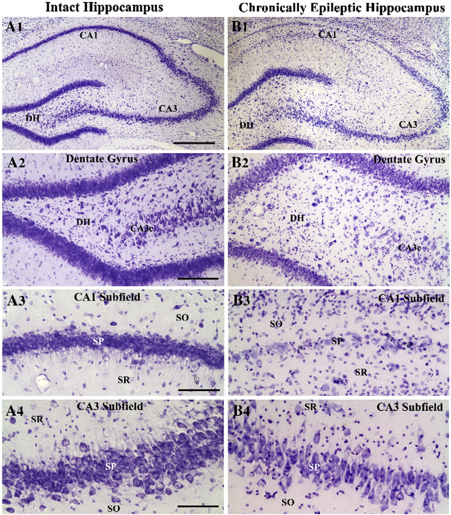
Histological examination of brains at 6 months post-KA injections (i.e. at 2 months post-grafting in animals receiving HFC grafts) demonstrated similar pattern of hippocampal neurodegeneration in all groups. This comprised apparent loss of neurons in the dentate hilus and the CA1 pyramidal cell layer and some intermittent loss of neurons in the CA3 pyramidal cell layer (Fig. 1 [A1–B4]). Thus, the overall hippocampal neurodegeneration in all chronically epileptic rats chosen for different groups in this study is characterized by considerable loss of neurons in the dentate hilus and CA1 pyramidal cell layer and modest loss of neurons in CA3 pyramidal cell layer.
Fig. 1.
Cytoarchitecture of the hippocampus in an intact control rat (A1) and a chronically epileptic rat at 6 months post-status epilepticus (B1). A2–B4 illustrate magnified views of regions of dentate hilus (A2, B2), CA1 subfield (A3, B3) and CA3 subfield (A4 and B4) from A1 and B1. Note that, in comparison to the control hippocampus, the hippocampus of a chronically epileptic rat exhibits considerable loss of neurons in the dentate hilus (DH) and CA1 pyramidal cell layer and moderate loss of neurons in the CA3 pyramidal cell layer. DG, dentate gyrus; DH, dentate hilus; SO, stratum oriens; SP, stratum pyramidale; SR, stratum radiatum. Scale bar, A1 and B1=500 µm; A2 and B2=100 µm; A3, B3, A4, B4=50 µm.
Evolution of chronic epilepsy in epilepsy-only rats and epileptic rats receiving different HFC grafts between 4 and 6 months post-KA injections
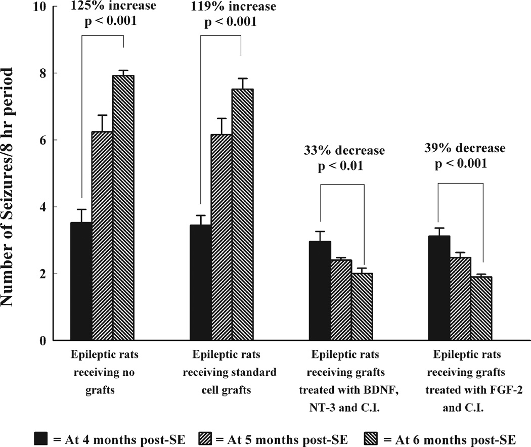
We quantified the frequency of SRMS for 2 months after grafting (equivalent to 4th and 5th month after KA injections), which comprised 8 h of direct observation per week (32 h/month, 64 h in total). The epilepsy-only controls were also scored during the same period for comparison. In epilepsy-only controls, the frequency of SRMS significantly increased between 4 and 6 months post-KA injections (Fig. 2). The overall increase was 76% at 5 months post-KA (p<0.001) and 125% (p<0.001) at 6 months post-KA (Fig. 2). Thus, in the absence of any therapeutic intervention, the extent of chronic epilepsy increases substantially between 4 and 6 months post-KA. In epileptic rats receiving standard HFC grafts, the overall trend of SRMS between 4 and 6 months post-KA injections was very similar to epilepsy-only controls (Fig. 2). The increase in SRMS was 80% at 5 months post-KA (i.e. at 1 month post-grafting; p<0.001) and 119% at 6 months post-KA (i.e. at 2 months post-grafting; p<0.001; Fig. 2). Thus, standard HFC grafts fail to check the evolution of chronic epilepsy as the frequency of SRMS at 5 and 6 months post-KA in epileptic rats receiving HFC grafts paralleled that of epilepsy-only rats.
Fig. 2.
Progression of the frequency of spontaneous recurrent motor seizures (SRMS) in different groups of chronically epileptic rats at 4, 5 and 6 months post-status epilepticus (SE). Note that, in epileptic rats receiving no grafts and epileptic rats receiving standard hippocampal fetal cell (HFC) grafts, the frequency of SRMS increases dramatically during this period. In contrast, in epileptic rats receiving grafts treated with BDNF, NT-3 and caspase inhibitor (CI) or grafts treated with FGF-2 and CI, the frequency of SRMS declines significantly. Comparison of the frequency of SRMS between different groups at 4, 5 and 6 months post-SE reveals that the frequency of SRMS in epileptic rats was similar in all groups at 4 months post-SE. However, at 1–2 months post-grafting, the frequency of SRMS in rats receiving grafts treated with BDNF, NT-3 and CI or grafts treated with FGF-2 and CI is considerably less than the frequency observed in epileptic rats receiving either no grafts or standard HFC grafts.
In contrast, in rats receiving HFC grafts treated and transplanted with BNC or FC, the frequency of SRMS was lower than their pretransplant scores (Fig. 2). In epileptic rats receiving BNC-treated HFC grafts, the reduction of SRMS was 18% at 1 month post-grafting and 33% at 2 months post-grafting (p<0.05; Fig. 2). However, in epileptic rats receiving FC-treated HFC grafts, the reduction was 21% at 1 month post-grafting (p<0.05) and 39% at 2 months post-grafting (p<0.01; Fig. 2). Thus, HFC grafts treated with BNC or FC are efficacious for blocking the progression of epilepsy between 4 and 6 months post-KA injections. In addition, they also considerably decreased the frequency of SRMS present at the time of grafting. Moreover, our qualitative observations suggested that, in the lesion-only group, individual seizures often progressed into stage V seizures throughout the period studied. Similar pattern was seen in animals receiving standard HFC grafts suggesting that standard HFC grafting has no effect on the intensity of seizures. Interestingly, in animals receiving either BNC or FC-treated HFC grafts, individual seizures were more often restricted to stages III and IV (i.e. unilateral forelimb clonus progressing into bilateral forelimb clonus) at 2 months post-grafting, in comparison to individual seizures that often progressed into stage V seizures prior to grafting in the same animals.
Comparison of the frequency of SRMS between epilepsy-only rats and rats receiving different HFC grafts
Comparison of the frequency of SRMS at 5 and 6 months post-KA (i.e. at 1 and 2 months post-grafting in animals receiving transplants) across different groups revealed that animals receiving BNC- or FC-treated HFC grafts exhibit significantly reduced number of SRMS, in comparison to both epilepsy-only rats and epileptic rats receiving standard HFC grafts (Fig. 2). The overall decrease in animals receiving BNC- or FC-treated HFC grafts was 60–62% (p<0.001) at 5 months post-KA and 67–75% (p<0.001) at 6 months post-KA (Fig. 2). Thus, therapeutic intervention in the form of bilateral grafting of HFCs pretreated with BNC or FC into the hippocampi considerably reduces the frequency of SRMS in chronic TLE within a short period after grafting.
Location and cytoarchitecture of grafts in different transplant groups
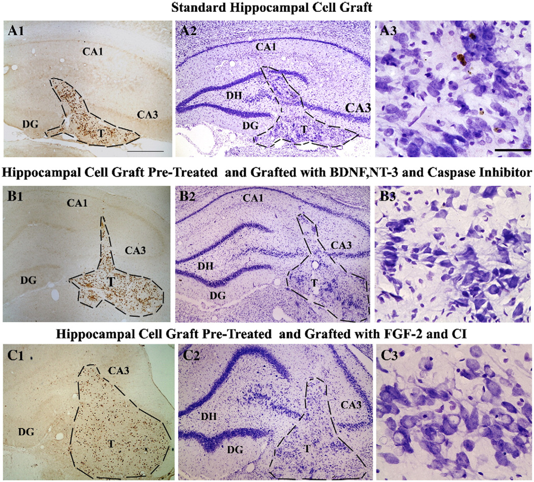
Examination of Nissl stained as well as BrdU immunostained sections from transplanted animals revealed that, in all groups, majority of grafts were located partly inside the hippocampus and partly below the hippocampal CA3 cell layer with projections into the lateral ventricle. However, a minority of grafts were located predominantly in the corpus callosum (i.e. above the CA1 cell layer) or in the lateral ventricle and thalamus. Fig. 3 illustrates examples of grafts from different groups that are located partly inside the hippocampus and partly below the hippocampal CA3 cell layer. The overall area of grafts in individual sections appeared smaller in animals receiving standard HFC grafts (Fig. 3 [A1, A2]), suggesting reduced yield surviving cells and minimal dispersion of grafted cells in this group. In addition, surviving cells and neurons were closely clustered, and sometimes, debris of degenerated tissue could be seen in the core of the transplant mass. In contrast, in both BNC- and FC-treated HFC grafts, the areas of transplants in individual sections as well as the densities of surviving grafted cells and neurons were greater in comparison to standard HFC grafts (Fig. 3 [B1, B2, C1, C2]). Larger CA3 pyramidal-like neurons in these treated transplants were arranged in smaller clusters separated by regions containing smaller neurons (presumably CA1 pyramidal and non-pyramidal neurons), glia and neuropil (Fig. 3 [B3, C3]). Majority of BrdU+ cells within all grafts exhibited dense BrdU immunoreaction product suggesting that transplanted cells did not undergo significant cell division after grafting. Thus, increased yields of surviving cells and neurons were apparent from HFC grafts pretreated and transplanted with BNC or FC in the chronically epileptic hippocampus, in comparison to standard HFC grafts.
Fig. 3.
Location and cytoarchitecture of transplants placed into the chronically epileptic hippocampus at 4 months post-status epilepticus and analyzed at 2 months post-transplantation, visualized with 5-bromodeoxyuridine (BrdU) immunostaining (A1, B1, and C1) and Nissl staining (A2, B2, and C2). In rats receiving standard HFC grafts (A1), the overall graft area in individual sections is smaller (A1, A2) and the transplant core displayed the debris of degenerated tissue (A3). In contrast, in rats receiving grafts treated with BDNF, NT-3 and caspase inhibitor (CI) or FGF-2 and CI, graft areas in individual sections are much larger (B1, B2, C1 and C2) and transplants contain large number of surviving cells and neurons (B1–C3). DG, dentate gyrus; DH, dentate hilus. Scale bar, A1–A2, B1–B2 and C1–C2=500 µm; A3, B3 and C3=100 µm.
Extent of hippocampal injury inflicted by grafting and placement of grafts
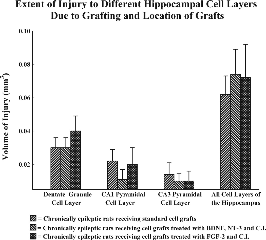
As typical treatment for intractable epilepsy is surgical resection of the hippocampus (which is analogous to considerable hippocampal injury to some extent), we questioned whether greater extent of hippocampal injury exacted by the grafting procedure and intrahippocampal placement of grafts underlies the reduced frequency of SRMS observed in animals receiving BNC and FC-treated transplants, in comparison to animals receiving standard grafts exhibiting SRMS that are equivalent to epilepsy-only controls. Measurement of hippocampal injury using Neurolucida and serial Nissl stained sections through the entire hippocampus demonstrated that the transplantation procedure and placement of grafts inside the hippocampus cause some damage to different hippocampal cell layers. However, the extent of damage to different cell layers inflicted by transplants was similar across the three transplant groups when compared for both individual hippocampal cell layers and the entire hippocampus (Fig. 4). As similar damage in epileptic rats receiving standard grafts does not result in reduced frequency of SRMS, it is clear that diminished frequency of SRMS observed in animals receiving BNC or FC-treated transplants is linked to the presence of these transplants and not to the extent of hippocampal injury inflicted by grafts.
Fig. 4.
Comparison of the extent of injury in different hippocampal cell layers and the entire hippocampus inflicted by the grafting procedure and placement of grafts inside the hippocampus. The bars denote the volumes of cell layers that are damaged. Note that the extent of injury is similar between the three transplant groups for all cell layers of the hippocampus as well as for the entire hippocampus.
Neuronal phenotypes within transplants
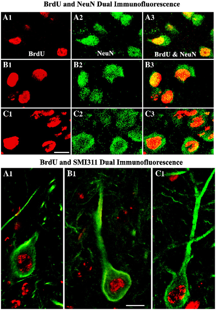
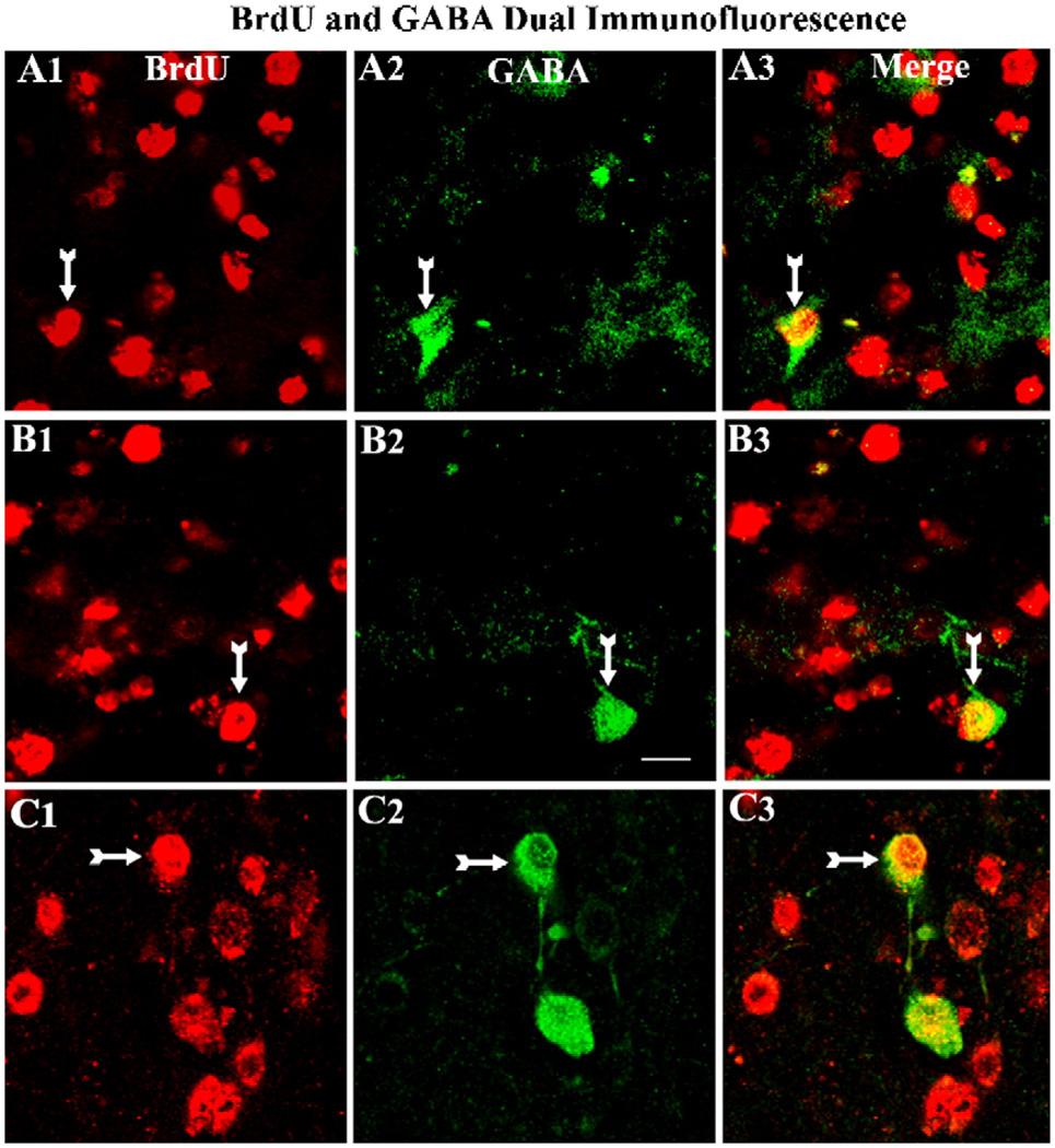
Examination of grafted cells using BrdU and NeuN dual immunofluorescence and confocal microscopy demonstrated the presence of neurons within grafts in all transplant groups (Fig. 5 [upper panels]). Grafted cells that were immunoreactive for both BrdU and NeuN displayed BrdU immunofluorescence in the nucleus and NeuN expression in both nucleus and cytoplasm of the cell body (Fig. 5). Quantification of the percentages of BrdU+ grafted cells expressing NeuN in different groups demonstrated that great majority of grafted cells are neurons in all groups (standard HFC grafts, 90.4±1.1%; BNC-treated HFC grafts, 92.9±0.5%; FC-treated HFC grafts, 90.8±0.7). Although BrdU+ cells counted at the time of grafting likely comprised both neuronal and glial precursors, E19 hippocampi (the age of hippocampi used in this study for collecting donor cells) mostly comprise neuronal precursors based on earlier cell culture analyses (Shetty et al., 1994; Zaman et al., 2000). This may be one of the reasons for most of the BrdU cells encountered within grafts are neurons. In addition, neuronal precursors (i.e. cells from E19 hippocampus) being post-mitotic at the time of grafting retain the BrdU label permanently, whereas glia (even though fewer in number at the time of grafting) likely divide after grafting and lose their BrdU label over time. Examination of grafted cells using BrdU and NPNFP dual immunofluorescence and confocal microscopy demonstrated the presence of CA3 pyramidal-like neurons within grafts in all transplant groups (Fig. 5 [lower panel]). The percentages of such neurons among BrdU+ grafted cells varied from 25 to 27% (standard HFC grafts, 27.0±2.0%; BNC-treated HFC grafts, 24.6±1.7%; FC-treated HFC grafts, 26.8±1.2%). Assessment of GABA-ergic neurons via BrdU and GABA double immunofluorescence methods and confocal microscopy revealed the occurrence of GABA-ergic interneurons within grafts in all transplant groups (Fig. 6). The fractions of GABA-ergic interneurons among BrdU+ grafted cells ranged from 8 to 10% (standard HFC grafts, 7.9±0.4%; BNC-treated HFC grafts, 9.1±0.9%; FC-treated HFC grafts, 10.1±0.7%).
Fig. 5.
Differentiation of grafted cells into neurons (upper panel) and CA3 pyramidal-like neurons (lower panel). The upper panel (A1–C3) illustrates confocal images of grafted neurons that are positive for 5′-bromodeoxyuridine (BrdU) and neuron-specific nuclear antigen (NeuN) in standard hippocampal fetal cell (HFC) grafts (A1–A3), HFC grafts treated with BDNF, NT-3 and caspase inhibitor (B1–B3) and HFC grafts treated with FGF-2 and caspase inhibitor (C1–C3). Note that a vast majority of transplanted cells differentiate into NeuN positive neurons in all groups. The lower panel shows representative confocal images of grafted neurons that are positive for BrdU and a CA3 pyramidal neuron marker (the non-phosphorylated neurofilament protein) from standard HFC grafts (A1), HFC grafts treated with BDNF, NT-3 and caspase inhibitor (B1) and HFC grafts treated with FGF-2 and caspase inhibitor (C1). Scale bar, upper panel (A1–C3) = 10 µm; lower panel (A1–C1)=10 µm.
Fig. 6.
Differentiation of grafted cells into GABA-ergic interneurons. A1–C3 illustrate confocal images of grafted neurons that are positive for 5′-bromodeoxyuridine (BrdU) and GABA (arrows) in standard hippocampal fetal cell (HFC) grafts (A1–A3), HFC grafts treated with BDNF, NT-3 and caspase inhibitor (B1–B3) and HFC grafts treated with FGF-2 and caspase inhibitor (C1–C3). Note that only smaller fractions of transplanted cells differentiate into GABA-ergic interneurons in all groups. Scale bar, 10 µm.
Yields of surviving cells and neurons from standard and treated grafts
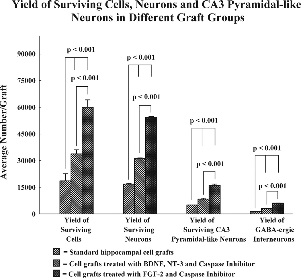
We transplanted 60,000 live cells into each of the 4 locations on both sides of the hippocampus in all transplant groups. However, based on the BrdU labeling index (92.1%), the average number of BrdU-labeled live cells implanted per site is 55,260. We quantified the total yield of surviving grafted cells (i.e. BrdU+ cells) in individual transplants belonging to different groups (n=5/group) using serial sections (every 10th) immunostained for BrdU and the optical fractionator cell counting method. This revealed significant variation in the yield of surviving grafted cells between different transplant groups. The number of BrdU+ recovered per transplant was 18,590±4000 in standard HFC grafts, 33,680±2321 in BNC-treated HFC grafts and 59,885±4216 in FC-treated HFC grafts (Fig. 7). In comparison to the number of BrdU-labeled cells initially grafted (i.e. 55,260 live cells), the average yield of surviving grafted cells was equivalent to 34% of injected cells in standard HFC grafts, 61% of injected cells in BNC-treated HFC grafts and 108% of injected cells in FC-treated HFC grafts.
Fig. 7.
Yields of 5′-bromodeoxyuridine (BrdU) positive surviving cells, neuron-specific nuclear antigen (NeuN) positive neurons, CA3 pyramidal-like (non-phosphorylated neurofilament protein positive) neurons and GABA-ergic interneurons in different graft groups. Note that the yields of surviving grafted cells are much greater when they are pretreated and transplanted with a combination of either BDNF, NT-3 and caspase inhibitor (BNC) or FGF-2 and caspase inhibitor (FC), in comparison to grafted cells that are transplanted with no pretreatment (i.e. standard hippocampal cell grafts). The overall yields of surviving neurons, CA3 pyramidal-like neurons and GABA-ergic interneurons are also substantially higher in grafts pretreated and transplanted with either BNC or FC than in standard hippocampal cell grafts. In addition, between the two treated groups (i. e. BNC and FC groups), the yields of surviving cells, neurons, CA3-pyramidal-like neurons and GABA-ergic interneurons are much greater when donor cells were pretreated and grafted with FC.
Through extrapolation of the absolute numbers of BrdU+ cells recovered from different grafts with the percentages of NeuN expressing neurons among BrdU+ cells (90–93%), we obtained the yield of neurons in different transplant groups. The average yield of neurons per graft was 16,797±208 in standard HFC grafts, 31,313±182 in BNC-treated grafts and 54,352±419 in FC-treated grafts (Fig. 7). In comparison to the number of BrdU-labeled cells initially grafted (i.e. 55,260 live cells), the yield of surviving neurons was equivalent to 30% of injected cells in standard HFC grafts, 57% in HFC grafts treated with BNC and 98% in HFC grafts treated with FC. Furthermore, through extrapolation of the absolute numbers of BrdU+ cells recovered from different grafts with the percentages NPNFP expressing neurons among BrdU+ cells (24–27%), we obtained the yield of CA3 pyramidal-like neurons in different transplant groups (Fig. 7). The average yield of CA3 pyramidal-like neurons per graft was 5015±37 in standard HFC grafts, 8299±573 in BNC-treated grafts and 16,049±719 in FC-treated grafts (Fig. 7). In comparison to the number of BrdU-labeled cells initially grafted (i.e. 55,260 live cells), the average yield of surviving CA3 pyramidal-like neurons was equivalent to 9% of injected cells in standard HFC grafts, 15% of injected cells in BNC-treated HFC grafts and 29% of injected cells in FC-treated HFC grafts. Additionally, extrapolating the total numbers of BrdU+ cells recovered from different transplants with fractions of GABA-ergic interneurons among BrdU+ cells (8–10%), we obtained the yield of GABA-ergic interneurons in different transplant groups (Fig. 7). The average yield of GABA-ergic interneurons per graft was 1469±15 in standard HFC grafts, 3031±21 in BNC-treated grafts and 6060±30 in FC-treated grafts (Fig. 7). In comparison to the number of BrdU-labeled cells initially grafted (i.e. 55,260 live cells), the average yield of surviving CA3 pyramidal-like neurons was equivalent ~3% of injected cells in standard HFC grafts, ~6% of injected cells in BNC-treated HFC grafts and ~11% of injected cells in FC-treated HFC grafts. Collectively, our results demonstrate that the overall yields of surviving grafted cells, neurons, CA3 pyramidal-like neurons and GABA-ergic interneurons are considerably greater when HFCs are pretreated and grafted with BNC or FC. Of the two graft augmentation strategies, FC treatment is more efficacious than BNC treatment however.
Discussion
This study provides the first evidence for the ability of appropriately treated HFC grafts to restrain SRMS in a rat model of chronic TLE. Specifically, chronically epileptic rats receiving HFC grafts pretreated and transplanted with either BNC or FC into their hippocampi exhibited greater numbers of graft-derived neurons and GABA-ergic inhibitory interneurons in hippocampi and reduced frequency of SRMS. In sharp contrast, chronically epileptic rats receiving standard HFC grafts into their hippocampi displayed fewer graft-derived neurons and GABA-ergic interneurons in hippocampi and a frequency of SRMS that was comparable to epilepsy-only controls. Furthermore, as the extent of damage to different hippocampal cell layers inflicted by the grafting procedure and location of grafts was similar in the three transplant groups, it appears that diminished frequency of SRMS observed in animals receiving BNC or FC-treated transplants is linked to the addition of a large number of new neurons (including GABA-ergic interneurons) from these grafts to chronically epileptic hippocampi. These results have implications for developing a suitable cell transplantation therapy for reducing SRMS in patients with drug-resistant TLE.
Importance of the chosen model and grafting strategies for studying the anti-seizure effects of HFC grafts
A series of grafting studies have been conducted earlier in the hippocampus of rats injured via unilateral intracerebroventricular (ICV) KA, a model of focal hippocampal CA3 region injury and milder TLE, using different types of fetal hippocampal cells (mixed hippocampal, CA3 or CA1 cells) as donor cells. These studies, performed at early time-points after the injury, have demonstrated excellent graft cell survival, appropriate differentiation of grafted cells and correct integration of grafted neurons into the host hippocampal circuitry (Shetty and Turner, 1995a, 1997a; Zaman et al., 2000; Shetty et al., 2000). Grafts that are specific to the injured hippocampal region (such as grafts containing CA3 cells) were also efficacious for reversing several pathological consequences of hippocampal injury. These include suppression/reversal of aberrant sprouting of host mossy fibers into the dentate supragranular layer by providing appropriate target neurons and specific afferent innervation to the inner molecular layer of the dentate gyrus (Shetty and Turner, 1997b; Shetty et al., 2005; Hattiangady et al., 2006), normalization of the numbers of GABA synthesizing interneurons in different regions of the hippocampus likely through graft-mediated afferent innervation and trophic support to deactivated host interneurons (Shetty and Turner, 2000, 2001) and re-expression of the calcium binding protein calbindin in dentate granule cells and CA1 pyramidal cells possibly via suppression of hippocampal hyperexcitability (Shetty and Hattiangady, in press). Nevertheless, the potential therapeutic benefits of HFC grafting in terms of reducing the frequency of seizures could not be evaluated in the above studies as animals treated with unilateral ICV KA do not exhibit significant SRMS (Hattiangady et al., 2004). As a result, an issue that remained unanswered was whether the structural repair accomplished through HFC grafting would have any consequence on the evolution of chronic epilepsy that typically follows hippocampal injury or SE (Shetty et al., 2005). Moreover, in the studies discussed above, grafting was performed relatively early after the hippocampal injury (i.e. at 4 or 45 days post-injury). When grafting was performed at 2 or 4 months after injury, there was a steep decline in the yield of surviving cells from HFC grafts, necessitating the adoption of graft augmentation strategies such as pretreatment and grafting of donor cells with specific neurotrophic factors or neurotrophic factors and CI (Zaman and Shetty, 2003b; Hattiangady et al., 2006). Although it is not clear when exactly hippocampal cell loss occurs in TLE patients, it is highly probable that any cell therapy will be considered relatively late in the disease process, and certainly after the onset of chronic seizures (Nilsen and Cock, 2004). Furthermore, at this time, the host hippocampus is likely afflicted with extensive abnormal synaptic reorganization and changes in microenvironment which may be detrimental for survival of grafted cells (Turner and Shetty, 2003).
Additionally, the thought of HFC therapy for reducing or suppressing epilepsy is somewhat contentious in view of observations in earlier studies that solid tissue or cell suspension grafts from E15/E16 hippocampi exhibit increased excitability and spontaneous seizures when placed into the hippocampi that are deafferented through fimbria-fornix lesions (Buzsaki et al., 1989, 1991). Thus, a number of concerns currently exist regarding the effectiveness of neural grafting therapy for chronic epilepsy, particularly in comparison to the established efficacy of surgical resection of hippocampal foci in TLE patients (Turner and Shetty, 2003; Nilsen and Cock, 2004). Considering these, we carefully chose host rats that are chronically epileptic at the time of grafting to facilitate the evaluation of anti-seizure effects of HFC grafts in this study. Furthermore, as the yield of surviving cells from grafts placed into the hippocampus is generally poor when grafts are placed at extended time-points after the injury, we employed three different grafting strategies for evaluating the yields of surviving cells and neurons and anti-seizure effects of HFC grafts in this study. The findings in our recent study that HFC grafts treated with appropriate neurotrophic factors and CI give rise to increased yields of surviving cells and neurons and disentangle some of the epileptogenic circuitry such as the aberrant mossy fiber sprouting in the chronically injured hippocampus prompted us to use similar graft augmentation strategies in this study as well (Hattiangady et al., 2006).
Relationship between the progression of chronic epilepsy and the extent of graft cell survival
Characterization of the frequency of SRMS in epilepsy-only control rats revealed that, in the absence of any therapeutic intervention, the extent of chronic epilepsy progressively increases between 4 and 6 months post-SE. The increase was 76% at 5 months post-SE and 125% at 6 months post-SE. Interestingly, grafting of standard HFCs into the hippocampus at 4 months post-SE does not have any effect on the progression of chronic epilepsy as the extent of increase (80–119%) in the frequency of SRMS at 4–6 months post-SE in this group was comparable to the frequency observed in epilepsy-only control rats. In contrast, HFC grafts treated with BNC or FC were efficacious not only for blocking the progression of chronic epilepsy but also for reducing the frequency of seizures that were present at the time of grafting. In comparison to both epilepsy-only controls and chronically epileptic rats receiving standard HFC grafts, the frequency of SRMS in chronically epileptic rats receiving BNC- or FC-treated HFC grafts was 60–62% less at 5 months post-SE and 67–75% less at 6 months post-SE. When compared to the status at the time of grafting, the frequency of SRMS exhibited 18–21% reduction at 1 month post-grafting and 33–39% reduction at 2 months post-grafting in chronically epileptic animals receiving BNC- and FC-treated grafts.
Histological analyses at 2 months post-grafting revealed that graft-mediated reductions in the frequency of SRMS in chronically epileptic rats were linked to the presence of large numbers of graft-derived neurons as epileptic animals that received either BNC- or FC-treated HFC grafts exhibited greater numbers of graft-derived new neurons in the hippocampus (equivalent to 57–98% of injected cells) in contrast to animals that received standard HFC grafts where graft-derived neurons were fewer (equivalent to 30% of injected cells) in the hippocampus. Based on the yield of surviving cells, neurons among surviving cells and the number of grafts placed per hippocampus, the number of new neurons added to each epileptic hippocampus was ~ 67,000 through standard HFC grafts, ~125,000 through HFC grafts treated with BNC and ~217,000 through HFC grafts treated with FC in this study. Likewise, the number of graft-derived GABA-ergic interneurons added to each of the epileptic hippocampus was ~6000 through standard HFC grafts, ~12,000 through HFC grafts treated with BNC and ~24,000 through HFC grafts treated with FC. Thus, grafts treated with BNC or FC gave rise to greater numbers of new neurons and GABA-ergic interneurons to the chronically epileptic hippocampus. However, between the two treated groups, the yield of neurons (including GABA-ergic interneurons) was greater from grafts treated with FC than grafts treated with BNC. This finding differs from our previous grafting study where the yield of neurons derived from grafts placed into the hippocampus at 4 months post-injury was robust and highly similar between grafts treated with BNC and grafts treated with FC (Hattiangady et al., 2006). The discrepancy between the two studies pertaining to the yield of neurons from grafts treated with BNC likely reflects the effects of SRMS on neuron survival within BNC-treated grafts because our previous study used an injury model of hippocampus with minimal SRMS whereas our current study utilized a model that exhibits significant SRMS (Hattiangady et al., 2004). Overall, these results suggest that treatment of donor HFCs with FC is highly efficacious for enhancing the yields of neurons and GABA-ergic interneurons from grafts placed into the chronically epileptic hippocampus.
From the above, it also appears that addition of a critical number (~125,000) of new neurons and GABA-ergic interneurons (~12,000) to each of the epileptic hippocampus is necessary for inducing anti-seizure effects through HFC grafting. Any further increase in the addition of new neurons and GABA-ergic interneurons does not seem to enhance the anti-seizure effects because the overall reductions in the frequency of seizures were similar between animals that received grafts treated with BNC (amounting to addition of ~125,000 new neurons and ~12,000 new GABA-ergic interneurons to each hippocampus) and animals that received grafts treated with FC (amounting to addition of ~217,000 new neurons and ~24,000 new GABA-ergic interneurons to each hippocampus). However, additional studies at prolonged time-points after grafting are required to clearly assess differences between these two transplant groups for their ability to restrain seizures on a long-term basis. Nevertheless, considerable survival of grafted neurons in the hippocampus appears to be an important prerequisite for reducing the frequency of SRMS following grafting. Indeed, an earlier grafting study has shown complete failure to diminish seizures in a kainate model of TLE with grafting of 40,000–80,000 Layton Bioscience (LBS) neurons per hippocampus (Hasegawa et al., 2004). The donor cells in this study were derived from the NT2/D1 human precursor cell line and were induced to differentiate into neurons prior to grafting using retinoic acid. Although the quantification of survival of grafted neurons was not done rigorously in this study, histological analyses suggested poor survival in the hippocampus of rats immunosuppressed with cyclosporine A (Hasegawa et al., 2004).
Reduced yields of surviving cells and neurons from standard HFC grafts are not surprising as our earlier studies have shown that the microenvironment of the hippocampus is less conducive for supporting the survival of immature fetal cells at extended post-lesion time-points (Zaman and Shetty, 2003b; Hattiangady et al., 2006). Diminished conduciveness of the milieu of chronically lesioned hippocampus likely reflects reduced concentration of neurotrophic factors such as BDNF, FGF-2 and insulin-like growth factor-1 (Shetty et al., 2003; Hattiangady et al., 2004) and lack of adequate vacant synaptic sites due to resolution of neosynaptogenesis at 4 months after injury or SE (Shetty, 2002; Shetty et al., 2003, 2005). Improved survival of HFC grafts treated with BNC or FC also supports the above possibility. The selection of combined neurotrophic supplementation (BDNF and NT-3 or FGF-2) and caspase inhibition for improving the yields of surviving cells and neurons in the hippocampus of chronically epileptic rats was based on results of both our earlier study (Hattiangady et al., 2006) and many other studies on graft augmentation in acutely injured adult or aged brain (Schierle et al., 1999; Hansson et al., 2000; Mundt-Petersen et al., 2000; Helt et al., 2001; Cicchetti et al., 2002; Zaman and Shetty, 2002a, 2003a,b; Karlsson et al., 2005; Rao et al., 2006b). Considering the functions of neurotrophic factors (BDNF, NT-3 and FGF-2) and CI, it appears that enhanced yields of surviving grafted cells and neurons observed after combined neurotrophic supplementation and caspase inhibition are a result of rapid differentiation of grafted cells and rescue of grafted cells from grafting trauma-related apoptosis at early post-grafting periods (Maisonpierre et al., 1990; Friedman et al., 1991; Ernfors et al., 1992; Friedman et al., 1998; Zawada et al., 1998; Sortwell et al., 2001). Furthermore, though both graft augmentation strategies significantly improved the yields of surviving cells and neurons from grafts, the overall yields were much greater when donor cells were treated and grafted with FGF-2 and CI than when treated and grafted with BDNF, NT-3 and CI. It is plausible that, at the concentrations used in this study, the FGF-2 has greater ability for providing neuroprotection and inducing rapid neuronal differentiation of immature hippocampal neurons than BDNF and NT-3. This is not astounding since the ability of FGF-2 to support the survival and differentiation of developing hippocampal neurons under multiple adverse conditions is well documented (Eves et al., 2001; Tretter et al., 2000; Szebenyi et al., 2001). Thus, pretreatment and grafting of donor HFCs with FGF-2 and CI appear ideal for augmenting the addition of new neurons (including GABA-ergic interneurons) into the chronically epileptic hippocampus via HFC grafts.
Significance and prospective mechanisms of reduced frequency of SRMS in rats receiving HFC grafts treated with BNC or FC
The ability to restrain seizures through grafting of different types of cells into different regions of the brain has been observed in earlier studies. However, the long-term efficacy of these cell grafts for reducing chronic seizures was not clear because of analyses of seizures only during the early post-grafting period or the application of grafting prior to the occurrence of spontaneous seizures. Thompson and Suchomelova (2004) bilaterally grafted conditionally immortalized neurons engineered to produce GABA into the substantia nigra of rats at 45–65 days after the SE induced by lithium-pilocarpine and measured the effects on spontaneous seizures at 1–10 days post-grafting. They found that animals that received GABA-producing cells exhibited fewer spontaneous seizures than animals that received only control cells but the survival of grafted GABA-producing cells was limited. Although these results suggest that cells genetically engineered to secrete GABA have the ability to diminish spontaneous seizures when transplanted into seizure-modulating nuclei, long-term efficacy is uncertain because of poor survival of grafted cells in the host brain. The effects of GABA-producing cells on the development of seizures were also examined following grafting into the dentate gyrus (Thompson, 2005) or the substantia nigra (Castillo et al., 2006). Grafting of these cells raised GABA levels, elevated the local electrical seizure threshold and slowed the development of behavioral seizures in the kindling model of epilepsy (Thompson, 2005) and decreased the KA-induced seizures (Castillo et al., 2006). However, the approaches employed in these studies are not clinically practicable as grafting was done prior to the induction of seizures.
Another series of experiments have examined the effects of grafts of encapsulated fibroblasts engineered to release the brain’s endogenous anticonvulsant adenosine on seizures (Boison et al., 1999, 2002; Huber et al., 2001; Boison, 2005). Grafting of these cells into the ventricles of rats kindled in the hippocampus resulted in reduced behavioral seizures and after discharges. However, this positive effect did not extend beyond 2 weeks after grafting because of the poor survival of fibroblasts. The efficacy of grafts of mouse myoblasts engineered to release adenosine was also examined for long-term reductions in seizure frequency (Fedele et al., 2004; Guttinger et al., 2005). Although reduced seizures were maintained for a period of 3 weeks in 50% of the animals in this study, long-term efficacy of these transplants for reducing the frequency of chronic seizures remains uncertain. Overall, the transient nature of positive effects in the above studies suggests that either the grafted cells fail to exhibit enduring survival in an ectopic location or the amount of GABA/adenosine released by these cells becomes inadequate over time. Thus, studies on long-term effects of various cell transplants placed into the hippocampus of chronically epileptic animals are critical to gauze the usefulness of distinct grafting approaches for treating chronic drug-resistant epilepsy. Additionally, cell transplantation may be more favorable than single neurotrophic factor delivery for treating chronic epilepsy because grafted neurons that are specific to the injured area may secrete multitude of beneficial trophic factors, in addition to providing additional synapses and facilitating the repair of disrupted circuits (Shetty et al., 2000, 2005; Hattiangady et al., 2006). In this context, our current finding that appropriately treated HFC grafts placed into the hippocampi are efficacious for reducing the frequency of SRMS for up to 2 months in a rat model of chronic TLE is significant. As the frequency of SRMS progressively declined during the two post-grafting months examined in this study, it is likely that the frequency of SRMS may decline further with increasing post-grafting time. Therefore, long-term analyses following grafting are needed in the future to validate the usefulness of this approach for treating chronic TLE.
The mechanisms by which HFC grafts treated with BNC or FC reduce the frequency of seizures are not examined in this study. However, based on both improved yields of surviving grafted neurons (including GABA-ergic interneurons) with these strategies and the relationship between the extent of addition of graft-derived new neurons and reduced SRMS, it is likely that multiple changes mediated by HFC grafts underlie their anti-seizure effects. These may include reconstruction of the disrupted hippocampal circuitry by the CA1 and CA3 pyramidal neurons in grafts, strengthening of GABA-ergic neurotransmission in the host hippocampus through new inhibitory synapses on host principal neurons by the axons of GABA-ergic neurons in grafts, reactivation of GABA-deficient host interneurons through afferent innervation and trophic support from grafted CA3 pyramidal neurons, and reversal of aberrant sprouting of mossy fibers via diversion of host mossy fibers towards grafted CA3 pyramidal neurons. Indeed, our earlier grafting studies in the ICV KA model of hippocampal injury have shown the ability of fetal hippocampal neurons for inducing most of the above changes (Shetty and Turner, 1997a,b, 2000; Shetty et al., 2000, 2005; Hattiangady et al., 2006). However, to clearly ascertain the mechanisms of reduced frequency of seizures mediated by grafts in this model, detailed correlative analyses between the extent/intensity of SRMS and the degree of graft axon growth, graft–host synaptic connectivity, changes in aberrant mossy fiber sprouting, GABA-ergic interneuron numbers in different regions of the host hippocampus and grafts at extended time-points after grafting are needed in the future.
Conclusions and future directions
This study demonstrates for the first time that appropriately treated HFC grafts have the ability to restrain seizures in conditions such as chronic epilepsy. These results are promising towards developing cell therapy for suppressing spontaneous seizures in patients with drug-resistant epilepsy. However, ethical concerns and problems associated with obtaining large amounts of human fetal tissues preclude the routine use of fetal cells as donor cells for grafting in neurodegenerative diseases afflicting humans (Shetty and Turner, 1996; Turner and Shetty, 2003). Therefore, alternative sources of neural cells that allow both maintenance and expansion in vitro for prolonged periods and exhibit characteristics of primary fetal neurons with regard to neuronal differentiation, and structural and functional integration into the host following grafting are essential. From this perspective, multipotent stem cells derived from different regions of the brain including the hippocampus (Shetty and Turner, 1998, 1999; Shetty, 2004; Chu et al., 2004) and bone marrow derived multipotent progenitor cells (Yasuhara et al., 2006) may be useful. However, stem/progenitor cell grafting studies in neurodegenerative disease models are still in infancy and the efficacy of such grafts for repairing the disrupted circuitry and promoting long-term functional recovery in conditions such as epilepsy is unknown at this time but clearly needs to be addressed in future studies.
Acknowledgments
This research was supported by grants from the National Institute of Neurological Disorders and Stroke (RO1 NS 043507 and RO1 NS 54780 to A.K.S.) and Department of Veterans Affairs (VA Merit Review Award to A.K.S.). We thank Dr. Bing Shuai for excellent technical assistance in this study.
References
- Altman J, Bayer SA. Mosaic organization of the hippocampal neuroepithelium and the multiple germinal sources of dentate granule cells. J. Comp. Neurol. 1990;301:325–342. doi: 10.1002/cne.903010302. [DOI] [PubMed] [Google Scholar]
- Ang CW, Carlson GC, Coulter DA. Massive and specific dysregulation of direct cortical input to the hippocampus in temporal lobe epilepsy. J. Neurosci. 2006;26:11850–11856. doi: 10.1523/JNEUROSCI.2354-06.2006. [DOI] [PMC free article] [PubMed] [Google Scholar]
- Ben-Ari Y. Limbic seizure and brain damage produced by kainic acid: mechanisms and relevance to human temporal lobe epilepsy. Neuroscience. 1985;14:375–403. doi: 10.1016/0306-4522(85)90299-4. [DOI] [PubMed] [Google Scholar]
- Boison D. Adenosine and epilepsy: from therapeutic rationale to new therapeutic strategies. Neuroscientist. 2005;11:25–36. doi: 10.1177/1073858404269112. [DOI] [PubMed] [Google Scholar]
- Boison D, Scheurer L, Tseng JL, Aebischer P, Mohler H. Seizure suppression in kindled rats by intraventricular grafting of an adenosine releasing synthetic polymer. Exp. Neurol. 1999;160:164–174. doi: 10.1006/exnr.1999.7209. [DOI] [PubMed] [Google Scholar]
- Boison D, Huber A, Padrun V, Deglon N, Aebischer P, Mohler H. Seizure suppression by adenosine-releasing cells is independent of seizure frequency. Epilepsia. 2002;43:788–796. doi: 10.1046/j.1528-1157.2002.33001.x. [DOI] [PubMed] [Google Scholar]
- Brewer GJ, Torricelli JR, Evege EK, Price PJ. Optimized survival of hippocampal neurons in B27-supplemented Neurobasal, a new serum-free medium combination. J. Neurosci. Res. 1993;35:567–576. doi: 10.1002/jnr.490350513. [DOI] [PubMed] [Google Scholar]
- Buckmaster PS, Zhang GF, Yamawaki R. Axon sprouting in a model of temporal lobe epilepsy creates a predominantly excitatory feedback circuit. J. Neurosci. 2002;22:6650–6658. doi: 10.1523/JNEUROSCI.22-15-06650.2002. [DOI] [PMC free article] [PubMed] [Google Scholar]
- Buzsaki G, Ryan JP, Isaacson RL. The effects of unilateral destruction of fimbria-fornix and supracallosal pathways in the rat. Behav. Neural Biol. 1989;51:278–288. doi: 10.1016/s0163-1047(89)90932-1. [DOI] [PubMed] [Google Scholar]
- Buzsaki G, Hsu M, Slamka C, Gage FH, Horvath Z. Emergence and propagation of interictal spikes in the subcortically denervated hippocampus. Hippocampus. 1991;1:163–180. doi: 10.1002/hipo.450010205. [DOI] [PubMed] [Google Scholar]
- Castillo CG, Mendoza S, Freed WJ, Giordano M. Intranigral transplants of immortalized GABAergic cells decrease the expression of kainic acid-induced seizures in the rat. Behav. Brain Res. 2006;171:109–115. doi: 10.1016/j.bbr.2006.03.025. [DOI] [PubMed] [Google Scholar]
- Cendes F. Febrile seizures and mesial temporal sclerosis. Curr. Opin. Neurol. 2004;17:161–164. doi: 10.1097/00019052-200404000-00013. [DOI] [PubMed] [Google Scholar]
- Chu K, Kim M, Jung KH, Jeon D, Lee ST, Kim J, Jeong SW, Kim SU, Lee SK, Shin HS, Roh JK. Human neural stem cell transplantation reduces spontaneous recurrent seizures following pilacarpine-induced status epilepticus in adult rats. Brain Res. 2004;1023:213–221. doi: 10.1016/j.brainres.2004.07.045. [DOI] [PubMed] [Google Scholar]
- Cicchetti F, Costantini L, Belizaire R, Burton W, Isacson O, Fodor W. Combined inhibition of apoptosis and complement improves neural graft survival of embryonic rat and porcine mesencephalon in the rat brain. Exp. Neurol. 2002;177:376–384. doi: 10.1006/exnr.2002.8007. [DOI] [PubMed] [Google Scholar]
- Dalby NO, Mody I. The process of epileptogenesis: a pathophysiological approach. Curr. Opin. Neurol. 2001;14:187–192. doi: 10.1097/00019052-200104000-00009. [DOI] [PubMed] [Google Scholar]
- Detour J, Schroeder H, Desor D, Nehlig A. A 5-month period of epilepsy impairs spatial memory, decreases anxiety, but spares object recognition in the lithium-pilocarpine model in adult rats. Epilepsia. 2005;46:499–508. doi: 10.1111/j.0013-9580.2005.38704.x. [DOI] [PubMed] [Google Scholar]
- Devinsky O. Diagnosis and treatment of temporal lobe epilepsy. Rev. Neurol. Dis. 2004;1:2–9. [PubMed] [Google Scholar]
- Engel J., Jr Classifications of the international league against epilepsy: time for reappraisal. Epilepsia. 1998;39:1014–1017. doi: 10.1111/j.1528-1157.1998.tb01453.x. [DOI] [PubMed] [Google Scholar]
- Engel J., Jr Intractable epilepsy: definition and neurobiology. Epilepsia. 2001;42(Suppl. 6):3. doi: 10.1046/j.1528-1157.2001.0420s6003.x. [DOI] [PubMed] [Google Scholar]
- Ernfors P, Merlio JP, Persson H. Cells expressing mRNA for neurotrophins and their receptors during embryonic rat development. Eur. J. Neurosci. 1992;4:1140–1158. doi: 10.1111/j.1460-9568.1992.tb00141.x. [DOI] [PubMed] [Google Scholar]
- Eves EM, Skoczylas C, Yoshida K, Alnemri ES, Rosner MR. FGF induces a switch in death receptor pathways in neuronal cells. J. Neurosci. 2001;21:4996–5006. doi: 10.1523/JNEUROSCI.21-14-04996.2001. [DOI] [PMC free article] [PubMed] [Google Scholar]
- Fedele DE, Koch P, Scheurer L, Simpson EM, Mohler H, Brustle O, Boison D. Engineering embryonic stem cell derived glia for adenosine delivery. Neurosci. Lett. 2004;370:160–165. doi: 10.1016/j.neulet.2004.08.031. [DOI] [PubMed] [Google Scholar]
- Fisher PD, Sperber EF, Moshe SL. Hippocampal sclerosis revisited. Brain Dev. 1998;20:563–573. doi: 10.1016/s0387-7604(98)00069-2. [DOI] [PubMed] [Google Scholar]
- French JA, Williamson PD, Thadani VM, Darcey TM, Mattson RH, Spencer SS, Spencer DD. Characteristics of medial temporal lobe epilepsy: I. Results of history and physical examination. Ann. Neurol. 1993;34:774–780. doi: 10.1002/ana.410340604. [DOI] [PubMed] [Google Scholar]
- Friedman WJ, Olson L, Persson H. Cells that express brain-derived neurotrophic factor mRNA in the developing postnatal rat brain. Eur. J. Neurosci. 1991;3:688–697. doi: 10.1111/j.1460-9568.1991.tb00854.x. [DOI] [PubMed] [Google Scholar]
- Friedman WJ, Black IB, Kaplan DR. Distribution of the neurotrophins brain-derived neurotrophic factor, neurotrophin-3, and neurotrophin-4/5 in the postnatal rat brain: an immunocytochemical study. Neuroscience. 1998;84:101–114. doi: 10.1016/s0306-4522(97)00526-5. [DOI] [PubMed] [Google Scholar]
- Guttinger M, Padrun V, Pralong WF, Boison D. Seizure suppression and lack of adenosine A1 receptor desensitization after focal long-term delivery of adenosine by encapsulated myoblasts. Exp. Neurol. 2005;193:53–64. doi: 10.1016/j.expneurol.2004.12.012. [DOI] [PubMed] [Google Scholar]
- Hansson O, Castilho RF, Kaminski Schierle GS, Karlsson J, Nicotera P, Leist M, Brundin P. Additive effects of caspase inhibitor and lazaroid on the survival of transplanted rat and human embryonic dopamine neurons. Exp. Neurol. 2000;164:102–111. doi: 10.1006/exnr.2000.7406. [DOI] [PubMed] [Google Scholar]
- Harvey AS, Berkovic SF, Wrennall JA, Hopkins IJ. Temporal lobe epilepsy in childhood: clinical EEG, neuroimaging findings and syndrome classification in a cohort with new-onset seizures. Neurology. 1997;49:960–968. doi: 10.1212/wnl.49.4.960. [DOI] [PubMed] [Google Scholar]
- Hasegawa T, Kondziolka D, Choi SJ, Balzer J, Dixon EC, Fellows-Mayle W, Elder E. Hippocampal neurotransplantation evaluated in the rat kainic acid epilepsy model. Neurosurgery. 2004;55:191–198. doi: 10.1227/01.neu.0000126881.40748.93. [DOI] [PubMed] [Google Scholar]
- Hattiangady B, Rao MS, Shetty AK. Chronic temporal lobe epilepsy is associated with severely declined dentate neurogenesis in the adult hippocampus. Neurobiol. Dis. 2004;17:473–490. doi: 10.1016/j.nbd.2004.08.008. [DOI] [PubMed] [Google Scholar]
- Hattiangady B, Rao MS, Zaman V, Shetty AK. Incorporation of embryonic CA3 cell grafts into the adult hippocampus at 4-months after injury: effects of combined neurotrophic supplementation and caspase inhibition. Neuroscience. 2006;139:1369–1383. doi: 10.1016/j.neuroscience.2006.01.058. [DOI] [PubMed] [Google Scholar]
- Hellier JL, Patrylo PR, Buckmaster PS, Dudek FE. Recurrent spontaneous motor seizures after repeated low-dose systemic treatment with kainate: assessment of a rat model of temporal lobe epilepsy. Epilepsy Res. 1998;31:73–84. doi: 10.1016/s0920-1211(98)00017-5. [DOI] [PubMed] [Google Scholar]
- Helt CE, Hoernig GR, Albeck DS, Gerhardt GA, Ickes B, Reyland ME, Quissell DO, Stromberg I, Granholm AC. Neuroprotection of grafted neurons with a GDNF/caspase inhibitor cocktail. Exp. Neurol. 2001;170:258–269. doi: 10.1006/exnr.2001.7709. [DOI] [PubMed] [Google Scholar]
- Huber A, Padrun V, Deglon N, Aebischer P, Mohler H, Boison D. Grafts of adenosine-releasing cells suppress seizures in kindling epilepsy. Proc. Natl. Acad Sci. 2001;98:7611–7616. doi: 10.1073/pnas.131102898. [DOI] [PMC free article] [PubMed] [Google Scholar]
- Karlsson J, Petersen A, Gido G, Wieloch T, Brundin P. Combining neuroprotective treatment of embryonic nigral donor tissue with mild hypothermia of the graft recipient. Cell Transplant. 2005;14:301–309. doi: 10.3727/000000005783983089. [DOI] [PubMed] [Google Scholar]
- Litt B, Esteller R, Echauz J, D’Alessandro M, Shor R, Henry T, Pennell P, Epstein C, Bakay R, Dichter M, Vachtsevanos G. Epileptic seizures may begin hours in advance of clinical onset: a report of five patients. Neuron. 2001;30:51–64. doi: 10.1016/s0896-6273(01)00262-8. [DOI] [PubMed] [Google Scholar]
- Maisonpierre PC, Belluscio L, Friedman B, Wiegand RF, Furth SJ, Lindsay ME, Yancopoulos RM. NT-3, BDNF, and NGF in the developing rat nervous system: parallel as well as reciprocal patterns of expression. Neuron. 1990;5:501–509. doi: 10.1016/0896-6273(90)90089-x. [DOI] [PubMed] [Google Scholar]
- McKeown MJ, McNamara JO. When do epileptic seizures really begin? Neuron. 2001;30:1–3. doi: 10.1016/s0896-6273(01)00253-7. [DOI] [PubMed] [Google Scholar]
- Mundt-Petersen U, Petersen A, Emgard M, Dunnett SB, Brundin P. Caspase inhibition increases embryonic striatal graft survival. Exp. Neurol. 2000;164:112–120. doi: 10.1006/exnr.2000.7407. [DOI] [PubMed] [Google Scholar]
- Nilsen KE, Cock HR. Focal treatment for refractory epilepsy: hope for the future? Brain Res. Brain Res. Rev. 2004;44:141–153. doi: 10.1016/j.brainresrev.2003.11.003. [DOI] [PubMed] [Google Scholar]
- Racine RJ. Modification of seizure activity by electrical stimulation. II. Motor seizure. Electroencephalogr. Clin. Neurophysiol. 1972;32:281–294. doi: 10.1016/0013-4694(72)90177-0. [DOI] [PubMed] [Google Scholar]
- Rao MS, Shetty AK. Efficacy of doublecortin as a marker to analyse the absolute number and dendritic growth of newly generated neurons in the adult dentate gyrus. Eur. J. Neurosci. 2004;19:234–246. doi: 10.1111/j.0953-816x.2003.03123.x. [DOI] [PubMed] [Google Scholar]
- Rao MS, Hattiangady B, Abdel-Rahman A, Stanley DP, Shetty AK. Newly born cells in the ageing dentate gyrus display normal migration, survival and neuronal fate choice but endure retarded early maturation. Eur. J. Neurosci. 2005;21:464–476. doi: 10.1111/j.1460-9568.2005.03853.x. [DOI] [PubMed] [Google Scholar]
- Rao MS, Hattiangady B, Reddy DS, Shetty AK. Hippocampal neurodegeneration, spontaneous seizures and mossy fiber sprouting in F344 rat model of temporal lobe epilepsy. J. Neurosci. Res. 2006a;83:1088–1105. doi: 10.1002/jnr.20802. [DOI] [PubMed] [Google Scholar]
- Rao MS, Hattiangady B, Shetty AK. Fetal hippocampal CA3 cell grafts enriched with FGF-2 and BDNF exhibit robust long-term survival and integration and suppress aberrant mossy fiber sprouting in the injured middle-aged hippocampus. Neurobiol. Dis. 2006b;21:276–290. doi: 10.1016/j.nbd.2005.07.009. [DOI] [PubMed] [Google Scholar]
- Schierle GS, Hansson O, Leist M, Nicotera P, Widner H, Brundin P. Caspase inhibition reduces apoptosis and increases survival of nigral transplants. Nat. Med. 1999;5:97–100. doi: 10.1038/4785. [DOI] [PubMed] [Google Scholar]
- Shetty AK. Entorhinal axons exhibit sprouting in CA1 subfield of the adult hippocampus in a rat model of temporal lobe epilepsy. Hippocampus. 2002;12:534–542. doi: 10.1002/hipo.10031. [DOI] [PubMed] [Google Scholar]
- Shetty AK. Progenitor cells from the CA3 region of the embryonic day 19 rat hippocampus generate region-specific neuronal phenotypes in vitro. Hippocampus. 2004;14:595–614. doi: 10.1002/hipo.10206. [DOI] [PubMed] [Google Scholar]
- Shetty AK, Turner DA. Enhanced cell survival in fetal hippocampal suspension transplants grafted to adult rat hippocampus following kainate lesions: a three-dimensional graft reconstruction study. Neuroscience. 1995a;67:561–582. doi: 10.1016/0306-4522(95)00025-e. [DOI] [PubMed] [Google Scholar]
- Shetty AK, Turner DA. Non-phosphorylated neurofilament protein immunoreactivity in adult and developing rat hippocampus: specificity and application in grafting studies. Brain Res. 1995b;676:293–306. doi: 10.1016/0006-8993(95)00109-4. [DOI] [PubMed] [Google Scholar]
- Shetty AK, Turner DA. Development of fetal hippocampal grafts in intact and lesioned hippocampus. Prog. Neurobiol. 1996;50:597–653. doi: 10.1016/s0301-0082(96)00048-2. [DOI] [PubMed] [Google Scholar]
- Shetty AK, Turner DA. Development of long-distance efferent projections from fetal hippocampal grafts depends upon pathway specificity and graft location in kainate-lesioned adult hippocampus. Neuroscience. 1997a;76:1205–1219. doi: 10.1016/s0306-4522(96)00413-7. [DOI] [PubMed] [Google Scholar]
- Shetty AK, Turner DA. Fetal hippocampal cells grafted to kainate-lesioned CA3 region of adult hippocampus suppress aberrant supragranular sprouting of host mossy fibers. Exp. Neurol. 1997b;143:231–245. doi: 10.1006/exnr.1996.6363. [DOI] [PubMed] [Google Scholar]
- Shetty AK, Turner DA. In vitro survival and differentiation of neurons derived from epidermal growth factor-responsive postnatal hippocampal stem cells: inducing effects of brain-derived neurotrophic factor. J. Neurobiol. 1998;35:395–425. doi: 10.1002/(sici)1097-4695(19980615)35:4<395::aid-neu7>3.0.co;2-u. [DOI] [PubMed] [Google Scholar]
- Shetty AK, Turner DA. Neurite outgrowth from progeny of epidermal growth factor-responsive hippocampal stem cells is significantly less robust than from fetal hippocampal cells following grafting onto organotypic hippocampal slice cultures: effect of brain-derived neurotrophic factor. J. Neurobiol. 1999;38:391–413. doi: 10.1002/(sici)1097-4695(19990215)38:3<391::aid-neu8>3.0.co;2-4. [DOI] [PubMed] [Google Scholar]
- Shetty AK, Turner DA. Fetal hippocampal transplants containing CA3 Cells restore host hippocampal interneuron numbers in a rat model of temporal lobe epilepsy. J. Neurosci. 2000;20:8788–8801. doi: 10.1523/JNEUROSCI.20-23-08788.2000. [DOI] [PMC free article] [PubMed] [Google Scholar]
- Shetty AK, Turner DA. Glutamic acid decarboxylase-67-positive hippocampal interneurons undergo a permanent reduction in number following kainic acid-induced degeneration of ca3 pyramidal neurons. Exp. Neurol. 2001;169:276–297. doi: 10.1006/exnr.2001.7668. [DOI] [PubMed] [Google Scholar]
- Shetty, Hattiangady Restoration of calbindin after fetal hippocampal CA3 cell grafting into the injured hippocampus in a rat model of temporal lobe epilepsy, Hippocampus. doi: 10.1002/hipo.20311. in press. [DOI] [PMC free article] [PubMed] [Google Scholar]
- Shetty AK, Madison RD, Bradley J, Turner DA. Quantitative graft integration of fetal hippocampal transplants labeled with 5’ bromodeoxyuridine into normal adult hippocampus. Exp. Neurol. 1994;126:205–224. doi: 10.1006/exnr.1994.1059. [DOI] [PubMed] [Google Scholar]
- Shetty AK, Zaman V, Turner DA. Pattern of long-distance projections from fetal hippocampal field CA3 and CA1 cell grafts in lesioned CA3 of adult hippocampus follows intrinsic character of respective donor cells. Neuroscience. 2000;99:243–255. doi: 10.1016/s0306-4522(00)00178-0. [DOI] [PubMed] [Google Scholar]
- Shetty AK, Zaman V, Shetty GA. Hippocampal neurotrophin levels in a kainate model of temporal lobe epilepsy: a lack of correlation between brain-derived neurotrophic factor content and progression of aberrant dentate mossy fiber sprouting. J. Neurochem. 2003;87:147–159. doi: 10.1046/j.1471-4159.2003.01979.x. [DOI] [PubMed] [Google Scholar]
- Shetty AK, Zaman V, Hattiangady B. Repair of the injured adult hippocampus through graft-mediated modulation of the plasticity of the dentate gyrus in a rat model of temporal lobe epilepsy. J. Neurosci. 2005;25:8391–8401. doi: 10.1523/JNEUROSCI.1538-05.2005. [DOI] [PMC free article] [PubMed] [Google Scholar]
- Sortwell CE, Camargo MD, Pitzer MR, Gyawali S, Collier TJ. Diminished survival of mesencephalic dopamine neurons grafted into aged hosts occurs during the immediate postgrafting interval. Exp. Neurol. 2001;169:23–29. doi: 10.1006/exnr.2001.7644. [DOI] [PubMed] [Google Scholar]
- Sutula T, Cascino G, Cavazos J, Parada I, Ramirez L. Mossy fiber synaptic reorganization in the epileptic human temporal lobe. Ann. Neurol. 1989;26:321–330. doi: 10.1002/ana.410260303. [DOI] [PubMed] [Google Scholar]
- Szebenyi G, Dent EW, Callaway JL, Seys C, Lueth H, Kalil K. Fibroblast growth factor-2 promotes axon branching of cortical neurons by influencing morphology and behavior of the primary growth cone. J. Neurosci. 2001;21:3932–3941. doi: 10.1523/JNEUROSCI.21-11-03932.2001. [DOI] [PMC free article] [PubMed] [Google Scholar]
- Thompson KW. Genetically engineered cells with regulatable GABA production can affect afterdischarges and behavioral seizures after transplantation into the dentate gyrus. Neuroscience. 2005;133:1029–1037. doi: 10.1016/j.neuroscience.2005.03.003. [DOI] [PubMed] [Google Scholar]
- Thompson KW, Suchomelova LM. Transplants of cells engineered to produce GABA suppress spontaneous seizures. Epilepsia. 2004;45:4–12. doi: 10.1111/j.0013-9580.2004.29503.x. [DOI] [PubMed] [Google Scholar]
- Tretter YP, Hertel M, Munz B, Bruggencate G, Werner S, Alzheimer C. Induction of activin A is essential for the neuroprotective action of basic fibroblast growth factor in vivo. Nat. Med. 2000;6:812–815. doi: 10.1038/77548. [DOI] [PubMed] [Google Scholar]
- Turner DA, Shetty AK. Clinical prospects for neural grafting therapy for hippocampal lesions and epilepsy. Neurosurgery. 2003;52:632–644. doi: 10.1227/01.neu.0000047825.91205.e6. [DOI] [PubMed] [Google Scholar]
- Wieser HG, Hane A. Antiepileptic drug treatment in seizure-free mesial temporal lobe epilepsy patients with hippocampal sclerosis following selective amygdalohippocampectomy. Seizure. 2004;13:534–536. doi: 10.1016/j.seizure.2003.12.010. [DOI] [PubMed] [Google Scholar]
- Yasuhara T, Matsukawa N, Yu G, Xu L, Mays RW, Kovach J, Deans RJ, Hess DC, Carroll JE, Borlongan CV. Behavioral and histological characterization of intrahippocampal grafts of human bone marrow-derived multipotent progenitor cells in neonatal rats with hypoxic–ischemic injury. Cell Transplant. 2006;15:231–238. doi: 10.3727/000000006783982034. [DOI] [PubMed] [Google Scholar]
- Zaman V, Shetty AK. Fetal hippocampal CA3 cell grafts transplanted to lesioned CA3 region of the adult hippocampus exhibit long-term survival in a rat model of temporal lobe epilepsy. Neurobiol. Dis. 2001;8:942–952. doi: 10.1006/nbdi.2001.0440. [DOI] [PubMed] [Google Scholar]
- Zaman V, Shetty AK. Combined neurotrophic supplementation and Caspase inhibition enhances survival of fetal hippocampal CA3 cell grafts in lesioned CA3 region of the aging hippocampus. Neuroscience. 2002a;109:537–553. doi: 10.1016/s0306-4522(01)00478-x. [DOI] [PubMed] [Google Scholar]
- Zaman V, Shetty AK. Survival of fetal hippocampal CA3 cell grafts in the middle-aged and aged hippocampus: effect of host age and deafferentation. J. Neurosci. Res. 2002b;70:190–199. doi: 10.1002/jnr.10401. [DOI] [PubMed] [Google Scholar]
- Zaman V, Shetty AK. Pretreatment of donor cells with FGF-2 enhances survival of fetal hippocampal CA3 cell transplants in the chronically lesioned young adult hippocampus. Exp. Neurol. 2003a;183:11–24. doi: 10.1016/s0014-4886(03)00167-5. [DOI] [PubMed] [Google Scholar]
- Zaman V, Shetty AK. Fetal hippocampal CA3 cell grafts enriched with fibroblast growth factor-2 exhibit enhanced neuronal integration into the lesioned aging rat hippocampus in a kainate model of temporal lobe epilepsy. Hippocampus. 2003b;13:618–632. doi: 10.1002/hipo.10091. [DOI] [PubMed] [Google Scholar]
- Zaman V, Turner DA, Shetty AK. Survival of grafted fetal neural cells in kainic acid lesioned CA3 region of adult hippocampus depends upon cell specificity. Exp. Neurol. 2000;161:535–561. doi: 10.1006/exnr.1999.7304. [DOI] [PubMed] [Google Scholar]
- Zaman V, Turner DA, Shetty AK. Prolonged post-lesion transplantation delay adversely influences survival of both homotopic and heterotopic fetal hippocampal cell grafts in kainate-lesioned CA3 region of adult hippocampus. Cell Transplant. 2001;10:41–52. [PubMed] [Google Scholar]
- Zawada WM, Zastrow DJ, Clarkson ED, Adam FS, Bell KP, Freed CR. Growth factors improve immediate survival of embryonic dopamine neurons after transplantation into rats. Brain Res. 1998;786:96–103. doi: 10.1016/s0006-8993(97)01408-x. [DOI] [PubMed] [Google Scholar]


