Abstract
Immunoglobulin (Ig) E has been shown to be a major contributing factor for the development of bronchial hyperresponsiveness in asthma. An elevation in serum IgE levels contributes to asthma and is considered a potent predictor of the development of asthma. The objectives of the present study were to estimate the levels of total serum IgE in asthmatic and healthy control subjects and to investigate the relationship of various demographic and clinical characteristics with the total serum IgE level in asthmatics. We measured the levels of total serum IgE using the ELISA kits (AccuBind, Monobind Inc., USA). The relevant demographic and clinical data were obtained using the questionnaire. The results showed that asthmatic children had significantly elevated level of total serum IgE compared to that of the healthy controls. The levels of total IgE and IL-4 in sera of 44 asthmatic children showed a significant positive correlation. Total serum IgE >150 IU/mL was found to be significantly associated with the age, exposure to cigarette smoke, and raised eosinophil count in asthmatic children. In conclusion, the elevated level of total serum IgE may demonstrate the allergic etiology of asthma in the subjects studied.
Keywords: Immunoglobulin E, Asthma, Children, Eosinophil, Interleukin-4
Introduction
Asthma is the most common chronic disorder in childhood, characterized by reversible airway obstruction, bronchial hyperresponsiveness (BHR) and atopy [1]. Total IgE level estimation provides evidence in support of atopy. Atopy is a nearly universal finding in children with asthma which is described as a tendency to produce excess amount of immunoglobulin (Ig) E antibodies when exposed to allergens [2]. Patients with asthma tend to have increase airway reactivity to a variety of stimuli such as allergens, irritants, exercise, cold air, and viruses [3]. The concentration of IgE in serum is age dependent and normally remains at levels less than 10 IU/mL in most infants during the first year of life [4].
Various population studies have shown an association between the prevalence of asthma/BHR and the total serum IgE levels, independent of specific reactivity to common allergens or symptoms of allergy [5, 6]. Burrows and his colleagues found a close correlation between serum IgE levels and the self-reported asthma [2].
The objectives of the present study were to estimate the levels of total serum IgE in asthmatic and healthy control subjects and to investigate the relationship of various demographic and clinical characteristics with the total serum IgE level in asthmatics.
Materials and Methods
Subjects and Collection of Blood Samples
In the present study, a total of 140 (70 asthmatic and 70 control) subjects were included. Children of the age group 3–12 years with asthma but free of other ailments such as parasitic infection, etc., diagnosed by the physicians were registered for the study. Each patient was thoroughly examined by the physician and the proforma was filled accordingly. Age and sex matched healthy subjects with no history of respiratory disorder, other atopic signs and symptoms, helminths or parasitic infection, were considered as control subjects. The registration of participants, data collection, and blood sample collection were done in the Out-Patient Department of Pediatrics, North Bengal Medical College & Hospital, Siliguri, West Bengal, India. All the laboratory investigations were performed in the Cellular Immunology Laboratory, Department of Zoology, University of North Bengal, Siliguri, West Bengal, India. The blood samples were collected by vein puncture method at appropriate conditions. Sera were separated and stored in aliquots at −70 °C until analysis.
Measurement of Total Serum IgE
Total serum IgE level was measured using Immunoenzymometric sequential assay (Type 4), ELISA kits (AccuBind, Monobind Inc., USA). The principle of the method involves the immobilization of the biotinylated monoclonal anti-IgE antibody on the surface of a microplate well on interaction with the streptavidin coated on the well. On addition of serum containing the native antigen, antibody–antigen complex is formed. Another antibody (directed to a different epitope) labeled with an enzyme is added which results in the formation of an enzyme labeled antibody–antigen–biotinylated–antibody complex on the surface of the wells. On addition of the substrate color is formed which is measured using a microplate spectrophotometer. The concentration of the unknown samples is determined from the standard curve created using reference samples with known antigen concentration.
The assay procedure was followed as per the manufacturer’s instruction. The absorbance was measured at 450 nm in the ELISA plate reader (Bio rad). The sensitivity of the IgE AccuBind™ ELISA test system was 1.0 IU/mL with the intra- and inter-assay precisions of 1.95–5.87 % and 3.52–8.42 %, respectively.
Determination of Serum Level of Interleukin-4
In our previous study, we investigated serum levels of IL-4 and IFN-γ in 48 asthmatic and 32 control subjects [7]. A total of 44 asthmatics whose sera were used for both IL-4 and IgE estimation were considered for determining the correlation between IL-4 and total serum IgE.
Ethics
This study was approved by the “Institutional Human Ethics Committee”, University of North Bengal, Siliguri, West Bengal, India. The written informed consent was obtained from the guardians/parents for their children to participate in this study.
Statistical Analysis
The data were compiled and tabulated in MS Excel 2007. Statistical analyses were done by the statistical computer software SPSS 16.0. First, means and SDs were calculated for the variables and t tests were applied for the comparison of means. For attributes, the percentages were calculated and χ2 test was used for the comparison. Pearson’s Chi-square test was used for analyzing the correlation between the total serum IgE and IL-4 in asthmatic subjects. A p value of <0.05 was considered to be statistically significant. The scatter diagram was plotted using the OriginLab v8.5.
Results and Discussion
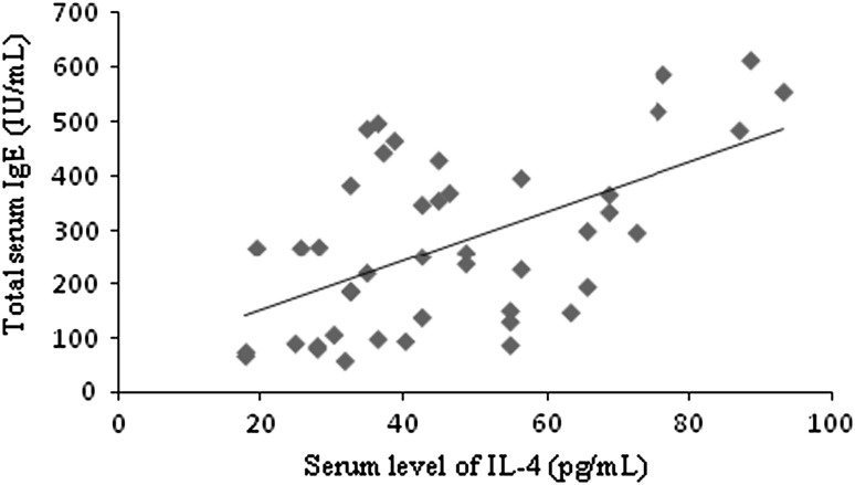
The demographic and biochemical profile of asthmatics and controls are presented in Table 1. Table 2 shows the relationship of demographic and clinical characteristics of asthmatic subjects with the elevated level of total serum IgE. The results showed no significant associations of gender, family history of asthma/atopy, exclusive breastfeeding up to 6 months and residential set up with the elevated level of total serum IgE. The higher age group, exposure to cigarette smoke and the raised eosinophil count showed the significant associations with the elevated levels of total serum IgE in asthmatics. Further, there was a significant positive correlation (r = 0.56, p < 0.001***) between the total serum IgE and IL-4 in 44 asthmatic children (Fig. 1).
Table 1.
Demographic and biochemical profile of asthmatic and control subjects
| Asthmatic subjects (n = 70) | Control subjects (n = 70) | p value | ||
|---|---|---|---|---|
| Age (years) | 6.93 ± 2.63 | 7.02 ± 2.29 | t = −0.211 | 0.833 |
| Sex | ||||
| Male/female (%) | 37/33 (52.86/47.14) | 39/31 (55.71/44.29) | χ2 = 0.115 | 0.734 |
| Height (cm) | 116.03 ± 17.16 | 119.59 ± 13.28 | t = −1.372 | 0.172 |
| Weight (kg) | 18.74 ± 6.01 | 19.80 ± 5.57 | t = −1.079 | 0.268 |
| Study community | ||||
| Bengali/non-Bengali | 54/16 (77.14/22.86) | 46/24 (65.71/34.29) | χ2 = 2.24 | 0.134 |
| Level of total serum IgE (IU/mL) | 269.21 ± 150.97 | 146.89 ± 77.32 | t = −6.03 | <0.001*** |
*** Significant at p < 0.001
Table 2.
Relationship of demographic and clinical characteristics of asthmatic children with elevated level of total serum IgE (>150 IU/mL)
| Characteristics | Total no. of asthmatic subjects (n = 70) | Total serum IgE, >150 IU/mL (n = 50) (%) | χ2 | p value |
|---|---|---|---|---|
| Age group | ||||
| 3–7 years | 40 | 24 (60.0) | 5.973 | 0.015* |
| 8–12 years | 30 | 26 (86.7) | ||
| Sex | ||||
| Male | 37 | 29 (78.4) | 1.857 | 0.173 |
| Female | 33 | 21 (63.6) | ||
| Eosinophil count | ||||
| Raised | 45 | 37 (82.2) | 7.193 | 0.007** |
| Normal | 25 | 13 (52.0) | ||
| FHA | ||||
| Yes | 22 | 19 (86.4) | 3.507 | 0.061 |
| No | 48 | 31 (64.6) | ||
| EBF up to 6 months | ||||
| Given | 52 | 37 (71.2) | 0.007 | 0.931 |
| Not given | 18 | 13 (72.2) | ||
| Exposure to cigarette smoke | ||||
| Exposed | 24 | 21 (87.5) | 4.622 | 0.032* |
| Not exposed | 46 | 29 (63.0) | ||
| Residential set up | ||||
| Rural | 56 | 41 (73.2) | 0.438 | 0.508 |
| Urban | 14 | 09 (64.3) | ||
FHA family history of asthma/atopy; EBF exclusive breastfeeding
* Significant at p < 0.05, ** Significant at p < 0.01
Fig. 1.
Correlation between serum levels of IL-4 and total IgE in 44 asthmatic subjects. The correlation coefficient was 0.56 and was statistically significant (p < 0.001***)
It was observed that out of 70 asthmatics, 50 (71.43 %) subjects had total serum IgE > 150 IU/mL. The mean total serum IgE level was significantly higher in asthmatic subjects compared to that of the control subjects, 269.21 ± 150.97 IU/mL versus 146.89 ± 77.32 IU/mL; p < 0.001*** (Fig. 2).
Fig. 2.
Comparison of total serum IgE levels (IU/mL) between asthmatic and control subjects
In the present study, the higher age group, exposure to cigarette smoke, and the raised eosinophil count showed the significant association with the elevated level of total serum IgE in asthmatic children. These findings are consistent with the findings of several earlier studies. Cline et al. [8] reported the higher total serum IgE levels in the age group of 8–14 years. Similarly, Strachan and Cook [9] showed the potential role of passive smoking on IgE in a study conducted in children. Satwani et al. [10] showed eosinophilia along with raised serum IgE levels to be a significant allergic marker. Peripheral blood eosinophil counting has tremendously important clinical implication in order to demonstrate the allergic etiology of the disease, to monitor its clinical course and to address the choice of therapy [11].
The major finding of the present study confirmed that 71.43 % of the asthmatic subjects had total serum IgE levels >150 IU/mL. The mean total serum IgE level in asthmatic group was 269.21 ± 150.97 and 146.89 ± 77.32 IU/mL in control group. The difference was statistically significant (p < 0.001***). Several studies have reported the elevated levels of total serum IgE in asthmatics [12, 13]. Therefore, it is in accordance with the well known fact that IgE plays a central role in the pathophysiology of allergic disorder such as asthma.
In the present study, it was also observed that there was a significant correlation between total IgE and IL-4 in sera of 44 asthmatic subjects. This finding is consistent with the finding of Afshari et al. [14] who reported considerably higher levels of serum IgE and IL-4 in asthmatics than non-asthmatic controls. IL-4 is one of the two cytokines known to cause switching in B-cells, a prerequisite for elevated IgE synthesis [15].
This study is a preliminary investigation and it has of course certain limitations. Further study investigating the prevalent allergens and the specific IgE estimation is warranted to strengthen the present study.
In conclusion, the elevated level of total serum IgE may demonstrate the allergic etiology of asthma in the subjects studied. Further, it also reveals the significant association of higher age, exposure to cigarette smoke and raised eosinophil count with the elevated level of total serum IgE in asthmatics.
Acknowledgments
Authors are thankful to the University Grants Commission (UGC), New Delhi, for the financial support provided to carry out this study. The authors are indebted to all the participants and their parents for their participation and cooperation. Further, authors would like to extend sincere thanks to medical officers of Pediatric Department, North Bengal Medical College and Hospital, Siliguri, for their kind help throughout the study.
References
- 1.Leung TF, Wong GWK, Ko FWS, Lam CWK, Fok TF. Clinical and atopic parameters and airway inflammatory markers in childhood asthma: a factor analysis. Thorax. 2005;60:822–826. doi: 10.1136/thx.2004.039321. [DOI] [PMC free article] [PubMed] [Google Scholar]
- 2.Burrows B, Martinez FD, Halonen M, Barbee RA, Cline MG. Association of asthma with serum IgE levels and skin test reactivity to allergens. New Engl J Med. 1989;320:270–277. doi: 10.1056/NEJM198902023200502. [DOI] [PubMed] [Google Scholar]
- 3.Borish L, Chipps B, Deniz Y, Gujrathi S, Zheng B, Dolan CM. Total serum IgE levels in a large cohort of patients with severe or difficult-to-treat asthma. Ann Allergy Asthma Immunol. 2005;95:247–253. doi: 10.1016/S1081-1206(10)61221-5. [DOI] [PubMed] [Google Scholar]
- 4.Anupama N, Sharma MV, Nagaraja HS, Bhat MR. The serum immunoglobulin E level reflects the severity of bronchial asthma. Thai J Physiol Sci. 2005;18:35–40. [Google Scholar]
- 5.Freidhoff LR, Marsh DG. Relationship among asthma, serum IgE levels, and skin test reactivity to inhaled allergens. Int Arch Allergy Appl Immunol. 1993;100:355–361. doi: 10.1159/000236438. [DOI] [PubMed] [Google Scholar]
- 6.Sears MR, Burrows B, Flannery EM, Herbison GP, Hewitt CJ, Holdaway MD. Relation between airway responsiveness and serum IgE in children with asthma and in apparently normal children. N Engl J Med. 1991;325:1067–1071. doi: 10.1056/NEJM199110103251504. [DOI] [PubMed] [Google Scholar]
- 7.Lama M, Chatterjee M, Nayak CR, Chaudhuri TK. Increased interleukin-4 and decreased interferon-γ levels in serum of children with asthma. Cytokine. 2011;55:335–338. doi: 10.1016/j.cyto.2011.05.011. [DOI] [PubMed] [Google Scholar]
- 8.Cline MG, Burrows B. Distribution of allergy in a population sample residing in Tucson, Arizona. Thorax. 1989;44:425–431. doi: 10.1136/thx.44.5.425. [DOI] [PMC free article] [PubMed] [Google Scholar]
- 9.Strachan DP, Cook DG. Parental smoking and allergic sensitisation in children. Thorax. 1998;53:117–123. doi: 10.1136/thx.53.2.117. [DOI] [PMC free article] [PubMed] [Google Scholar]
- 10.Satwani H, Rehman A, Ashraf S, Hassan A. Is serum IgE levels a good predictor of allergies in children? J Pak Med Assoc. 2009;59:698–702. [PubMed] [Google Scholar]
- 11.Mesinga TT, Schouten JP, Rijcken B, Weiss ST, van des Lende R. Host factors and environmental determinants associated with skin test reactivity and eosinophilia in a community-based population study. Ann Epidemiol. 1994;4:382–392. doi: 10.1016/1047-2797(94)90073-6. [DOI] [PubMed] [Google Scholar]
- 12.Sandeep T, Roopakala MS, Silvia CRWD, Chandrashekara S, Rao M. Evaluation of serum immunoglobulin E levels in bronchial asthma. Lung India. 2010;27:138–140. doi: 10.4103/0970-2113.68312. [DOI] [PMC free article] [PubMed] [Google Scholar]
- 13.Sharma S, Kathuria PC, Gupta CK, Nordling K, Ghosh B, Singh AB. Total serum immunoglobulin E levels in a case–control study in asthmatic/allergic patients, their family members, and healthy subjects from India. Clin Exp Allergy. 2006;36:1019–1027. doi: 10.1111/j.1365-2222.2006.02525.x. [DOI] [PubMed] [Google Scholar]
- 14.Afshari JT, Hosseini RF, Farahabadi SH, Heydarian F, Boskabady MH, Khoshnavaz R, et al. Association of the expression of IL-4 and IL-13 genes, IL-4 and IgE serum levels with allergic asthma. Iran J Allergy Asthma Immunol. 2007;6:67–72. [PubMed] [Google Scholar]
- 15.Del Prete G, Maggi E, Parronchi P, Chretien I, Tiri A, Macchia D, et al. IL-4 is an essential factor for the IgE synthesis induced in vitro by human T cell clones and their supernatants. J Immunol. 1988;140:4193–4198. [PubMed] [Google Scholar]


