Abstract
Background:
Direct antiglobulin test (DAT) is the most common test done in immunohematology lab, which detects immunoglobulin and fragments of complement attached to the red blood cells. These coated red blood cells are difficult to accurately phenotype, which may be required for selection of appropriate unit of red blood cells for transfusion.
Aims:
We have studied the efficacy of various elution methods in removing the antibodies coating the red cells and their impact on different blood group antigen activity.
Materials and Methods:
Patient samples sent for serological evaluation of autoimmune hemolysis were included in the study. DAT and Indirect antiglobulin test (IAT) were performed using gel cards (ID system, DiaMed Switzerland). Antibody coated red cells, either by in-vivo or in-vitro sensitization, were used to assess the outcome of three elution methods.
Results:
Out of 93 DAT positive samples already sensitized in vivo, 28 (30 %) samples became DAT negative post elution using either of three methods, while 36 (38.8%) showed reduction in strength of reaction, whereas in 29 (31.2%) there was no change in strength of reaction. Similarly, out of the 17 samples prepared by in vitro sensitization, 12 samples became completely negative after glycine-HCl/EDTA elution, 9 and 5 samples became negative after heat elution and chloroquine diphosphate elution methods, respectively.
Conclusion:
On comparative analysis glycine-HCl/EDTA elution method was better than the other two methods and can be used for eluting immunoglobulins from intact red cells.
Keywords: Elution, phenotype, red cells
Introduction
Direct antiglobulin test (DAT) is the most common test done in immunohematology lab, which detects immunoglobulin and fragments of complement attached to the red blood cells. It is usually performed for evaluation of patients with suspected hemolysis and positive result signifies in vivo coating of red blood cells. The red cells can be coated with IgG or complement alone or with a combination.[1] These coated red cells are difficult to accurately phenotype, which may be required for selection of appropriate unit of blood for transfusion in these patients.[2]
Saline reactive antisera, chemically modified antisera and IgM monoclonal antibodies are available for some of the red cell antigens but; antigens detected by indirect antiglobulin test are difficult to phenotype.[3] It is therefore necessary to remove antibodies from in vivo sensitized red cells to phenotype them. Various elution procedures are used for dissociating antibodies from red cells. Many of the elution procedures either cause total hemolysis of red cells, as seen with ether chloroform or xylene elution methods or cause denaturation of Kell; Duffy and MNS system antigens as seen with ZZAP (dithiothreitol and papain).[4,5]
We have studied the efficacy of various elution methods in removing the antibodies coating the red cells and their impact on different blood group antigen activity.
Materials and Methods
Patient samples sent for serological evaluation of autoimmune hemolysis were included in the study. DAT and IAT were performed using gel cards (ID system, DiaMed Switzerland). Antibody coated red cells, either by in-vivo or in-vitro sensitization, were used to assess the outcome of three elution methods.
Glycine-HCl/EDTA, Heat elution and Chloroquine diphosphate elution methods were performed on all DAT positive samples and their efficacy in removal of autoantibodies was compared.
Sensitization of red cells
Samples of red cells sensitized in vivo were obtained from patients with warm reactive autoantibodies in their sera. Red cells were washed six times with normal saline before elution. The supernatant of last wash was preserved and used as a negative control. A total of 93 samples which were positive by gel cards (polyspecific AHG), were subjected to three elution methods.
For in vitro sensitization, pooled group O red cells obtained from healthy donors were incubated with the appropriate sera. Sera containing alloantibodies (Anti D: 7, Anti D+C: 3, Anti E: 2, Anti Jka: 2, Anti M: 2, Anti Fya: 1) were obtained from alloimmunized patients. All alloantibodies used for in vitro sensitization were clinically significant and were IgG type. Doubling dilution method was used to dilute the antibodies in sera to get a strongest possible DAT without causing red cell agglutination. One volume of diluted sera was incubated with one volume of washed packed red cells for 45 minutes at 37°C. The sensitized red cells were washed six times with normal saline and were then tested by gel card.
Direct antiglobulin testing
DAT was performed by gel technique using commercially available gel cards (ID system DiaMed, Switzerland) containing poly specific antiglobulin reagent.[6] The agglutination reaction was graded according to the manufacturer instructions from ± to 4+. The scores were determined as follows: ± (questionable) =1; 1+ (weak)=3; 2+ (moderate)=6; 3+(strong) =9; 4+ (very strong) = 12.[7]
Elution methods
The following elution methods were used: glycine-HCl/EDTA elution, heat elution at 56°C for 10 minutes, and chloroquine diphosphate dissociation.[8–10]
EDTA (10%) was prepared by adding 10gm of Na2EDTA (Qualigens fine chemicals, Pvt Ltd, India) to 100 ml of distilled water. Glycine-HCl (0.1 M at pH 1.5) was prepared by adding 0.75gm glycine (Sisco research laboratories Pvt Ltd, India) to 100 mL 0.9% NaCl (Qualigens, fine chemicals, Pvt Ltd, India) and pH was adjusted to 1.5 with concentrated HCl (Qualigens fine chemical, Pvt Ltd, India). Glycine-HCl/EDTA solution was prepared by mixing 4 volumes of 0.1 M glycine-HCl buffer (pH 2.0) with 1 volume of 10 % disodium dihydrate EDTA.
1M Tris-NaCl was prepared by dissolving 12.1gm of Tris (hydroxymethyl) amino methane (S.D. fine chemicals Pvt Ltd, India) and 5.25gm of sodium chloride in 100 ml distilled water. The elution procedure was carried out by thoroughly mixing 1 volume of freshly prepared Glycine-HCl/EDTA solution with equal volume of the 50 % suspension of sensitized red cells. After 2 minutes, one volume of 1M Tris-NaCl was added to the tube containing the cell suspension. The tube was centrifuged at 3000 rpm for 1 minute. The red cells obtained were washed three times in normal saline. DAT was performed on the treated cells to check the outcome.
The chloroquine diphosphate solution was prepared by dissolving 20g of chloroquine diphosphate (Sigma Chemical Co., St. Louis) in 100 mL of phosphate buffer saline. The pH was adjusted to 5 by using 5 N NaOH. One volume of sensitized packed red cells was mixed with four volumes of chloroquine solution for dissociation. A fraction of red cells from the suspension was removed after ≈30 minute and checked for removal for antibodies. After 2 hours at room temperature, the red cells were washed three times in normal saline.
Heat elution was performed by mixing equal volumes of 50% sensitized red cells and normal saline containing 6% bovine serum albumin (Tulip diagnostics Pvt Ltd, India). The suspension was incubated for 10 minutes at 56°C in water bath and immediately centrifuged for 1 minute at 3000rpm. The red cells obtained were washed three times and tested.
Effect of elution methods on activity of red cell antigen
Fresh uncoated red cells from normal healthy blood donors were phenotyped both before and after elution procedures. Commercially available specific antisera were used to type the blood group antigens (DiaMed, Switzerland). Twenty donor samples for each blood group system were studied.
Statistical analysis
We analyzed the data using commercially available statistical software (SPSS version 13). The results are showed as mean ± SD. The Krushal-Willis was used to test between the groups. Statistical test used in analysis was two tailed and p value less than 0.05 were considered significant.
Results
Reduction in strength of DAT positivity
Out of 93 DAT positive samples already sensitized in vivo, 28 (30 %) samples became DAT negative post elution using either of three methods, while 36 (38.8%) showed reduction in strength of reaction whereas in 29 (31.2%) there was no change in strength of reaction.
On analyzing the outcome of three elution methods on 93 DAT positive samples, Glycine-HCl/EDTA elution showed decrease in reaction in 59 samples (p < 0.001) of which 22 samples were DAT negative. With Heat Elution, there was decrease in strength of reaction in 36 samples of which 11 samples were DAT negative, whereas after Chloroquine diphosphate treatment only 29 samples showed decrease in strength of reaction [Table 1].
Table 1.
DAT Agglutination score before and after Glycine HCl/EDTA elution, heat elution and chloroquine elution of DAT positive cells

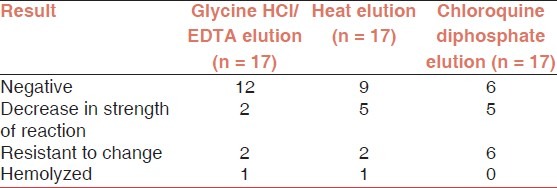
Similarly, out of the 17 samples prepared by in vitro sensitization, 12 samples became completely negative after glycine HCl/EDTA elution, 9 and 5 samples became negative after heat elution and chloroquine diphosphate elution methods, respectively.
On performing elution on in vitro sensitized red cells, Glycine-HCl/EDTA elution and heat elution was found to be more effective in removing the antibodies attached to the red cells compared to chloroquine diphosphate. (p < 0.001) [Tables 2 and 3].
Table 2.
Effect of elution methods on in vitro sensitized red blood cells

Table 3.
Comparison of agglutination score of DAT positive red cells before and after the three-elution methods. Glycine HCl/EDTA elution and heat elution was significantly better than chloroquine diphosphate in removing the alloantibodies. (*p < 0.001)

Effect of elution on red cell antigens
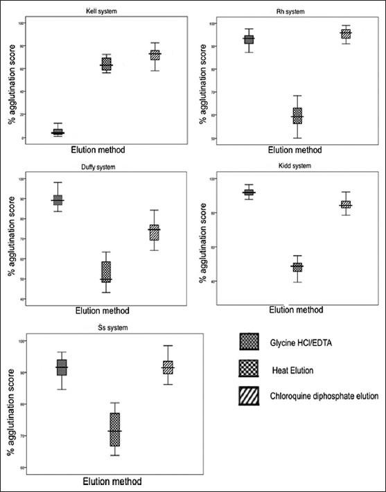
Red cell antigens within Kell, Kidd, Duffy, S s and Rh system were treated with all the three-elution methods. Kell was almost completely rendered non-reactive by Glycine-HCl/EDTA method though it did not affect other antigen system. Heat elution caused marked decrease in expression of the antigens in all the blood group systems studied. It was also associated with more hemolysis when compared to the other two methods. Chloroquine diphosphate caused minimal damage to the red cell antigen [Figure 1].
Figure 1.

Reduction in percent agglutination score in fresh red cells after treating the red cells with three elution methods. Pretreated red cells agglutination was taken as 100 percent
Discussion
The phenotyping of red blood cells is helpful for the use of phenotypically matched red cells for transfusion in patients with warm autoantibodies.[11] Elution methods such as citric acid, glycine-HCl/EDTA and cysteine activated papain and dithiothreitol (ZZAP) have been routinely used in removal of such autoantibodies from intact red cells.[2,12]
Heat elution of DAT positive red cells has been used to detect Rh and other antigens.[13] Chloroquine diphosphate dissociation of immunoglobulin is a commonly used technique that allows more complete phenotyping of red cells coated with warm reacting antibodies.[10]
On comparing three elution methods, glycine-HCl/EDTA appeared to be significantly better than the other two procedures and so it can be used for typing red cells coated with antibodies. Our results were in concurrence with the previously published studies.[7]
Chloroquine was effective in removing antibodies attached from sensitized intact red cells. Although, it was not as potent as glycine-HCl/EDTA and heat elution, but it caused significant decrease in DAT positivity. Quinoline derivatives have been reported to split immune-complexes and to inhibit antigen-antibody reaction.[14,15] Elution by chloroquine diphosphate of red blood cells sensitized in vivo and in vitro has been reported and our results are in agreement with the published studies.[10,16]
Heat elution procedure, done at 56 ° C for 10 minutes, effectively removed antibodies attached to the red cells. Although it was equally potent as glycine-HCl/EDTA in removing antibodies from in vitro sensitized red cells, decrease in DAT positivity was not as effective on in vivo-sensitized red cells. Glycine-HCl/EDTA elution was clearly superior to the other two methods for removing IgG alloantibodies and autoantibodies. Similar findings have been reported in other studies.[7]
Effect on red cell antigen system
Heat elution denatured most clinically relevant blood group antigens, unlike chloroquine dissociation or acid/EDTA elution. However, marked hemolysis was observed following heating for 10 minutes at 56°C. Nevertheless, it was possible to phenotype RBC antigens accurately. Glycine HCl/EDTA method almost completely damaged the Kell system antigens, though, with minimal effect on other systems.
Overall, Glycine-HCl/EDTA elution method was more effective in reducing strength of reaction in both in vivo and in vitro sensitization. Although, heat elution was equally effective in removing antibodies from in vitro sensitized cells when compared to acid/EDTA elution but not in red cells coated with autoantibodies.
Chloroquine diphosphate method also caused statistically significant decrease in DAT positivity.
Conclusion
Based on our results, it can be concluded that an elution method, which requires small amount of red cells, provides quick and reliable results with little damage to the red cell antigens should be selected in patients with DAT positivity. On comparative analysis glycine-HCl/EDTA elution method was better than the other two methods and can be used for eluting immunoglobulins from intact red cells.
Footnotes
Source of Support: Nil
Conflict of Interest: None.
References
- 1.Yazer MH, Triulzi DJ. The role of the elution in antibody investigations. Transfusion. 2009;49:2395–9. doi: 10.1111/j.1537-2995.2009.02304.x. [DOI] [PubMed] [Google Scholar]
- 2.lssitt PD. Applied blood group serology. 3rd ed. Miami: Montgomery Scientific Publications; 1985. p. 387. [Google Scholar]
- 3.Strobel E, Wullenweber J. Suitability of monoclonal test sera for determination of blood group markers in positive direct coombs test. Infusionsther Transfusionsmed. 1995;22:117–27. [PubMed] [Google Scholar]
- 4.Judd WJ. Elution dissociation of antibody from the red blood cells: Theoretical and practical considerations. Transfus Med Rev. 1999;13:297–310. doi: 10.1016/s0887-7963(99)80059-5. [DOI] [PubMed] [Google Scholar]
- 5.Branch DR, Petz LD. A new reagent (ZZAP) having multiple applications in immunohemotolgy. Am J Clin Pathol. 1982;78:161–7. doi: 10.1093/ajcp/78.2.161. [DOI] [PubMed] [Google Scholar]
- 6.Lapierre Y, Rigal D, Adam J, Josef D, Meyer F, Greber S, et al. The gel test: A new way to detect red cell antigen-antibody reactions. Transfusion. 1990;30:109–13. doi: 10.1046/j.1537-2995.1990.30290162894.x. [DOI] [PubMed] [Google Scholar]
- 7.Burin des Rosiers N, Squalli S. Removing IgG antibodies from intact red cells: Comparison of acid and EDTA, heat and chloroquine elution methods. Transfusion. 1997;37:497–501. doi: 10.1046/j.1537-2995.1997.37597293880.x. [DOI] [PubMed] [Google Scholar]
- 8.Byrne PC. Use of a modified acid/EDTA elution technique. Immunohematology. 1991;2:46–7. [PubMed] [Google Scholar]
- 9.Walker RH, editor. Technical manual. 41th ed. Bethesda: American Association of Blood Banks; 2003. [Google Scholar]
- 10.Edwards JM, Moulds JJ, Judd WJ. Chloroquine dissociation of antigen-antibody complexes. A new technique for typing red blood cells with a positive direct antiglobulin test. Transfusion. 1982;22:59–61. doi: 10.1046/j.1537-2995.1982.22182154219.x. [DOI] [PubMed] [Google Scholar]
- 11.Petz LD, Garratty G. Acquired Immune Hemolytic Anemias. New York: NY: Churchill Livingstone; 1980. pp. 37–50. [Google Scholar]
- 12.Louie JE, Jieng AF, Szearoulis CG. Preparation of intact antibody free red blood cells in autoimmune hemolytic anemia (abstract) Transfusion. 1986;26:550. [Google Scholar]
- 13.Walker RH, editor. Technical manual. 11th ed. Arlington: American Association of Blood Banks; 1990. [Google Scholar]
- 14.Szilagyi T, Kavai M. The effect of chloroquine on the antigen- antibody reaction. Acta Physiol Acad Sci Hung. 1970;38:411–7. [PubMed] [Google Scholar]
- 15.Domokos V, Aszody L. The effect of chloroquine on the antigen-antibody reaction of red blood cells. Haemat Hung. 1964;41:79–86. [Google Scholar]
- 16.Mantel W, Holtz G. Characterisation of autoantibodies to erythrocytes in auto-immune haemolytic anemia by chloroquine. Vox Sang. 1976:30453–63. doi: 10.1111/j.1423-0410.1976.tb02851.x. [DOI] [PubMed] [Google Scholar]


