Abstract
Upon his death in 1955, Albert Einstein’s brain was removed, fixed and photographed from multiple angles. It was then sectioned into 240 blocks, and histological slides were prepared. At the time, a roadmap was drawn that illustrates the location within the brain of each block and its associated slides. Here we describe the external gross neuroanatomy of Einstein’s entire cerebral cortex from 14 recently discovered photographs, most of which were taken from unconventional angles. Two of the photographs reveal sulcal patterns of the medial surfaces of the hemispheres, and another shows the neuroanatomy of the right (exposed) insula. Most of Einstein’s sulci are identified, and sulcal patterns in various parts of the brain are compared with those of 85 human brains that have been described in the literature. To the extent currently possible, unusual features of Einstein’s brain are tentatively interpreted in light of what is known about the evolution of higher cognitive processes in humans. As an aid to future investigators, these (and other) features are correlated with blocks on the roadmap (and therefore histological slides). Einstein’s brain has an extraordinary prefrontal cortex, which may have contributed to the neurological substrates for some of his remarkable cognitive abilities. The primary somatosensory and motor cortices near the regions that typically represent face and tongue are greatly expanded in the left hemisphere. Einstein’s parietal lobes are also unusual and may have provided some of the neurological underpinnings for his visuospatial and mathematical skills, as others have hypothesized. Einstein’s brain has typical frontal and occipital shape asymmetries (petalias) and grossly asymmetrical inferior and superior parietal lobules. Contrary to the literature, Einstein’s brain is not spherical, does not lack parietal opercula and has non-confluent Sylvian and inferior postcentral sulci.
Keywords: Albert Einstein, Broca’s area, parietal lobules, inferior third frontal gyrus, prefrontal cortex
Introduction
Albert Einstein was born at 11:30 a.m. on 14 March 1879 and died shortly after 1 a.m. on 18 April 1955, at the age of 76 (Isaacson, 2007). Within hours of his death at Princeton (N.J.) Hospital, from a ruptured abdominal aortic aneurysm, his brain was removed, weighed (1230 g), measured (40 measurements), and immersed and perfused with 10% formalin. Permission to preserve and study the brain was obtained from Einstein’s son, Hans Albert, and executor, Otto Nathan, who attended the post-mortem examination. The examining pathologist, Thomas S. Harvey, MD, used an Exakta 35 mm camera to take dozens of black and white photographs of the whole and partially dissected brain before partitioning the hemispheres by a modification of the technique of Bailey and von Bonin (1951) into 240 blocks. Cerebellum, brainstem and cerebral arteries were also preserved. The blocks were embedded in celloidin; 5 to 12 sets of 100 or 200 (accounts vary) histological slides were sectioned and stained with cell body and myelin stains. At present there is no consensus as to the particular stains used. At the time the brain was sectioned into the 240 blocks, a ‘road map’ was prepared to illustrate the locations in the brain of each block and, hence, the locations of the histological slides subsequently prepared from them. With the exception of his brain and eyes, Einstein’s body was cremated on the day of his death. The eyes were removed by his ophthalmologist and remain in private hands. The autopsy report (Report 33 for 1955 at Princeton Hospital) has been missing for >18 years.
The subsequent fate of the brain blocks and slides over the next five decades included travel with Dr Harvey from Princeton to the Midwest and return to The University Medical Centre at Princeton in 1996. No less than 18 investigators received brain tissue or photographs from Dr Harvey. Six peer-reviewed publications have resulted from analysis of tissue blocks, microscope slides or photographs. Diamond et al. (1985) found a higher glia:neuron ratio in the left inferior parietal lobule (cf. Hines, 1998). Anderson and Harvey (1996) found greater neuronal density in the right frontal lobe. Kigar et al. (1997) reported increased glia:neuron ratio in the bilateral temporal neocortices. Witelson et al. (1999b) observed a larger expanse of the bilateral inferior parietal lobules. Colombo (2006) found larger astrocytic processes and more numerous interlaminar terminal masses. Falk’s study (2009) documented unusual gross anatomy ‘in and around the primary somatosensory and motor cortices’.
Although the results are unpublished in the scientific literature, DNA sequencing was performed by Charles Boyd in 1988. The DNA extracted from a celloidin block of Einstein’s brain ‘had completely fragmented, completely denatured’ (cited in Abraham, 2002, p. 230). Currently the largest collection of celloidin-embedded brain blocks (180 out of the original 240) is at The University Medical Centre at Princeton, and the largest known aggregation of microscope slides (n = 567) is at the National Museum of Health and Medicine. With the exception of a few scattered blocks of tissue in Ontario, California, Alabama, Argentina, Japan, Hawaii and Philadelphia, the location(s) of the remaining portions of Einstein’s brain are unknown. Similarly, the majority of the microscope slides are unaccounted for. The largest collection of Dr Harvey’s photographs of Einstein’s brain (last seen intact in 1955), a subset of the histological slides, and the road map that identifies the locations in the brain of the specific blocks that yielded the slides were donated by Dr Harvey’s Estate and curated by the National Museum of Health and Medicine in 2010. Except for those mentioned in the report by Witelson et al. (1999b), the location of other extant photographs is unknown or unacknowledged.
The materials were physically acquired in June of 2010 and are cared for by members of the staff of the National Museum of Health and Medicine, then a component of the Armed Forces Institute of Pathology on the grounds of Walter Reed Army Medical Centre in Washington, DC. They were accessioned into the Museum’s permanent collection as Accession Number 2010.0010. The donation was executed formally by the Estate of Thomas Harvey, MD, and includes a number of items in addition to the images duplicated here. They are histologically prepared material, correspondence, publication clippings and other complete publications that include information about the work of Dr Harvey and his management and care of this material. No blocks of brain tissue were included in the accession, and no such material exists elsewhere at the Museum. Individually, these objects represent investigations performed or planned by Harvey. Collectively, they represent the long commitment on the part of Dr Harvey to facilitate the study of this most challenging anatomical structure. Interest in this collection is such that its most robust study may be achieved when as much material as possible, natural and archival, is reassembled in one place, physically or virtually. The National Museum of Health and Medicine has an interest in providing appropriate curation for such materials and related items in order to achieve that potential.
For the first time, the photographs that have recently come to light permit detailed identifications of sulci and other features of the external morphology of Einstein’s entire cerebral cortex, which we provide in this report. The morphology of Einstein’s frontal and parietal lobes is analysed in light of known neurological substrates for uniquely human cognitive abilities associated with specific parts of these lobes, and previously published misinformation about Einstein’s brain (made clear by the newly available material) is corrected. Although our interpretations focus on frontal and parietal morphology, interesting features throughout the brain are described and correlated with corresponding blocks on the roadmap of Einstein’s brain. It is our hope that these identifications will be of use to future researchers who wish to examine the histological slides from Einstein’s brain. Although it is beyond the scope of this article, we also hope that our identifications will be useful for workers interested in comparing Einstein’s brain with preserved brains from other gifted individuals, such as the German mathematician Carl Friedrich Gauss (1777–1855) and the Russian physiologist Ivan Pavlov (1849–1936) (Vein and Maat-Schieman, 2008).
Materials and methods
Cortical sulci and other features are identified (by D.F.) on 14 high-quality photographs of Albert Einstein’s cerebral cortex (reproduced in Figs 1–9) that were taken by Thomas Harvey in 1955 before the brain was sectioned (Lepore, 2001). The photographs of Einstein’s brain were taken from various angles that imaged all external surfaces of the cerebral cortex, the medial surface of each hemisphere and (after dissection of the overlying opercula) the insula of the right hemisphere. Although some sulci were labelled on the photographs, some of the identifications are incorrect or based on archaic terminology, and most sulci were not identified (it is not known who provided these earlier identifications). Here we provide identifications of most of the sulci and some other features on traced illustrations of the photographs and, to the extent possible, compare their configurations to those described for 60 human brains (120 hemispheres) by Connolly (1950) and 25 human brains (50 hemispheres) by Ono et al. (1990). Because the research by Ono et al. (1990) was undertaken at the University of Zurich’s Institute of Anatomy, it is reasonable to assume that most of their specimens are from Europeans, although ages and sex are not reported. Thirty of the brains studied by Connolly (1950) were from white Germans; the other half were from black Americans (Connolly, 1950, p. 181). Connolly (1950) reports race and sex, but not age, for specimens. The 60 brains he describes are not included in his chapter about children’s brains, therefore most are likely from adults. The heavily European (especially German) origin of the specimens in both studies is fortuitous, because Einstein was born in Germany.
Figure 1.
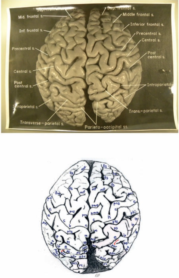
Top: Dorsal photograph of Einstein’s brain with original labels. Bottom: Our identifications. a2 = angular; a3 = anterior occipital; c = central; e = processus acuminis; fm = midfrontal; fs = superior frontal; inp = intermediate posterior parietal; ip = intraparietal; m = marginal; mf = medial frontal; ocs = superior occipital; otr = transverse occipital; par = paroccipital; pci = precentral inferior; pcs = precentral superior; pma = marginal precentral; pme = medial precentral; po = parieto-occipital; prc = paracentral; ps = superior parietal; pst = transverse parietal; pti = postcentral inferior; pts = postcentral superior; rc = retrocalcarine; u = unnamed. k = presumed motor cortex for right hand; K = ‘knob’ representing motor cortex for left hand. In both hemispheres, e limits anteriorly the first annectant gyrus, a pli de passage of Gratiolet that connects the parietal and occipital lobes, indicated by red arrows (see also Fig. 7). This figure is reproduced with permission from the National Museum of Health and Medicine.
Figure 2.
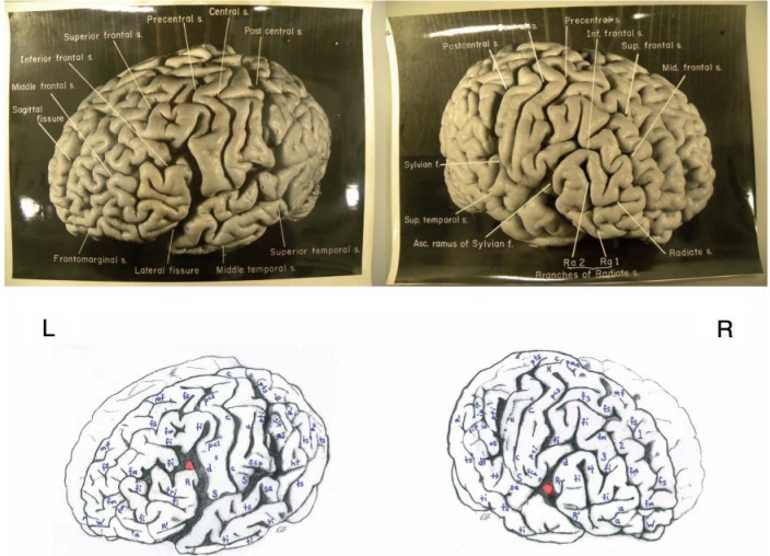
Top: Photographs of the left (L) and right (R) lateral surfaces of Einstein’s brain taken with the front of the brain rotated toward the viewer, with original labels. Bottom: Our identifications. Numbers 1–4 indicate four gyri in Einstein’s right frontal lobe, rather than three as is typical; K = ‘knob’ representing motor cortex for left hand. Submerged gyri are shaded red near the diagonal sulcus on each side. It is clear from the left hemisphere that the posterior ascending limb of the Sylvian fissure and the postcentral inferior sulcus are not confluent, contrary to the literature. Sulci: a = additional inferior frontal; a1 = ascending branch of the superior temporal sulcus; a2 = angular; aS = posterior ascending limb of the Sylvian; c = central; d = diagonal; dt = descending terminal branch of the Sylvian; fi = inferior frontal; fm = midfrontal; fs = superior frontal; ht = posterior terminal horizontal branch of the Sylvian; ip = intraparietal; mf = medial frontal; pci = precentral inferior; pcs = precentral superior; pma = marginal precentral; pti = postcentral inferior; pts = postcentral superior; R = ascending ramus of anterior Sylvian fissure; R’ = horizontal ramus of anterior Sylvian fissure; S = Sylvian fissure; sa = sulcus acousticus; sca = subcentral anterior; scp = subcentral posterior; sip = intermedius primus of Jensen; ti = inferior temporal; tri = triangular; ts = superior temporal; tt = transverse temporal; u = unnamed; W = fronto-marginal of Wernicke. 1 = superior frontal gyrus; 2 = atypical superior middle frontal gyrus; 3 = atypical inferior middle frontal gyrus; 4 = inferior frontal gyrus (usually the ‘inferior third frontal gyrus’). The figure reproduced with permission from the National Museum of Health and Medicine.
Figure 3.
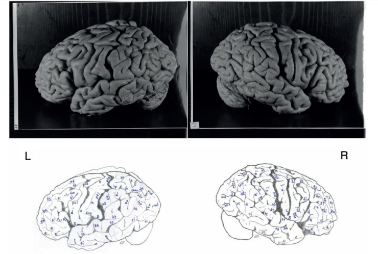
Top: Photographs of the left (L) and right (R) lateral surfaces of Einstein’s brain taken from a traditional view, which lack original labels. Bottom: Our identifications. Numbers 1–4 on the right hemisphere indicate four gyri in Einstein’s right frontal lobe, rather than three as is typical. Sulci: a = additional inferior frontal; a1 = ascending branch of the superior temporal sulcus; a2 = angular; a3 = anterior occipital; aS = posterior ascending limb of the Sylvian; c = central; d = diagonal; dt = descending terminal branch of the Sylvian; e = processus acuminis; fi = inferior frontal; fm = midfrontal; fs = superior frontal; ht = posterior terminal horizontal branch of the Sylvian; inp = intermediate posterior parietal; ip = intraparietal; mf = medial frontal; ocl = lateral occipital; ocs = superior occipital; otr = transverse occipital; par = paroccipital; pci = precentral inferior; pcs = precentral superior; ps = superior parietal; pti = postcentral inferior; pts = postcentral superior; R = ascending ramus of anterior Sylvian fissure; R’ = horizontal ramus of anterior Sylvian fissure; S = Sylvian fissure; sa = sulcus acousticus; sca = subcentral anterior; scp = subcentral posterior; sip = intermedius primus of Jensen; ti = inferior temporal; tri = triangular; ts = superior temporal; tt = transverse temporal; u = unnamed. 1 = superior frontal gyrus; 2 = atypical superior middle frontal gyrus; 3 = atypical inferior middle frontal gyrus; 4 = inferior frontal gyrus (usually the ‘inferior third frontal gyrus’). K = ‘knob’ representing motor cortex for left hand. The figure is reproduced with permission from the National Museum of Health and Medicine.
Figure 4.
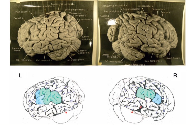
Top: Photographs of the left (L) and right (R) lateral surfaces of Einstein’s brain taken with the back of the brain rotated towards the viewer, with original labels. Bottom: Our identifications. The arrows indicate the pre-occipital notch at the inferolateral border of each hemisphere, which indicate the approximate inferior boundary between the lateral surfaces of the temporal and occipital lobes; on the right, an apparent artificial cut severed the rostral tip (shaded red) of a gyrus in the posterior part of the inferior temporal lobe. This cut appears to be a lateral extension of that observed on the right side of the base of the brain (Fig. 6). Typically, the supramarginal gyrus surrounds the posterior ascending limb of the Sylvian, and the angular gyrus surrounds the upturned end(s) of superior temporal sulcus. These gyri are separated approximately at the level of the intermedius primus sulcus of Jensen and together form the inferior parietal lobule. The supramarginal gyri are shaded blue; the angular gyri are aqua. In the left hemisphere, part of the cortical region above posterior terminal horizontal branch of the Sylvian is shaded an inbetween colour because it could arguably belong to either gyrus. Einstein’s inferior parietal lobules have different shapes in the two hemispheres, and appear to be relatively larger on the left side. Sulci: a1 = ascending branch of the superior temporal sulcus; a2 = angular; a3 = anterior occipital; aS = posterior ascending limb of the Sylvian; c = central; dt = descending terminal branch of the Sylvian; e = processus acuminis; ht = posterior terminal horizontal branch of the Sylvian; i = inferior polar; inp = intermediate posterior parietal; ip = intraparietal; lc = lateral calcarine; oci = inferior occipital; ocl = lateral occipital; ocs = superior occipital; otr = transverse occipital; par = paroccipital; ps = superior parietal; pti = postcentral inferior; pts = postcentral superior; rc = retrocalcarine; S = Sylvian fissure; scp = subcentral posterior; sip = intermedius primus of Jensen; ti = inferior temporal; ts = superior temporal; u = unnamed. The figure is reproduced with permission from the National Museum of Health and Medicine.
Figure 5.
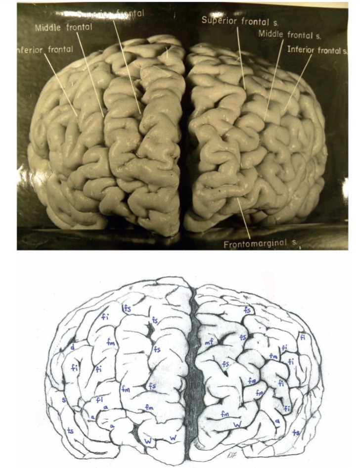
Top: Photograph of a frontal view of Einstein’s brain in an unconventional orientation, with original labels. Bottom: Our identifications of sulci. a = additional inferior frontal; fi = inferior frontal; fm = midfrontal; fs = superior frontal; mf = medial frontal; S = Sylvian fissure; ts = superior temporal; W = fronto-marginal of Wernicke. The figure is reproduced with permission from the National Museum of Health and Medicine.
Figure 6.
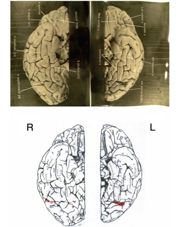
Top: Separate photographs of the right (R) and left (L) basal views of Einstein’s bisected brain with cerebellum removed and original labels. Bottom: Our identifications. The two photographs are not to the same scale and the right hemisphere is rotated slightly laterally compared with the left, as suggested by a published basal photograph of the entire brain with its cerebellum attached (Witelson et al., 1999b). The base of Einstein’s brain appears to have been accidentally cut, perhaps with a scalpel, as indicated in red shading. This may have occurred during removal of the dura mater (tentorium cerebelli) that separates the dorsum of the cerebellum from the inferior surface of the occipital lobes. Magnifying the photographs on a computer screen should facilitate observation of these cuts. See Fig. 4 for an extension of this cut that reached the right lateral surface of the temporal lobe where it severed the tip of a gyrus (shaded in red). Sulci: arc = arcuate orbital; col = collateral; fi = inferior frontal; i = inferior polar; mo = medial orbital; oa = anterior orbital; oal = lateral anterior orbital; oci = inferior occipital; oct = occipito-temporal; op = posterior orbital; opl = lateral posterior orbital; os = olfactory; R’ = horizontal ramus of anterior Sylvian fissure; rh = rhinal; ti = inferior temporal. Abbreviations of other features: los = lateral olfactory stria; mb = mammillary body; mos = medial olfactory stria; ob = olfactory bulb; on = optic nerve; ot = olfactory tract. The figure is reproduced with permission from the National Museum of Health and Medicine.
Figure 7.
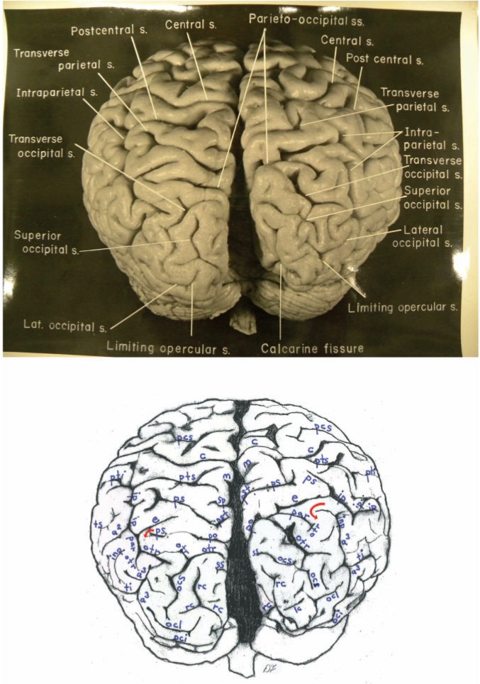
Top: Photograph of an occipital view of Einstein’s brain in an unconventional orientation, with original labels. Bottom: Our identifications. In both hemispheres, a processus acuminis limits anteriorly the first annectant gyrus, a pli de passage of Gratiolet that connects the parietal and occipital lobes, indicated by red arrows (see also Fig. 1). See Fig. 10B for shading of the superior and inferior parietal lobules and the occipital lobe on this image. Sulci: a2 = angular; a3 = anterior occipital; c = central; cu = cuneus; e = processus acuminis; inp = intermediate posterior parietal; ip = intraparietal; lc = lateral calcarine; m = marginal; oci = inferior occipital; ocl = lateral occipital; ocs = superior occipital; otr = transverse occipital; par = paroccipital; pcs = precentral superior; po = parieto-occipital; ps = superior parietal; pst = transverse parietal; pti = postcentral inferior; pts = postcentral superior; rc = retrocalcarine; sp = subparietal; ss = superior sagittal; ti = inferior temporal; ts = superior temporal. The figure is reproduced with permission from the National Museum of Health and Medicine.
Figure 8.
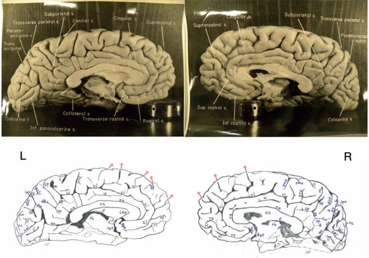
Top: Photographs of the left (L) and right (R) medial surfaces of Einstein’s brain with original labels. Bottom: Our identifications. Arrows indicate sulci that extend onto the dorsolateral surface of the brain. Sulci: ac = anterior calcarine; apo = anterior parolfactory; c = central; ca = callosal; cal = calcarine; ci = cingulate; cu = cuneus; li = lingual; lp = limiting sulcus of precuneus; m = marginal; mf = medial frontal; otr = transverse occipital; pc = paracalcarine; pma = marginal precentral; pme = medial precentral; po = parieto-occipital; prc = paracentral; pst = transverse parietal; rc = retrocalcarine; ri = inferior rostral; rs = superior rostral; si = inferior sagittal; sp = subparietal; ss = superior sagittal; u = unnamed. Other abbreviations: cc = corpus callosum; f = fornix; hpt = hypothalamus; ipo = parieto-occipital incisure; sep = septum pellucidum; th = thalamus. See text for discussion. The figure is reproduced with permission from the National Museum of Health and Medicine.
Figure 9.
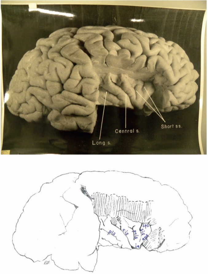
Top: Photograph of Einstein’s right insula after removal of the opercula, with original labels. Bottom: Our identifications of sulci: aps = anterior periinsular; cis = central insular; pcis = precentral insular; pis = postcentral insular; sis = short insular; sps = superior periinsular; Other identification: ia = apex of insula. The figure is reproduced with permission from the National Museum of Health and Medicine.
Per convention, sulci that appear superficially united but, upon closer inspection (including in other views), are actually separated below the surface (e.g. by a submerged gyrus) are sometimes indicated on our tracings by two dots (Connolly, 1950). For comparative purposes, our traced and labelled images are provided alongside the original labelled photographs. The reader may wish to magnify the photographs on a computer screen to better observe submerged gyri and other 3D features.
Most of the photographs of Einstein’s brain are in unconventional views. Below, we describe the cerebral cortex from photographs of a standard dorsal view (Fig. 1), right and left lateral views with the frontal lobes rotated ∼45° toward the viewer (Fig. 2), right and left lateral views in a traditional orientation (Fig. 3), and right and left lateral views with the frontal lobes rotated ∼45° away from the viewer (Fig. 4). A frontal view of the brain is rotated with the frontal poles tilted somewhat ventrally compared with standard frontal views (Fig. 5). Basal photographs show both hemispheres with the cerebellar hemispheres removed (Fig. 6). A posterior view shows the occipital poles rotated somewhat over the cerebellum (Fig. 7). Additional photographs reveal the medial surfaces of the left and right hemispheres (Fig. 8) and the right insula after its opercular covering was removed (Fig. 9). Particularly unusual or interesting features revealed in Figs 1–9 are presented in a summary illustration (Fig. 10). Witelson et al. (1999a, b) included photographs that are similar to those in our Figs 1, 3 and 8L as part of their composite Fig. 1 (Witelson et al., 1999b, p. 2150), but identified very few sulci on these images. As far as we know, this is the first publication of the other photographs reproduced in this article.
Figure 10.
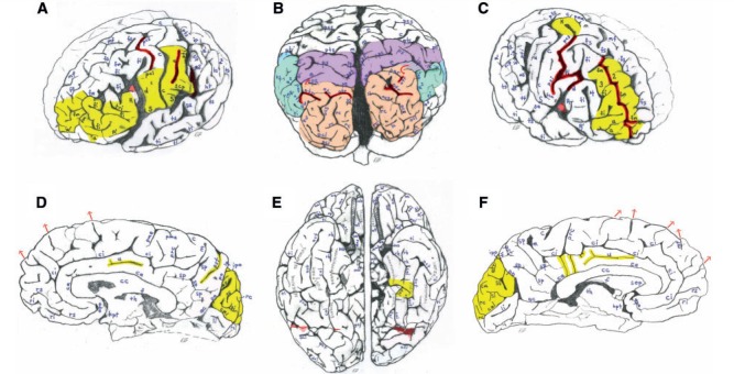
Highlights of Einstein’s brain. (A) Figure 2 of the left lateral surface of Einstein’s brain highlighted to summarize interesting features, which have been darkened. These include a connected precentral superior and inferior sulcus, a long unnamed sulcus in the inferior primary somatosensory cortex, and a posterior ascending limb of the Sylvian fissure that, contrary to the literature, is not confluent with the postcentral inferior sulcus. Unusually expanded primary somatosensory (posterior to the central sulcus) and primary motor cortices (rectangular region below the precentral inferior sulcus) are highlighted in yellow, as are the unusually convoluted surface of the pars triangularis (part of Broca’s speech area) and the frontal polar region. (B) Figure 7 of an occipital view of Einstein’s brain coloured to indicate the approximate boundaries of the superior parietal lobule (purple), inferior parietal lobule (aqua/blue) and occipital lobes (salmon). Presence of four transverse occipital sulci (darkened) is extremely rare, if not unique. Parts of the posterior temporal lobes are uncoloured below the inferior parietal lobules and rostral to the occipital lobes. Although the small striped patch between the superior and inferior parietal lobules on the right belongs with the superior parietal lobule rather than the angular gyrus of the inferior parietal lobule, its relationship with the bordering intraparietal sulcus is usually associated with a location in the angular gyrus. It would therefore be interesting to study the cytoarchitecture of this enigmatic patch of cortex. Notice that the inferior parietal lobule is favoured on the left (and see Fig. 4), while the superior parietal lobule is relatively greater on the right. There is also an asymmetry that favours the right posterior temporal region, and the right occipital lobe is shifted forward relative to the left. (C) Figure 2 of the right lateral surface of Einstein’s brain highlighted to summarize interesting features, including sulci that are darkened. Unusual sulcal patterns include a connected precentral superior and inferior sulcus, a caudal segment of the inferior frontal sulcus that is connected with both the diagonal and precentral inferior sulci, and a long midfrontal sulcus that terminates in the fronto-marginal sulcus of Wernicke. The midfrontal sulcus divides the middle frontal region into two distinct gyri (highlighted in yellow), which causes Einstein’s right frontal lobe to have four rather than the typical three gyri. The enlarged ‘knob’ that probably represents motor cortex for the left hand and the highly convoluted frontal polar region are also highlighted in yellow. (D) Figure 8 of the right medial surface of Einstein’s brain with unusual features highlighted in yellow. The cingulate gyrus has a long unnamed sulcus, the transverse parietal sulcus seems relatively elongated and the cuneus appears to be unusually convoluted. (E) Figure 6 of the basal surface of Einstein’s brain highlighted to show that the left collateral sulcus is divided into two segments, and that part of the fusiform gyrus bridges between these segments to merge with the parahippocampal gyrus. (F) Figure 8 of the left medial surface of Einstein’s brain with unusual features highlighted in yellow. The cingulate gyrus has a long unnamed sulcus, and the cingulate sulcus gives off four inferiorly directed branches (two of which are tiny), which suggest that the cingulate gyrus may be relatively convoluted. The cuneus appears to be unusually convoluted. The figures are reproduced with permission from the National Museum of Health and Medicine.
The terminology for sulci can be confusing because several have been known by different names over the years. Table 1 provides the names and abbreviations of features that we identify on photographs of Einstein’s brain, some of which are classical terms (Connolly, 1950). Many identifications, however, are more contemporary alternatives, e.g. the inferior temporal sulcus recognized here is equivalent to the middle temporal sulcus in Connolly (1950); our identification of the occipito-temporal sulcus is the modern label for the sulcus Connolly (1950) recognizes as the inferior temporal sulcus. The terminology for sulci of the human occipital lobe, in particular, has been influenced by an erroneous historical claim that human brains manifest a so-called lunate sulcus that is homologous to the Affenspalte (‘ape sulcus’) that forms the rostral boundary of the primary visual cortex [Brodmann area (BA) 17] on the lateral surface of the brain in apes and some monkeys (Smith, 1904, 1925). However, BA 17 of humans may, or may not, extend onto the external surface of the occipital lobe. When it does, its rostral border is located far posterior to the normal position for ape brains and is rarely bordered by a sulcus (Allen et al., 2006). Despite the fact that recent gross morphological and cytoarchitectural studies refute the assertion that humans have a lunate sulcus that is homologous with the Affenspalte (Allen et al., 2006; see Falk, 2012 for a discussion of the evolutionary implications), contemporary authors continue to use a variety of criteria to identify different sulci as so-called lunate sulci in humans (Duvernoy et al., 1999; Iaria and Petrides, 2007). The classical terminology used by Connolly (1950) for the occipital lobe is also grounded on the mistaken notion that humans have lunate sulci that are homologous to those of apes. For example, Connolly (1950) identifies a prelunate sulcus, which we identify with its modern name of the lateral occipital sulcus (Table 1). For these reasons, we do not recognize a lunate sulcus in Einstein’s brain. To minimize confusion, Table 1 lists alternative identifications for some sulci.
Table 1.
Abbreviations used for Einstein’s brain
| a, additional fi (Connolly, 1950; earlier termed radiate sulcus) |
| a1, ascending branch of ts |
| a2, angular sulcus; often a branch of ts |
| a3, anterior occipital sulcus; may be a branch of ts or ti (pre-occipital sulcus of Connolly, 1950) |
| ac, anterior calcarine sulcus |
| apo, anterior parolfactory sulcus |
| aps, anterior periinsular sulcus |
| arc, arcuate orbital sulcus |
| aS, posterior ascending limb of Sylvian fissure |
| c, central sulcus |
| ca, callosal sulcus |
| cal, calcarine sulcus |
| cc, corpus callosum |
| ci, cingulate sulcus |
| cis, central insular sulcus |
| col, collateral sulcus |
| cu, cuneus sulcus |
| d, diagonal sulcus |
| dt, descending terminal branch of Sylvian fissure |
| e, processus acuminis sulcus (usually a branch of par) |
| f, fornix |
| fi, inferior frontal sulcus |
| fm, midfrontal sulcus (intermediate frontal sulcus of Ono et al., 1990) |
| fs, superior frontal sulcus |
| hpt, hypothalamus |
| ht, posterior terminal horizontal branch of Sylvian fissure |
| i, inferior polar sulcus |
| ia, insular apex |
| inp, intermediate posterior parietal sulcus |
| ip, intraparietal sulcus |
| ipo, parieto-occipital incisure |
| k, motor cortex for right hand |
| K, knob representing motor cortex for left hand |
| lc, lateral calcarine sulcus |
| li, lingual sulcus (intralingual sulcus of Ono et al., 1990) |
| los, lateral olfactory stria |
| lp, limiting sulcus of precuneus |
| m, marginal sulcus |
| mb, mammillary body |
| mf, medial frontal sulcus |
| mo, medial orbital sulcus |
| mos, medial olfactory stria |
| oa, anterior orbital sulcus |
| oal, lateral anterior orbital sulcus |
| ob, olfactory bulb |
| oci, inferior occipital sulcus |
| ocl, lateral occipital sulcus (prelunate sulcus of Connolly, 1950) |
| ocs, superior occipital sulcus |
| oct, occipito-temporal sulcus (ti of Connolly, 1950; lateral oct of Duvernoy et al., 1999) |
| on, optic nerve |
| op, posterior orbital sulcus |
| opl, lateral posterior orbital sulcus |
| os, olfactory sulcus |
| ot, olfactory tract |
| otr, transverse occipital sulcus |
| par, paroccipital sulcus |
| pc, paracalcarine sulcus |
| pci, precentral inferior sulcus |
| pcis, precentral insular sulcus |
| pcs, precentral superior sulcus |
| pis, postcentral insular sulcus |
| pma, marginal precentral sulcus |
| pme, medial precentral sulcus |
| po, parieto-occipital sulcus |
| prc, paracentral sulcus |
| ps, superior parietal sulcus |
| pst, transverse parietal sulcus (superior transverse parietal of Connolly, 1950) |
| pti, postcentral inferior sulcus |
| pts, postcentral superior sulcus |
| R, ascending ramus of anterior Sylvian fissure |
| R’, horizontal ramus of anterior Sylvian fissure |
| rc, retrocalcarine sulcus |
| rh, rhinal sulcus |
| ri, inferior rostral sulcus |
| rs, superior rostral sulcus |
| sa, sulcus acousticus (Duvernoy et al., 1999) |
| S, Sylvian fissure |
| sca, subcentral anterior sulcus |
| scp, subcentral posterior sulcus |
| sep, septum pellucidum |
| si, inferior sagittal sulcus |
| sip, intermedius primus sulcus (of Jensen) [intermedius anterior (ina) of Connolly, 1950] |
| sis, short insular sulcus |
| sp, subparietal sulcus |
| sps, superior periinsular sulcus |
| ss, superior sagittal sulcus |
| th, thalamus |
| ti, inferior temporal sulcus (tm of Connolly, 1950) |
| tri, triangular sulcus (Keller et al., 2009) |
| ts, superior temporal sulcus (parallel sulcus) |
| tt, transverse temporal sulcus (Duvernoy et al., 1999) |
| u, unnamed sulcus |
| W, fronto-marginal sulcus of Wernicke |
| 1, superior frontal gyrus |
| 2, atypical superior middle frontal gyrus |
| 3, atypical inferior middle frontal gyrus |
| 4, inferior frontal gyrus (usually the ‘inferior third frontal gyrus’) |
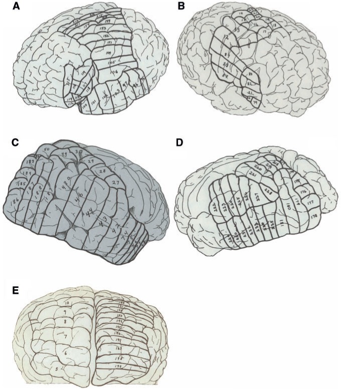
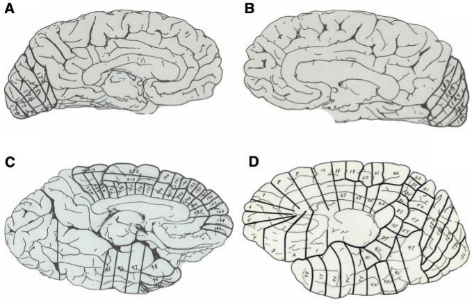
The ‘road map’ that was produced when Einstein’s brain was sectioned into 240 blocks has nine parts that are reproduced in Fig. 11A–E, which correspond with Figs 2, 4 and 5, and Fig. 12A–D, which corresponds with Fig. 8. Particularly interesting features of Einstein’s cerebral cortex are correlated with specific blocks of the road map as a guide for future researchers who may wish to access corresponding histological slides.
Figure 11.
Part of the original ‘road map’ to the 240 blocks sectioned from Einstein’s brain. A and B correspond with Fig. 2; C and D correspond with Fig. 4, and E with Fig. 5. The figure is reproduced with permission from the National Museum of Health and Medicine.
Figure 12.
The remainder of the original ‘road map’ to the 240 blocks sectioned from Einstein’s brain. A–D correspond with Fig. 8. The figure is reproduced with permission from the National Museum of Health and Medicine.
Here we describe the external surfaces of the frontal, parietal, temporal and occipital lobes. As a rule, sulci are first described for the left hemisphere, and then for the right. The medial surfaces of frontal, parietal and occipital lobes are described separately in a section on the medial and internal surfaces of Einstein’s brain.
Results
External surfaces of the brain
Frontal lobes
The newly disclosed photographs provide details about the frontal lobe of Einstein’s brain that were not visible in previously published photographs of standard views (Witelson et al., 1999a, b; Falk, 2009). As in all normal people, the central sulcus separates the primary somatosensory cortex at the front (rostral end) of the parietal lobes from the primary motor cortex at the back (caudal end) of the frontal lobes (Figs 1–4). There is a large ‘knob’-shaped fold (the ‘knob’, known to surgeons as the sign of omega) in the right hemisphere that represents enlarged motor representation for the left hand. This is an unusual feature that is seen in some long-time right-handed violinists, such as Einstein (Bangert and Schlaug, 2006; Falk, 2009). It is interesting to compare this ‘knob’ to the smaller motor representation that likely represents Einstein’s right hand, in Fig. 1. Intriguingly, magnification of the photograph in Fig. 1 underscores the expansion of the ‘knob’ over the surface of the postcentral gyrus. Another unusual feature is that, in both hemispheres, the precentral superior sulcus is continuous with the precentral inferior sulcus so that the precentral is one long sulcus rather than separated into two or more segments, unlike any of the 48 hemispheres scored for this trait by Ono et al. (1990, p. 43) (Falk, 2009, p. 3) (Figs 1–3). Similarly, of the 60 hemispheres illustrated by Connolly (1950, pp. 186–202), only two had the precentral superior sulcus continuous with the precentral inferior sulcus (Connolly, 1950, p. 192 and 202).
In the left (but not right) hemisphere of Einstein’s brain, the precentral inferior sulcus terminates extraordinarily high above the Sylvian fissure, which is not shown as a variation by Ono et al. (1990) or in the 60 frontal lobes illustrated by Connolly (1950) (left hemispheres in Figs 2 and 3). It is possible that this truncation of the lateral part of the precentral inferior sulcus is due to expansion of the motor cortex for (lower) face and tongue (i.e. which occupies at least part of the rectangular patch of cortex bordered in the left hemisphere by the precentral inferior sulcus, central sulcus, Sylvian fissure and the diagonal sulcus) (left hemispheres in Figs 2 and 3) (Penfield and Rasmussen, 1968).
Although the morphology of the diagonal sulcus is highly variable, it is estimated to be present in roughly one-half of human brains (Keller et al., 2009, p. 35) and is frequently located just behind the ascending ramus of the anterior segment of the Sylvian fissure, with which it is closely associated (Connolly, 1950; Ono et al., 1990; Keller et al., 2007, 2009). This is the case for both of Einstein’s hemispheres (Figs 2 and 3), despite the fact that Fig. 2 alone might give the impression that the ascending rami of the Sylvian fissure have been misidentified as diagonal sulci. As shown in Fig. 3, however, each hemisphere manifests a pars triangularis [BA 45, which in the left hemisphere forms part of classical Broca’s speech area (Broca, 1861)] that is bordered partly by anterior ascending and horizontal rami of the anterior segment of the Sylvian fissure. On each side, the pars triangularis is located rostral to, but in close association with, a long diagonal sulcus that extends from the Sylvian fissure to the posterior end of the inferior frontal sulcus [for similar sulcal patterns, see Connolly (1950, p. 199) and Ono et al. (1990, p. 57)].
The diagonal sulcus may be separated from the ascending branch of the Sylvian fissure by a submerged (or partially submerged) gyrus (Connolly, 1950, p.193), which is the case for part of the diagonal sulcus in Einstein’s left hemisphere (Figs 2 and 3). The right hemisphere reveals the tip of a submerged gyrus near the inferior end of the diagonal sulcus (Fig. 2), although its precise origin cannot be determined from the available photographs. Like the partially submerged gyrus on the left, however, this gyrus (which seems too near the surface to be part of the insula) is likely to represent expansion of the pars opercularis. It is interesting that Einstein has diagonal sulci in both hemispheres because it is often present only unilaterally, particularly in the left hemisphere (Galaburda, 1980; Keller et al., 2007, 2009). The truncation of the precentral inferior sulcus on the left (Fig. 2), mentioned above, may be associated with an increased volume of cortex within the depths of the diagonal sulcus, which is frequently located within the pars opercularis (BA 44) of Broca’s speech area rostral to the primary motor cortex (Keller et al., 2009, p. 34 and footnote 7). Presence of a diagonal sulcus and a nearby submerged gyrus on the right (Fig. 2) may also be associated with expansion of BA 44. Further details regarding Einstein’s speech area are given in the ‘Discussion’ section.
On the left, Einstein’s horizontal branch of the Sylvian fissure is slightly above the orbital margin (similar to 28% of the left hemispheres in Ono et al., 1990), whereas on the right, the horizontal branch of the Sylvian courses at the orbital margin [as do 16% of comparable sulci in Ono et al. (1990, p. 142)] (Figs 2 and 3). Our identification of the horizontal and ascending branches of the anterior segment of the Sylvian fissure and of the diagonal sulci are, of necessity, based on superficial relationships among sulci, so must be regarded as hypotheses that, hopefully, will be explored by studying histological slides from Einstein’s brain (refer to the ‘Discussion’ section).
The superior frontal sulcus is interrupted into two segments on the left [as in 36% of left hemispheres in Ono et al. (1990, p. 49)] (Figs 2 and 5). The superior frontal sulcus courses in one long segment on the right (found in 40% of the comparable sample investigated by Ono et al. (1990)], although it appears to be interrupted before coursing caudolaterally towards the precentral superior sulcus by a small triangular piece of cortex ripped out of the adjacent gyrus by adherent pia mater during the removal of the meninges (Figs 1 and 2R). The superior frontal gyri contain other small sulci, some of which course from the medial surface of the brain, as well as two longer medial frontal sulci on the left (Fig. 2), and one long and one short medial frontal sulcus on the right (Fig. 1). Interestingly, the two ends of the superior frontal sulcus are shifted rostrally in the right compared with left hemisphere (Figs 1 and 5) (see the discussion of Einstein’s petalia pattern below).
The midfrontal sulcus (the intermediate frontal sulcus of Ono et al., 1990) in each hemisphere merges rostrally with the fronto-marginal sulcus of Wernicke (Figs 2 and 5), which occurs in a number of frontal lobes illustrated by Connolly (1950). On the left hemisphere, the largest segment of the midfrontal sulcus forks rostrally into two branches, while its stem curves to join the inferior frontal sulcus caudally (Figs 2 and 3). A small separate segment of the midfrontal sulcus is located some distance caudal to its main stem on the left (Figs 2 and 3). On the right, the midfrontal sulcus courses lateral to the superior frontal sulcus in a long continuous sulcus (Figs 2 and 3). A similar midfrontal sulcus does not occur in any of the right hemispheres studied by Ono et al. (1990, p. 59). Connolly (1950), however, illustrates a number of hemispheres that have a relatively long single midfrontal sulcus, although these rarely terminate in the fronto-marginal sulcus of Wernicke, as Einstein’s do. Rostrally, the right midfrontal sulcus gives off a branch above the fronto-marginal sulcus of Wernicke in the frontopolar region (Figs 2R and 5). The caudal end of the midfrontal sulcus in Einstein’s right hemisphere terminates in a short relatively transverse sulcus that communicates with the superior frontal sulcus (Figs 2, 3 and 5). This configuration of long continuous superior frontal and midfrontal sulci, and the branch that connects them, is not described by Ono et al. (1990) and is similar to the pattern in only one (left) hemisphere of the 60 illustrated by Connolly (1950, p. 192).
Einstein’s left hemisphere has a long inferior frontal sulcus that sends out numerous branches and connects with the diagonal sulcus (Figs 2 and 3). Ono et al. (1990, p. 57) note that a connection exists between the diagonal sulcus and inferior frontal sulcus in 24% of the left hemispheres in their sample. One of the terminal branches of the inferior frontal sulcus courses between the ascending and horizontal rami of the Sylvian fissure that border the pars triangularis, as does a shorter separate sulcus known as the triangular sulcus (Keller et al., 2009) (Figs 2 and 3). It is unusual for both a terminal branch of the inferior frontal sulcus and an additional triangular sulcus to be located in the pars triangularis, which is shown for only one of the 60 hemispheres illustrated by Connolly (1950, p. 191). As noted, a partially submerged gyrus is visible near the intersection of the inferior frontal sulcus and the diagonal sulcus (Fig. 2), which is not unusual (Connolly, 1950, p. 193). In Einstein’s case, the submerged gyrus continues to the lateral surface, surrounds the superior end of the ascending ramus of the Sylvian fissure and continues onto the pars triangularis (Fig. 2).
Two transversely oriented inferior frontal sulci are located rostral to the long inferior frontal sulcus in the left hemisphere, and each gives off a branch that projects towards the orbital margin. The posterior of these two courses over the orbital margin onto the orbital surface (Figs 2 and 3) (these rostral branches of the inferior frontal sulcus were once called radiate sulci, but the term has been out of favour for more than half a century) (Connolly, 1950, p. 194). In Figs 2 and 3, the most rostral segment of the inferior frontal sulcus is labelled as an additional inferior frontal sulcus after Connolly (1950, p. 196), who notes a correlation between the presence of these rostral segments of the inferior frontal sulcus ‘and the degree of the development of the anterior part of the frontal lobe’ (p. 196).
The right hemisphere manifests two long and elaborate inferior frontal sulci in addition to a separate branch of the inferior frontal sulcus, which is located in the frontal polar region and labelled as an additional sulcus (Figs 2 and 3). The caudal segment of the inferior frontal sulcus is connected with both the diagonal sulcus and the precentral inferior sulcus, which occurs in only two of the 60 hemispheres illustrated by Connolly (1950, p. 188 and 199) (Figs 2 and 3). As noted, a submerged gyrus is associated with the diagonal sulcus in the right hemisphere, as is the case for the left (Fig. 2). On the right, a lower branch from the inferior frontal sulcus projects axially into the frontal operculum between the ascending and horizontal rami of the Sylvian fissure, which is not unusual. Near the ascending ramus of the Sylvian fissure, the right pars triangularis is creased by a dimple rather than the triangular sulcus, as in the opposite hemisphere.
As a rule, the superior frontal and inferior frontal sulci demarcate three large gyri that are more or less obvious depending on how extensive and continuous these sulci are. The superior frontal gyrus is above the superior frontal sulcus, the middle frontal gyrus is between the superior frontal sulcus and the inferior frontal sulcus, and the so-called ‘inferior third frontal gyrus’ (which, in the left hemisphere, includes Broca’s speech area) is below the inferior frontal sulcus. Rather than bordering gyri as the superior frontal sulcus and inferior frontal sulcus do, the midfrontal sulcus is usually a supplementary sulcus that consists of one or more short or medium length segments that are located within the middle frontal gyrus (Ono et al., 1990, p. 15). Indeed, this is the case for Einstein’s left frontal lobe (Figs 2 and 3). The pattern of gyri in Einstein’s right frontal lobe is highly unusual, however, because the relatively long single midfrontal sulcus separates the midfrontal region into two distinct gyri (Figs 2 and 3). This gives the impression that Einstein’s inferior frontal gyrus on the right is a fourth rather than the inferior third frontal gyrus (Gyri 1–4 in Figs 2 and 3). This pattern appears to result from expansion of midfrontal association cortex because in humans, ‘the greater part of the midfrontal appears … to be a new sulcus accompanying the expansion of the frontal association area’ and ‘its degree of development shows a fair correlation with the development of the frontal lobe as a whole’ (Connolly, 1950, p. 197). It is clear from illustrations and discussion of the midfrontal sulcus in 60 hemispheres by Connolly (1950) that Einstein’s middle frontal region is relatively expanded in both hemipsheres, including in the frontopolar region (Connolly, 1950, pp. 197–200).
The orbital surfaces of the frontal lobes appear in photographs of each hemisphere that were taken after the cerebellum was removed and the brain was bisected along the mid-sagittal plane. We have combined two images into one basal view so that the viewer may compare the morphology on both sides (Fig. 6). The photographs for the two hemispheres are scaled somewhat differently, and the right hemisphere is rotated slightly laterally compared with the left [for accurate relative scaling and orientation of the two basal views see Witelson et al. (1999b), which shows the base of the entire brain with the cerebellum in place]. The olfactory tract and bulb, and bisected optic chiasm are present in each hemisphere, and the medial and lateral olfactory stria are partly visible just lateral to each optic nerve. The orbital surfaces of both of Einstein’s frontal lobes manifest typical external orbital sulci (lateral anterior orbital and lateral posterior orbital sulci) and medial orbital sulci (anterior orbital and posterior orbital sulci) (Fig. 6) (Connolly, 1950, p. 183). In the left hemisphere, these four sulci form the arms of an H-shaped pattern, whereas they form two separate furrows in the right hemisphere. Neither of these patterns is unusual. Each orbital surface contains an additional sagittally oriented medial orbital sulcus, which is particularly long in the left hemisphere, and there is an arcuate sulcus on the right, which is shown to be connected with the posterior orbital sulcus in the slightly differently angled photograph of Witelson et al. (1999b). These kinds of variations in orbital sulci appear to be common (Duvernoy et al., 1999, p. 36). In the left hemisphere, a small anterior orbital sulcus ascends to the lateral surface and, on the lateral surface, an inferior branch of the inferior frontal sulcus, which is rostral to the horizontal branch of the Sylvian fissure, courses to the orbital surface (Figs 5 and 6).
The medial surfaces of the frontal lobes are described later in the text.
Parietal lobes
As noted above, in both hemispheres, the central sulcus borders the rostral boundary of the postcentral gyrus, which represents primary somatosensory cortex that is limited caudally by postcentral superior and postcentral inferior sulci (Figs 1 and 4). On the left, the postcentral gyrus is considerably wider at its lateral than medial end compared with a sample of 25 human brains (Ono et al., 1990, pp. 152–53; Falk, 2009) (Fig. 1). The wider part of the gyrus contains an unusual long unnamed sulcus that is bordered above by a side branch of the postcentral inferior sulcus, which suggests expansion of the depth and surface area of the regions that normally represent face and tongue (Penfield and Rasmussen, 1968; Falk, 2009). Traces of an unnamed sulcus appear in the right hemisphere, although the lateral part of the postcentral gyrus is not as expanded as on the left (compare the left and right hemispheres in Fig 2). The lateral end of the left postcentral inferior sulcus terminates noticeably high, as is the case for the precentral inferior sulcus in the left frontal lobe (Fig. 2). Together, these features suggest that the primary sensory, as well as motor representations of the face and tongue mentioned earlier in the text, may have been unusually expanded in Einstein’s left hemisphere.
Earlier reports that Einstein’s left Sylvian fissure is confluent with the postcentral inferior sulcus (Witelson et al., 1999b; Falk, 2009) are incorrect, as shown in Fig. 2. Instead, this photograph reveals the depths of the posterior ascending limb of the left Sylvian fissure and shows that it terminates behind the lateral end of the postcentral inferior sulcus and that the two sulci are separated by a submerged part of the supramarginal gyrus. The cortex directly caudal to the subcentral posterior sulcus is expanded so that it partially covers (opercularizes) the region (Figs 2 and 3). This new evidence confirms that Einstein’s insula was covered partly by parietal opercula (Galaburda, 1999; Falk, 2009), contrary to previous suggestions that were based on the incorrect inference that the postcentral inferior sulcus and posterior ascending limb of the Sylvian fissure were confluent, drawn from less optimal photographs (Witelson et al., 1999a, b). Although the deep morphology within the posterior ascending limb of the Sylvian fissure of the right hemisphere is not as clear from the comparable photograph (compare right and left hemispheres in Fig. 2), the two hemispheres’ similar external sulcal patterns in this region and the complex morphology of their supramarginal gyri (see below) suggest that the relationship between the postcentral inferior sulcus and posterior ascending limb of the Sylvian fissure is similar (non-confluent) in both hemispheres.
The supramarginal and angular gyri comprise the inferior parietal lobule. Typically, the supramarginal gyrus surrounds the posterior ascending limb of the Sylvian fissure. The cortex that surrounds Einstein’s left posterior ascending limb of the Sylvian fissure forms a clear supramarginal gyrus, the superior part of which is separated from the angular gyrus by the intermedius primus sulcus of Jensen (Connolly, 1950, p. 216; Duvernoy et al., 1999, p. 13) (Figs 2, 3 and 4). In addition to the posterior ascending limb of the Sylvian fissure, Einstein’s left Sylvian fissure has a terminal branch that courses horizontally, which borders the supramarginal gyrus inferolaterally (Falk, 2009) (Figs 2, 3 and 4) (see Duvernoy et al., 1999, p. 11 for a photograph of another brain with a similar posterior segment of the lateral fissure, which the authors note is ‘contrary to its usual vertical orientation’). The lateral surface of the left supramarginal gyrus is bisected by an unnamed rostrally coursing branch off of the ascending ramus of the superior temporal sulcus, and has a dimple in its lower section (Figs 2, 3 and 4). The cortex below the posterior portion of this branch could arguably be part of either the supramarginal gyrus or the angular gyrus, as indicated by its intermediate shading in Fig. 4. The newly recovered slides may be useful in resolving this issue.
In the right hemisphere, the intermedius primus sulcus of Jensen; portions of the superior temporal, intraparietal and posterior ascending limb of the Sylvian fissure; and the descending terminal branch of the Sylvian fissure border the external part of the supramarginal gyrus, which, like its counterpart on the left, is creased by additional unnamed sulci (Figs 2, 3 and 4). Superficially, the left supramarginal gyrus appears longer along its vertical (dorsoventral) axis than that on the right (compare right and left hemispheres in Figs 2, 3 and 4).
The angular gyrus typically surrounds the upward-turned posterior end of the superior temporal sulcus (called the ascending branch) and the angular sulcus that is posterior to it. The angular sulcus may, or may not, branch from the superior temporal sulcus. Einstein’s angular gyri are clearly visible in the photographs. On the left, the posterior segment of the superior temporal sulcus divides into two branches (Figs 2, 3 and 4). The rostral branch terminates in the ascending branch. Einstein’s angular gyrus curves around this branch and courses down and around the lateral end of the angular sulcus, which is separated from the superior temporal sulcus and intersects with the intraparietal sulcus (Figs 3 and 4). As is typical, the angular sulcus is axial to the central portion of the angular gyrus (Connolly, 1950, p. 216). The second terminal branch of the superior temporal sulcus in Einstein’s left hemisphere is caudal and inferior to the angular sulcus (Figs 3 and 4). A small superior terminal spur of this sulcus is surrounded by a continuation of the angular gyrus. Caudal to this, the angular gyrus surrounds an intermediate posterior parietal sulcus that may occupy a position between the angular sulcus and the anterior occipital sulcus (Connolly, 1950, p. 217), as it does in Einstein’s left hemisphere (Figs 3 and 4). Posterior to the intermediate posterior parietal sulcus in Einstein’s left hemisphere, the anterior occipital sulcus branches from the inferior temporal sulcus and part of it separates the angular gyrus from the occipital cortex caudal to it (Connolly, 1950, p. 217) (Figs 3 and 4).
In Einstein’s right hemisphere, the single branch of the superior temporal sulcus is not as extensive as the two branches on the left side, and the angular sulcus does not intersect with the intraparietal sulcus. The right angular gyrus is separated from the occipital cortex by a short segment of the anterior occipital sulcus and most of the intermediate posterior parietal sulcus with which it merges (compare the right and left hemispheres in Figs 3, 4 and 10B). The net effect is that the surface area of the angular gyrus, and indeed, the entire inferior parietal lobule, is shaped differently in the two hemispheres, and appears to be relatively expanded on the left side (compare shading in the left and right hemispheres in Fig. 4, and see Fig. 10B). This finding is consistent with a voxel-based analysis of 142 MRI scans that revealed a significantly greater volume of grey matter in the left than right angular gyrus (Watkins et al., 2001). In this context, it is interesting that Einstein’s left hemisphere has double terminal branches of the Sylvian fissure (i.e. its posterior ascending limb and posterior terminal horizontal branch) in addition to double branches of the superior temporal sulcus.
The superior parietal lobules, which are located rostral to the occipital lobes, are usually bordered inferiorly by the intraparietal sulcus and rostrally by the postcentral superior sulcus. Accordingly, Einstein’s left superior parietal lobule is separated from the inferior parietal lobule by a continuous intraparietal sulcus that stems from the postcentral inferior sulcus (Figs 1, 4 and 10B). A substantial superior parietal sulcus that ends laterally in a fork courses across the left lobule posterior to and approximately parallel with the postcentral superior sulcus, and a shorter superior parietal sulcus is located caudally (Figs 1 and 7). The longer superior parietal sulcus merges superficially with a small part of the left superior transverse parietal sulcus that courses onto the dorsal surface from the medial surface (Figs 1 and 7).
Caudal to the superior transverse parietal sulcus on the left side, the parieto-occipital sulcus also reaches the dorsal surface of the left hemisphere from the medial one (Figs 1 and 7). Lateral to the left parieto-occipital and short superior parietal sulci, the intraparietal sulcus gives off a processus acuminis sulcus and then continues caudally onto the occipital lobe as the paroccipital sulcus, which ends by forking into two branches of the transverse occipital sulcus (Figs 1, 4 and 7). The processus acuminis sulcus limits anteriorly the first annectant gyrus (also called the arcus parieto-occipital gyrus), a pli de passage (of Gratiolet) that connects the left superior parietal lobule and occipital lobes (red arrows in Figs 1, 7 and 10B). These variations in the left hemisphere are all common, as shown by numerous illustrations in Connolly (1950, p. 206).
The right superior parietal lobule is considerably wider than the left (Fig. 10B), although this may be due partly to the fact that the configuration of sulci differ in the left and right lobules. In the right hemisphere, the intraparietal sulcus consists of two partly parallel segments that are joined by a shallow connection (Figs 1, 3, 4 and 7), unlike any of the brains figured by Connolly (1950), but somewhat similar to the ‘double parallel pattern’ illustrated by Ono et al. (1990, p. 67) as representative of 12% of the right hemispheres in their sample. In a highly unusual feature, the united segments of the intraparietal sulcus on the right side course upward into the superior parietal lobule, rather than along its inferior border (compare both hemispheres in Figs 4 and 10B). On the right, the processus acuminis sulcus is aligned with, rather than located rostral to, the level of the parieto-occipital sulcus, and the paroccipital sulcus appears to intersect with it superficially, but may actually be separated from it by a submerged gyrus (Figs 1 and 7). The right hemisphere, like the left, has a pli de passage that bridges between the parietal and occipital lobes (red arrows in Fig. 7). As noted, the right superior parietal lobule is wider than the left (Fig. 10B), although the shapes of the superior parietal lobules differ in the two hemispheres because that on the right is shifted forward in conjunction with asymmetrical occipital and (posterior) temporal lobes (Figs 1, 4 and 10B). Refer to the discussion of Einstein’s petalia pattern below. The medial surfaces of the parietal lobes are described later, in the section on the medial and internal surfaces of the brain.
Temporal lobes
As Connolly (1950, p. 204 and 222) observes, it makes sense to discuss the temporal lobe along with the parietal lobe because the transition between them is gradual and there are no definite sulcal boundaries separating the caudal part of the temporal lobe from the parietal lobe (Fig. 4). We have already discussed the branching of the caudal portions of Einstein’s superior temporal sulcus in his parietal lobes. The superior and inferior temporal sulci (the latter of which is labelled the middle temporal sulcus by Connolly, 1950) divide the lateral surface of the temporal lobes into superior (above the superior temporal sulcus), middle (between the two sulci) and inferior (below the inferior temporal sulcus) gyri, the last of which continues onto the inferior surface of the brain (Figs 2, 3, 4 and 6). Falk (2009), in describing Einstein’s brain, used Connolly’s (1950) label of middle temporal sulcus, and not inferior temporal sulcus, as preferred here. Rather than indicating a different sulcus, this change in labels reflects a preference for more contemporary terminology for human brains (Table 1).
In both of Einstein’s temporal lobes, the anterior segment of the superior temporal sulcus appears to be continuous (Figs 2 and 3), which occurs in 28% and 36% of Ono et al.’s (1990, p. 75) left and right hemispheres, respectively. Connolly (1950, p. 222) regards this pattern as even more common, stating that ‘the superior temporal is most frequently a continuous furrow running more or less parallel to the lateral [Sylvian] fissure’. The superior temporal gyri in both hemispheres have a sulcus acousticus, which, on the right, is rostral to a transverse temporal sulcus (Duvernoy et al., 1999) (Figs 2 and 3). The superior surface of the posterior part of Einstein’s right temporal lobe appears to be more expanded than on the left side (Figs 4 and 10B). As far as one can tell from the available photographs, the configuration of the superior and inferior temporal sulci on the lateral surface of Einstein’s temporal lobes is otherwise unremarkable.
The inferior rostromedial surfaces of Einstein’s temporal lobes have parahippocampal gyri that are bordered rostrolaterally by rhinal sulci and caudolaterally by collateral sulci (Fig. 6). On the left hemisphere (Fig. 6), the rhinal sulcus is continuous with a rostral segment of the collateral sulcus, whereas these two sulci are separate on the right. Both of these are normal variations. In both hemispheres, lingual gyri are located caudal to the parahippocampal gyri, and are bordered laterally by collateral sulci that are bifurcated caudally, which is also a normal variation (Ono et al., 1990, p. 101). On the right hemisphere, the collateral sulcus appears to be one continuous, but branched, sulcus, as illustrated for the few basal views of brains in Connolly (1950) and the larger sample discussed by Ono et al. (1990). In Einstein’s left hemisphere, however, the collateral sulcus is divided into two separate segments (Fig. 6). Although the fusiform gyrus is typically separated from the parahippocampal and lingual gyri by a continuous collateral sulcus, Einstein’s segmented collateral sulcus on the left is associated with a fusiform gyrus that bridges between the two segments (and seems to opercularize the rostral end of the posterior segment; Fig. 6). At the level of this bridge, the medial part of the fusiform gyrus merges with the caudal end of the parahippocampal gyrus, whereas its lateral part continues in a caudal direction lateral to the lingual gyrus (Fig. 6).
In each of Einstein’s hemispheres, the lateral border of the fusiform gyrus is bordered by an occipito-temporal sulcus (called the inferior temporal sulcus by Connolly, 1950) that separates it from the inferior temporal gyrus, which, as noted earlier, continues from the lateral surface of the temporal lobes (Fig. 6). One cannot be sure whether there is more than one segment of occipito-temporal sulcus on the left (there may be) because of an artificial cut in the brain (reddened in Fig. 6) that may have been made during removal of the dura mater, especially the tentorium cerebelli, which separates the dorsum of the cerebellum from the inferior surface of the occipital lobes. A cut is also present on the right (reddened in Fig. 6), and its lateral extent seems to sever the rostral tip of a convolution in the inferior temporal gyrus located just above the cerebellum (Figs 3 and 4). On the right, it is apparent that the occipito-temporal sulcus is disrupted into two branched segments, similar to 32% of the right hemispheres scored by Ono et al. (1990, p. 106) (Fig. 6). The occipito-temporal sulcus takes a relatively medial course in each hemisphere, rather than the more typical lateral course near the infero-lateral margin of the temporal lobe, similar to 36% of the left hemispheres and 16% of the right hemispheres examined by Ono et al. (1990, p. 107) (Fig. 6). This suggests a relatively great development of Einstein’s inferior temporal gyri, which grow downward in humans and push the cortex medially (Connolly, 1950, p. 255).
Occipital lobes
The left occipital lobe is bordered (approximately) rostrolaterally by the parieto-occipital sulcus, a short superior parietal sulcus that is lateral to this sulcus, the paroccipital sulcus, the most lateral segment of the transverse occipital sulcus and part of the anterior occipital sulcus. The anterior border of the occipital lobe continues from the bottom of the vertical part of the anterior occipital sulcus in an imaginary line that courses inferiorly to the pre-occipital notch (Figs 4 and 10B). The right occipital lobe is bordered approximately by the parieto-occipital sulcus, the medial part of the processus acuminis sulcus, the posterior superior fork and vertical part of the intermediate posterior parietal sulcus and the relatively vertical part of the anterior occipital sulcus. As on the left, the anterior border of the right occipital lobe continues from the bottom of the vertical portion of the anterior occipital sulcus in an imaginary line that courses to the pre-occipital notch (Figs 4 and 10B). The locations of the anterior occipital sulcus in each hemisphere appear to be relatively caudal compared with those illustrated by Connolly (1950), which may be due to caudal expansion of the inferior parietal lobules, especially on the left (Figs 4 and 10B). In a description of Einstein’s brain, Falk (2009) misidentified a branch of the inferior temporal sulcus in the right hemisphere as the anterior occipital sulcus, which is corrected in Fig. 4, in light of information from the newly available photographs.
In addition to the normal variation of a forked transverse occipital sulcus at the caudal end of the paroccipital sulcus in both hemispheres (which is more forked on Einstein’s left hemisphere), Einstein’s brain has a separate medial segment of the transverse occipital sulcus on the left that crosses the superior margin of the hemisphere (Figs 7 and 8), which Ono et al. (1990, p. 73) report occurs in 0% of the left hemispheres in their sample. There is also a relatively large additional transverse occipital sulcus in the right hemisphere (Figs 4 and 7). Ono et al. (1990) do not include multiple segments of the transverse occipital sulcus as a variation among the 25 brains in his sample, nor does Connolly (1950) discuss or illustrate segments of the transverse occipital sulcus that are additional to those that fork from the caudal end of the paroccipital sulcus in the 60 brains studied. The unique configuration of multiple segments of the transverse occipital sulcus in Einstein’s brain suggests that the occipital lobes may be relatively wide near their dorsal rostral borders.
Each hemisphere has a lateral occipital sulcus that forks at its caudal end (a normal variation), which divides the latero-posterior region of the occipital lobes into superior and inferior parts (Duvernoy et al., 1999) (Figs 4 and 7). In Einstein’s case, the superior parts are quite convoluted. Each contains a substantial superior occipital sulcus (Fig. 7). On the left, the superior occipital sulcus forks at its rostral end into two of the aforementioned segments of the transverse occipital sulcus; on the right, the superior occipital sulcus appears as a continuation of the superior sagittal sulcus from the medial surface of the brain (Fig. 7), although the superior sagittal sulcus is not mentioned as one of the sulci that cross the medial hemisphere by Ono et al. (1990, p. 12) or Connolly (1950, pp. 253–55). Surprisingly large retrocalcarine sulci course from the medial to the lateral surface of both occipital lobes, and a small lateral calcarine sulcus appears on the right (Fig. 7). Both hemispheres have typical inferior occipital sulci, which appear in basal and lateral posterior views (Figs 4 and 6), in addition to inferior polar sulci (Figs 4 and 6).
Medial and internal surfaces of the brain
The medial surface of Einstein’s left hemisphere (Fig. 8) has a typical pattern of a cingulate sulcus that courses caudally and gives off a paracentral and then marginal sulcus, which bracket the paracentral lobule. This lobule contains extensions from the lateral surface of the pre- and postcentral gyri (Fig. 1) On the left, but not right, hemisphere (Fig. 1), the marginal sulcus ends at the superior medial border rather than extending for a short distance on the dorsolateral surface of the brain, which occurs in only 4% of the left hemispheres described by Ono et al. (1990, p. 115). The left hemisphere also has a second cingulate sulcus (sometimes called the paracingulate sulcus or the superior cingulate sulcus) (Yücel et al., 2001) that is separate from and rostral to the first (Fig. 8). This ‘double parallel’ pattern appears in 24% of the left hemispheres described by Ono et al. (1990, p. 113). Einstein’s right hemisphere, on the other hand, has a single uninterrupted cingulate sulcus, as do 60% of the right hemispheres in sample studied by Ono et al. (1990, p. 112) (Fig. 8R). Regretfully, Harvey's 2D photographs do not permit quantification of the extent of this asymmetry, which would be possible with MRI methods (e.g. Clark et al., 2010). The right cingulate sulcus also gives off paracentral and marginal sulci, but the paracentral sulcus does not reach the dorsal surface as it does on the left (Fig. 1). In dorsal view, the rostral boundary of the right paracentral lobule is bordered by the marginal precentral sulcus rather than the paracentral sulcus, like the left hemisphere (Fig. 1).
In the frontal lobes, the cingulate sulcus typically separates the cingulate gyrus from the medial frontal gyrus above it. The middle portion of Einstein’s left and right cingulate gyri each contain a long unnamed sulcus (Fig. 8). Near the caudal end of the unnamed sulcus on the left, the cingulate sulcus gives off four inferiorly directed branches (two tiny and two long, which cross the cingulate gyrus, Fig. 8), which occurs in only 8% of the comparable sample studied by Ono et al. (1990, p. 118). In the same region on the right, the cingulate sulcus gives off two small inferiorly directed side branches (Fig. 8), which is fairly typical. These features suggest that Einstein’s cingulate gyri may have been relatively convoluted, especially in the left hemisphere, which manifests the double parallel cingulate sulcus described above.
In each of Einstein’s frontal lobes, the rectus gyrus (which usually represents BA 11) is bordered dorsally by the inferior rostral sulcus, which is in two segments in the right hemisphere (Fig. 8). Each hemisphere also has a superior rostral sulcus, which usually separates BA 12 below from BA 10 above. On the left, the superior rostral sulcus stems from the anterior branch of the double cingulate sulcus, a pattern that Ono et al. (1990, p. 119) report for 12% of their left hemispheres. The superior frontal gyrus continues onto the medial surface of both hemispheres as the medial frontal gyrus, which, as noted, is above the cingulate sulcus (Fig. 8). On the left, four sulci extend from the medial frontal gyrus onto the lateral surface of the frontal lobe (arrows in Fig. 8). One of these is a terminal branch given off by the most caudal of the two cingulate sulci (Fig. 8). Rostrally, a fifth sulcus, which stems from the anterior branch of the double cingulate sulcus, extends onto the lateral frontal polar region (anterior arrow in Fig. 8). In Einstein’s right hemisphere, a branch given off by the cingulate sulcus courses rostrally and extends onto the dorsolateral surface of the frontal polar region (arrow in Fig. 8). Caudal to that, two small independent sulci course onto the lateral surface of the superior frontal convolution (arrows in Fig. 8). Ono et al. (1990) do not provide data for numbers and frequencies of rostrally directed branches of the anterior part of the cingulate sulcus that cross onto the lateral surface of the frontal lobe.
Sulci of Einstein’s parietal lobes also appear on the medial surfaces of the hemispheres (Fig. 8). In both hemispheres, the marginal sulcus is rostral to (and to some degree linked with) an H-shaped subparietal sulcus (Fig. 8), which is the most common pattern noted by Ono et al. (1990, p. 122). On the left, the rostral superior limb of the H-shaped subparietal sulcus courses to the dorsal surface as is common, and caudal to that a short transverse parietal sulcus crosses from the lateral surface onto the superior parietal lobule between the two superior limbs of the H-shaped subparietal sulcus (Figs 1 and 8). On the right side (Fig. 8), no limb of the H-shaped subparietal sulcus crosses the superior margin of the hemisphere, a pattern reported for only 4% of right hemispheres by Ono et al. (1990, p. 122). A transverse parietal sulcus that is considerably longer than its counterpart on the left crosses from the dorsal surface and courses between the superior limbs of the H-shaped subparietal sulcus (Fig. 8).
The medial surfaces of Einstein’s occipital lobes are also interesting. The cuneus of the left hemisphere contains inferior and superior sagittal sulci (Fig. 8), which are more ramified than comparable sulci shown by Ono et al. (1990) and are reported to occur together in only 4% of the left hemispheres in the sample described by Ono et al. (1990, p. 135). Einstein’s left superior sagittal sulcus is connected with the parieto-occipital sulcus, which occurs in 8% of the comparable sample investigated by Ono et al. (1990). In a normal variation, the left calcarine sulcus forks caudally into two branches of the retrocalcarine sulcus (Fig. 8). Just rostral to this, however, an unusual separate branch of the retrocalcarine sulcus courses upward and crosses the superior border of the occipital lobe (Fig. 8) where it ramifies into two substantial branches on the lateral surface (Fig. 7). A similar combination of dual branches of the retrocalcarine sulcus is not discussed or illustrated in Connolly (1950), Ono et al. (1990) or Iaria and Petrides (2007). The medial surface of the left occipital lobe also contains another sulcus, the cuneus and, as noted above, the most medial branch of the transverse occipital sulcus extends from the lateral to the medial surface of the brain, which is highly unusual (Figs 7 and 8).
Both of Einstein’s hemispheres reveal an unusual pattern in which the parieto-occipital incisure is distinct from and just caudal to a sulcus that is usually opercularized: the limiting sulcus of the precuneus (Fig. 8). On the right, a paracalcarine sulcus superficially appears to stem from the parieto-occipital sulcus (Fig. 8). Refer to Connolly (1950, pp. 253–55) for a discussion. In addition to having superior and inferior sagittal sulci in the right hemisphere, the calcarine sulcus forks caudally into the retrocalcarine sulcus, the dorsal branch of which crosses the superior margin and ends in a fork on the right dorsolateral surface of the occipital lobe (Figs 7 and 8).
The lingual gyrus (below the calcarine sulcus) in both of Einstein’s hemispheres contains a lingual sulcus (intralingual sulcus of Ono et al., 1990) (Fig. 8), which occurs in 40% and 28% of the left and right hemispheres, respectively, of the sample studied by Ono et al. (1990, p. 135). It thus appears that the medial surface of Einstein’s visual cortex is relatively convoluted in both hemispheres compared with normal brains surveyed by other authors, especially in the cuneus.
Insula
The newly discovered materials also contain a photograph of Einstein’s right (but not left) insula (island of Reil), which was taken after the frontal, parietal and temporal opercula were removed (Fig. 9). As is typical (Türe et al., 1999), a central insular sulcus divides the insula into anterior and posterior zones. Rostral to this sulcus, the precentral insular sulcus and short insular sulcus separate the anterior zone into anterior, middle and posterior short insular gyri. Caudal to the central insular sulcus, a postcentral insular sulcus divides the posterior zone into anterior and posterior long insular gyri. Figure 9 also reveals another sulcus caudal to the postcentral insular sulcus, but without more of the surrounding region, we cannot be confident of its identity. The anterior periinsular sulcus and a rostral portion of the superior periinsular sulcus are also visible. Although the apex of the pyramid-shaped insula is clear, the insular polar region that is located slightly anteroinferiorly to the apex (Türe et al., 1999) was apparently removed along with the opercula.
Petalia pattern
For both hemispheres, an outline of the perimeter of the brain and of the corpus callosum was traced from the photograph of the medial surface. The mirror image of the tracing from the right hemisphere was flipped and superimposed on that from the left hemisphere, matching the perimeters of their severed corpus callosums. The match was almost perfect, suggesting that the hemispheres were aligned in the same mid-sagittal plane when they were photographed. In the superimposed tracings, the occipital lobe of the left hemisphere droops lower and extends more posteriorly than that of the right hemisphere. The asymmetry in the caudal projections of the occipital lobes occurs partly because the midline of the right occipital lobe curves laterally compared with the relatively straight midline of its left counterpart, as seen in dorsal view (Figs 1 and 10B).
The left occipital lobe is also wider towards the posterior end than the right occipital lobe (Fig. 10B). The anterior end of the right frontal lobe, on the other hand, is wider than its counterpart on the left (Fig. 1), and it projects more rostrally than the left in the superimposed tracings, although not to the extent that the left occipital lobe projects caudally relative to the right. The right frontal projection petalia is illustrated best in the basal photograph reproduced by Witelson et al. (1999b) because the brain in that photograph still has the cerebellum attached, which suggests that the two hemispheres are in their natural positions relative to each other. Einstein’s brain, thus, manifests right frontal/left occipital width and projection petalias, which is the most typical pattern seen in humans (LeMay, 1976, 1977, 1992; Galaburda et al., 1978; Zilles et al., 1996; Toga et al., 2009). This information, together with the numerous asymmetries noted above [e.g. surface areas of the inferior and superior parietal lobules (Figs 4 and 10B)] indicate that Einstein’s brain was neither spherical or symmetrical, as suggested elsewhere (Witelson et al., 1999b).
Discussion
Although scientists and others have long been fascinated by geniuses (for discussion, see Witelson et al., 1999b), it must be kept in mind that people, in general, comprise the most intelligent species on the planet. The cognitive abilities and their neuroanatomical substrates for geniuses like Albert Einstein are, therefore, best evaluated within this context. The highly derived (advanced) cognitive abilities of humans have been attributed to an evolutionary increase in overall brain size (Finlay and Darlington, 1995) on the one hand, and evolved alterations in the relative volumes and connections of the brain’s internal components (‘neurological reorganization’) on the other (Teffer and Semendeferi, 2012). Despite the fact that individual researchers tend to emphasize the importance of one over the other, brain size and reorganization were both important and probably evolved in concert in hominins (Falk, 2012; Hofman, 2012). Expansion of the whole cerebral cortex across primates, including humans, is manifested externally in the addition of cortical sulci (Jerison, 1973), some of which occur regularly, rather than randomly. The evolution of non-random sulci has been attributed to alterations in internal connection patterns (i.e. the ‘tension-based theory’ of the formation of convolutions and sulci; Van Essen, 1997, 2007). Notably, human neurological reorganization is also manifested by a relatively marked asymmetry in the shapes and sizes of the cerebral hemispheres and some of their subareas (Geschwind and Levitsky, 1968; LeMay, 1976, 1977, 1992; Galaburda and Geschwind, 1981; Toga et al., 2009).
Ideally, Einstein’s brain should be evaluated in terms of its size as well as signs of neurological reorganization indicated by sulcal patterns, histological features and anatomical asymmetries. Some of this is possible now; some must await (hopefully productive) analyses of the histological slides from his brain. Einstein was 76 years old when he died in 1955, and his brain manifested certain normal signs of ageing such as a thinned cerebral cortex (Anderson and Harvey, 1996) and widened sulci (Magnotta et al., 1999) that probably contributed to a decrease in brain size, which is to be expected (Courchesne et al., 2000). His fresh brain weight at autopsy was 1230 g (Witelson et al., 1999b, Table 1), although details of how, exactly, this weight was measured are not available (e.g. whether it was weighed with or without meninges and CSF etc.). The fresh brain weight of a 76-year-old male (with CSF etc.) averages ∼9% less than its weight in adolescence (Courchesne et al., 2000), which corresponds with an age-corrected weight of 1352 g for Einstein’s brain. This is a conservative estimate, however, because, depending on how Einstein’s fresh brain weight was measured, the age-corrected brain weight could be considerably higher (Courchesne et al., 2000). However, as far as one can tell from available information, Einstein’s brain size was unexceptional (Witelson et al., 1999b).
On the other hand, neuroscientists are beginning to understand some of the derived features related to cortical reorganization that are associated with unique cognitive abilities (Gilbert and Wilson, 2007; Schenker et al., 2008; Semendeferi et al., 2011; Teffer and Semendeferi, 2012), and our findings regarding Einstein’s brain are particularly interesting within this context. Intriguingly, some of the derived features of external neuroanatomy that are associated with specific higher cognitive functions in humans appear to be especially marked in Einstein. Furthermore, there may be a possibility of examining the microscopic organization associated with these features from the recovered histological slides from Einstein’s brain, as indicated below.
All normal people are capable of speech, which is functionally associated with certain gross and microscopic features of Broca’s speech area in the left frontal lobe (see Keller et al., 2009 for review). The definitions of this area vary, with some researchers including wide areas of the left frontal lobe (Keller et al., 2009). Classical Broca’s speech area (Broca, 1861) (and its homologue in the right hemisphere), however, is a circumscribed region of the posterior left inferior third frontal gyrus, which includes cytoarchitectonic areas BA 44 (pars opercularis) and BA 45 (pars triangularis), the latter of which is usually bordered partly by the ascending and horizontal branches of the anterior Sylvian fissure (Keller et al., 2009). Today, many also include BA 47 (pars orbitalis) as part of Broca’s speech area. Despite these conventional definitions, the precise sulcal boundaries of cytoarchitectonic areas BA 44 and BA 45 vary somewhat relative to the horizontal and ascending branches of the anterior Sylvian fissure and the diagonal sulcus, particularly when one considers submerged gyri (Amunts et al., 1999). Nevertheless, it is safe to assume that the free surface of the pars triangularis is cytoarchitecturally BA 45 (Amunts et al., 1999). Functional neuroimaging studies in conjunction with cytoarchitectural studies suggest that BA 44 ‘may subserve an end-stage articulatory function, perhaps assisting in control over the muscles of articulation’ (Keller et al., 2009, p. 32) as well as syntactic processing, whereas BA 45 is more involved with semantic processing (see Keller et al., 2009 for review). The homologue of Broca’s speech area on the right is probably differentially involved with producing the emotional and prosodic aspects of speech (tone of voice).
Although it has been suggested that Broca’s area has a greater volume than its counterpart in the right hemisphere, most post-mortem and MRI studies that have measured overall volumes or surface areas of BA 44 and BA 45 find little support for this assertion (reviewed in Keller et al., 2009). Cytoarchitectonic studies, on the other hand, ‘broadly indicate that the various measures of area 44 in particular and area 45 are leftward asymmetric in the human brain … Leftward asymmetry appears to exist of the size of the large magnopyramidal neurons in layer III, but not consistently (albeit more often than not) of neuronal density, volume or number of neurons’ (Keller et al., 2009 p. 44; see also Amunts et al., 2003).
When viewed laterally, the pars triangularis is generally larger than the pars opercularis and pars orbitalis (Keller et al., 2009), which is true for Einstein’s brain (Figs 2 and 3). It is also noteworthy that Einstein’s left pars triangularis is unusually convoluted (with both a terminal branch of the inferior frontal sulcus and an additional triangular sulcus) and accompanied by a highly developed anterior part of the frontal lobe indicated by an additional branch of the inferior frontal sulcus (Figs 2, 3 and 10A). A partially submerged gyrus in the region of Broca’s area is visible in the left hemisphere (Fig. 2), which corresponds with Block 117 of the ‘road map’ (Fig. 11A). It would be interesting to examine the histological slides from Block 117 and nearby Blocks 116, 118 and 119 in addition to the rostral parts of Blocks 104–106 in order to learn details about the cytoarchitecture of Einstein’s Broca’s area, including the histological characteristics of and relationship between the walls of the diagonal sulcus, BA 44 and BA 45, and how Broca’s area in Einstein’s brain compares histologically with those of normal adults (Amunts et al., 1999). The homologue of Broca’s area in the right hemisphere is likely to be represented in Blocks 79–83 plus the rostral ends of Blocks 87 and 88 (Fig. 11B). A comparison of the slides from this region in both hemispheres might allow evaluation of the hypothesis that Einstein’s brain manifests a marked left over right volume asymmetry in Broca’s area, as suggested by the relative convolutedness of left compared with right pars triangularis.
BA 44 and BA 45 are part of the inferior frontal gyrus, which appears quite extensive in both of Einstein’s hemispheres (Figs 2, 3, 10A and C). As discussed above, the pattern of gyri in Einstein’s right frontal lobe is highly unusual because a relatively long single midfrontal sulcus separates the middle frontal region into two distinct gyri, which gives the impression that Einstein’s inferior frontal gyrus is a fourth rather than the typical inferior third frontal gyrus (see Gyri 1–4 in Figs 2, 3 and 10C). This pattern (Fig. 10C) and the complicated branching of the midfrontal sulcus in the rostral part of the left frontal lobe (Fig. 10A) suggest that Einstein had relatively expanded prefrontal association cortices, which is interesting because in humans, the greatest part of the midfrontal sulcus is an evolutionarily new sulcus associated with expansion in this region (Connolly, 1950, p. 197). In Einstein’s case, this cortex appears to be highly convoluted around the midfrontal sulcus in both hemispheres, including the most anterior (fronto-polar) part of the frontal lobes, which contains BA 10 (Fig. 10A and C). The convolutions of Einstein’s prefrontal lobe suggest that, volumetrically, his prefrontal association areas may have been on the high end of the range of variation for humans. This is consistent with the evolutionary hypothesis that an increase in a cortical region’s number of sulci may be associated with differential expansion of its volume accompanied by increased complexity of its internal wiring (Jerison, 1973; Van Essen, 2007; Hofman, 2012).
Significantly, within the frontal lobes and compared with apes, human BA 10 is extraordinarily large and, similar to Broca’s area, has a more complex Layer III, which has connections with neurons in other parts of the brain, believed to be critical for higher cognition (Semendeferi et al., 2011). The morphology of Layer III in BA 10 contrasts with that in other parts of the brain including visual cortex (BA 17), primary somatosensory cortex (BA 3) and primary motor cortex (BA 4), and is thought to have evolved in the human lineage after its split from chimpanzees (Semendeferi et al., 2011). In humans as well as other primates, BA 10 facilitates ‘executive functions’ including organizing sensory information, maintaining attention, monitoring working memory, and coordinating goal-directed behaviours (Teffer and Semendeferi, 2012). In addition to speech, the prefrontal cortex facilitates another uniquely human behaviour, namely imagining events and simulating their consequences (Gilbert and Wilson, 2007). Such ‘prospection’ is a way of pre-experiencing and planning for possible future events: ‘Alas, actually perceiving a bear is a potentially expensive way to learn about its adaptive significance, and thus evolution has provided us with a method for getting this information in advance of the encounter. When we preview the future and prefeel its consequences, we are soliciting advice from our ancestors’ (Gilbert and Wilson, 2007, p. 1354).
The highly evolved human prefrontal cortex, including BA 10, is just the sort of mental machinery that one would use for thought experiments like those Einstein famously (and productively) engaged in, such as imagining himself riding alongside a light beam, and being enclosed in an elevator accelerating up through space (Isaacson, 2007). Such imaginings might additionally recruit parietal and occipital association cortices (Kosslyn et al., 2001; Sack et al., 2002). Einstein’s prefrontal cortex (and—hopefully—its Layer III) could be investigated histologically by examining slides from Block 6 (and perhaps neighbouring Blocks 5 and 7) from the right frontal lobe and Block 198 (and neighbouring Blocks 163 and 199) from the left frontal lobe (Fig. 11E).
The newly recovered photographs also underscore the extraordinary expansion of the lateral part of Einstein’s left primary somatosensory (indicated by u in Figs 2 and 3) and left primary motor cortices (see the rectangular patch of cortex underneath the precentral inferior sulcus in Fig. 10A) (Falk, 2009). Slides from these parts of Einstein’s brain should be available in Blocks 104–106 of the left hemisphere (Fig. 11A) and Blocks 87–89 from the right hemisphere (Fig. 11B). It is difficult to interpret the expanded lower parts of the primary sensory and motor cortices, other than to note that these parts of the brain normally represent face and tongue (Penfield and Rasmussen, 1968; see Falk, 2009 for discussion). In this context, it is interesting that Einstein famously wrote that thinking entailed an association of images and ‘feelings’, and that, for him, the elements of thought were, not only visual, but also ‘muscular’ (Hadamard, 1945). Another interesting feature of Einstein’s primary motor cortex is the enlarged ‘knob’ that represents the left hand (Fig. 10C), which is probably associated with his early and extensive training on the violin (Bangert and Schlaug, 2006). This particular feature of Einstein’s gross neuroanatomy was likely the result of both nature and nurture (Falk, 2009). Slides from Blocks 14 and 15 (Fig. 11B) correspond with Einstein’s enlarged ‘knob’.
Earlier reports that Einstein’s posterior ascending limb of the Sylvian fissure is confluent with the postcentral inferior sulcus were based on visual inspection of photographs in traditional right and left lateral views (Witelson et al., 1999a, b; Falk, 2009). However, the newly recovered photographs that show the frontal lobes from non-traditional angles suggest that the two sulci are, in fact, separate (Figs 2 and 10A). The claim that Einstein lacked the anterior parts of the supramarginal gyri (or parietal opercula) (Witelson et al., 1999a, b) is questionable (Galaburda, 1999; Falk, 2009) because these photographs indicate not only the presence of complete supramarginal gyri (Figs 2, 3 and 4) but also that the lateral parts of the gyri are expanded to such an extent that they opercularize the posterior ascending limb of the Sylvian fissure when the brain is observed in traditional lateral views (Fig. 3). Histological information about the supramarginal gyri may be sought in slides from Block 222 (and possibly Block 223) from the left hemisphere (Fig. 11D) and superior parts of Blocks 42 and 43 from the right hemisphere (Fig. 11C). Einstein’s angular gyri (which normally corresponds with BA 39) are also extensive (Figs 3 and 4), and are represented in Blocks 224–227 (and possibly Block 228) on the left (Fig. 11D) and Blocks 44, 46 and 48a from the right hemisphere (Fig. 11C).
Despite an earlier claim that Einstein’s parietal lobes were atypically symmetrical (Witelson et al., 1999b), the surface areas and shapes of the inferior and superior parietal lobules appear highly asymmetrical, albeit in different directions (Fig. 10B). The surface area of the angular gyrus (BA 39) and, indeed, the entire inferior parietal lobule (usually consisting of BA 39 + BA 40), appears to be substantially expanded in Einstein’s left compared with the right hemisphere (Figs 4 and 10). This is intriguing in light of an earlier histological study that found a significantly greater ratio of glial cells to neurons in Einstein’s left BA 39 compared with the average for a sample of 11 older control males, whereas comparable ratios on the right side did not differ significantly between Einstein and the others (Diamond et al., 1985). The Diamond et al. (1985) study is interesting in light of recent indications that glial cells may play a critical role in regulating synaptic function and plasticity (Filosa et al., 2009). Einstein’s relatively large left angular gyrus is consistent with a study of 142 healthy adults (Watkins et al., 2001).
The inferior parietal lobule on the left is concerned with language, body image and mathematics, whereas that on the right focuses more on non-verbal visuospatial processing. More specifically, the angular gyrus is involved with the recognition of visual symbols and (especially on the left) sensory aspects of language such as those entailed in reading. The supramarginal gyrus on the left is a part of Wernicke’s area that is involved with speech comprehension and short-term maintenance of phonemes and syllables during linguistic tasks (Galaburda et al., 2002).
Einstein’s right superior parietal lobule appears wider than the left (Fig. 10C) and extends further rostrally and inferiorly, so that it is superior to the supramarginal gyrus on the right but not on the left (compare left and right hemispheres in Fig. 4). This suggests that Einstein’s right superior parietal lobule may have been substantially larger than the left. As discussed earlier, Einstein’s right intraparietal sulcus courses into the superior parietal lobule rather than remaining as its inferior border, which is highly unusual (Figs 4 and 10B). Consequently, a small portion of parietal cortex that would normally be part of the angular gyrus because of its relationship to the intraparietal sulcus, is located in the superior parietal lobule on the right side (striped patch in Fig. 10B). It would be extremely interesting to investigate the cytoarchitecture of this patch of cortex, which corresponds to the top of Block 48a and the inferior part of Block 49 (Fig. 11C).
The superior parietal lobules are involved in spatial attention, spatial orientation, localizing stimuli in space, and sensing the location of one’s body parts and guiding their movements. The right superior parietal lobule may be differentially involved in certain aspects of visuospatial imagery (Sack et al., 2002). Interestingly, the superior parietal lobules, especially their posterior parts, function during mental arithmetic, presumably because this activity co-opts parietal circuitry involved in spatial coding (Knops et al., 2009). Because number processing also involves regions within the depths of the intraparietal sulcus (Knops et al., 2009), histologists might wish to observe slides from the relevant parts of Blocks 221, 226 and 227 from Einstein’s left hemisphere (Fig. 11D) and Blocks 27–28, 44, 46 and 48a from his right hemisphere (Fig. 11C). Despite the fact that our anatomical analysis of Einstein’s parietal lobes differs in some details from that of Witelson et al. (1999a, b), we think the authors’ hypothesis that Einstein’s visuospatial and mathematical thinking may have been facilitated by extraordinary parietal substrates is reasonable.
In light of this, it would also be interesting to access slides from the medial surfaces of Einstein’s hemispheres that correspond to areas that process visual information. The medial surfaces of the occipital lobes contain primary visual area BA 17 as well as association visual areas BA 18 and BA 19. As described above, these surfaces of Einstein’s brain are very convoluted, which may be significant because visual cortex is activated when subjects close their eyes and visualize objects (Kosslyn et al., 1995, 2001). This region corresponds with the blocks illustrated in Fig. 12A and B. We have shown that Einstein’s inferior temporal gyrus is relatively expanded on the basal surface of each hemisphere (Fig. 6). This gyrus normally contains BA 20 and, caudal to that, BA 37, which on the left is involved in reading and has been called the ‘brain’s letterbox’ (Dehaene, 2009). In general, the inferior temporal gyrus is activated during high-level processing associated with the remembering, recognizing and naming of visual objects and forms (i.e. it facilitates the ventral ‘what’ visual stream). Slides representing the inferior temporal gyrus from the medial surfaces of the brain correspond with the inferior parts of Blocks 95–97 on the left (Fig. 12C) (there would be more blocks on this side if the ‘road map’ were not incomplete caudal to Block 95), and with the inferior portions of (from caudal to rostral) Blocks 47, 45, 43, 42, 74 and 75 on the right (Fig. 12D).
The medial surfaces of Einstein’s brain are also interesting in the region of the frontal lobes. The frontopolar region (BA 10), which is unusually large and histologically complex in humans, extends to the medial surface where it is typically bounded ventrally by the superior rostral sulcus (Fig. 8). Histological information about Einstein’s frontopolar region may be sought in the rostral portions of slides from Blocks 163 and 198 on the left (Fig. 12C) and Blocks 5 and 6 on the right (Fig. 12D). The caudal portions of Blocks 5 and 6 (and Block 205 on the left) also have potential for revealing histological information about Einstein’s anterior cingulate cortex, which is of interest because in humans this region has derived histological features [e.g. a high number of Von Economo neurons (Allman et al., 2010)] that may provide a neurological substrate for the enhanced ability in humans for resolving conflicting impulses, which is necessary to self-regulate behaviour (Posner et al., 2007). Although whether or not the human anterior cingulate cortex has evolved more generally to support enhanced cognitive flexibility is currently a topic of debate (Cole et al., 2010), it would be most interesting to learn about Einstein’s Von Economo neurons.
Summary and conclusions
Einstein’s brain is of unexceptional size and its combination of a relatively wide and forward-projecting right frontal lobe with a relatively wide and posteriorly protruding left occipital lobe is the most prevalent pattern seen in right-handed adult males. We have identified the sulci that delimit expansions of cortex (gyri or convolutions) on the external surfaces of all of the lobes of the brain and on the medial surfaces of both hemispheres. The morphology in some parts of Einstein’s cerebral cortex is highly unusual compared with 25 (Ono et al., 1990) and 60 (Connolly, 1950) human brains for which sulcal patterns have been thoroughly described. To the extent possible, the blocks of brain from particularly interesting areas are identified on the ‘roadmap’ that was prepared when Einstein’s brain was sectioned, as a guide for researchers who may wish to explore the histological correlates of Einstein’s gross cortical morphology.
Contrary to earlier reports, newly available photographs reveal that Einstein’s brain is not spherical in shape. The surface area of Einstein’s inferior parietal lobule is larger on the left than the right side, whereas that of his superior parietal lobule appears markedly larger in the right hemisphere. The photographs also suggest that the primary somatosensory and motor cortices representing the face and tongue are differentially expanded in the left hemisphere, that the posterior ascending limb of the Sylvian fissure is separate from (rather than confluent with) the postcentral inferior sulcus, and that parietal opercula are present. Nevertheless, our findings are concordant with the earlier suggestion that unusual morphology in Einstein’s parietal lobes may have provided neurological substrates for his visuospatial and mathematical abilities (Witelson et al., 1999a, b).
Our results also suggest that Einstein had relatively expanded prefrontal cortices, which may have provided underpinnings for some of his extraordinary cognitive abilities, including his productive use of thought experiments. From an evolutionary perspective, the specific parts of Einstein’s prefrontal cortex that appear to be differentially expanded are of interest because recent findings indicate that these same areas increased differentially in size and became neurologically reorganized at microanatomical levels during hominin evolution in association with the emergence of higher cognitive abilities (Semendeferi et al., 2011). It would be interesting therefore to investigate the histological correlates of these (as well as parietal) regions of Einstein’s brain from the newly available slides. We hope that future research on comparative primate neuroanatomy, paleoneurology and functional neuroanatomy will provide insight about some of the unusually convoluted parts of Einstein’s brain that we have described with little, if any, interpretation (e.g. the external neuroanatomy of the occipital lobes, posterolateral temporal cortex, and inferior temporal gyri).
Funding
Publication costs were provided by the College of Arts and Sciences at Florida State University, and travel support for DF was provided by the School for Advanced Research in Santa Fe, New Mexico.
Acknowledgements
The authors thank the estate of Thomas S. Harvey, MD, for donating the materials that form the basis for this article to the National Museum of Health and Medicine, Elizabeth Lockett and Emily Wilson for help in accessing materials, and Jessica Calzada for preparation of figures. Kurt Rockenstein is thanked for extensive technical support. We also received help from Eric Boyle, Tim Clarke, Jr., Laura Cutter, Elizabeth Eubanks, Albert Galaburda, Lois Hawkes, Sam Huckaba and Micah Vandegrift. The National Museum of Health and Medicine is acknowledged for permission to reproduce the 12 images that appear in this article. The views expressed are those of the authors and do not reflect the official policy or position of the Department of Defense or the United States Government.
Individuals who are interested in studying the newly emerged Harvey Collection should contact medicalmuseum@amedd.army.mil.
Glossary
Abbreviation
- BA
Brodmann area
References
- Abraham C. Possessing genius. New York: St. Martin’s Press; 2002. [Google Scholar]
- Allen JS, Bruss J, Damasio H. Looking for the lunate sulcus: a magnetic resonance imaging study in modern humans. Anat Rec A Discov Mol Cell Evol Biol. 2006;288:867–76. doi: 10.1002/ar.a.20362. [DOI] [PubMed] [Google Scholar]
- Allman JM, Tetreault NA, Hakeem AY, Manaye KF, Semendeferi K, Erwin JM, et al. The von Economo neurons in frontoinsular and anterior cingulate cortex in great apes and humans. Brain Struct Funct. 2010;214:495–517. doi: 10.1007/s00429-010-0254-0. [DOI] [PubMed] [Google Scholar]
- Amunts K, Schleicher A, Burgel U, Mohlberg H, Uylings HB, Zilles K. Broca's region revisited: cytoarchitecture and intersubject variability. J Comp Neurol. 1999;412:319–41. doi: 10.1002/(sici)1096-9861(19990920)412:2<319::aid-cne10>3.0.co;2-7. [DOI] [PubMed] [Google Scholar]
- Amunts K, Schleicher A, Ditterich A, Zilles K. Broca's region: cytoarchitectonic asymmetry and developmental changes. J Comp Neurol. 2003;465:72–89. doi: 10.1002/cne.10829. [DOI] [PubMed] [Google Scholar]
- Anderson B, Harvey T. Alterations in cortical thickness and neuronal density in the frontal cortex of Albert Einstein. Neurosci Lett. 1996;210:161–4. doi: 10.1016/0304-3940(96)12693-8. [DOI] [PubMed] [Google Scholar]
- Bailey P, Von Bonin G. The isocortex of man. Urbana, IL: University of Illinois Press; 1951. [Google Scholar]
- Bangert M, Schlaug G. Specialization of the specialized in features of external human brain morphology. Eur J Neurosci. 2006;24:1832–4. doi: 10.1111/j.1460-9568.2006.05031.x. [DOI] [PubMed] [Google Scholar]
- Broca P. Remarques sur le siége de la faculté du langage articulé, suivies d’une observation d’aphémie (Perte de la Parole) Bulletin de la Société Anatomique de Paris. 1861;6:330–57. [Google Scholar]
- Clark GM, Mackay CE, Davidson ME, Iversen SD, Collinson SL, James AC, et al. Paracingulate sulcus asymmetry; sex difference, correlation with semantic fluency and change over time in adolescent onset psychosis. Psychiatry Res. 2010;184:10–15. doi: 10.1016/j.pscychresns.2010.06.012. [DOI] [PubMed] [Google Scholar]
- Cole MW, Yeung N, Freiwald WA, Botvinick M. Conflict over cingulate cortex: between-species differences in cingulate may support enhanced cognitive flexibility in humans. Brain Behav Evol. 2010;75:239–40. doi: 10.1159/000313860. [DOI] [PubMed] [Google Scholar]
- Colombo JA, Reisin HD, Miguel-Hidalgo JJ, Rajkowska G. Cerebral cortex astroglia and the brain of a genius: a propos of A. Einstein's. Brain Res Rev. 2006;52:257–63. doi: 10.1016/j.brainresrev.2006.03.002. [DOI] [PMC free article] [PubMed] [Google Scholar]
- Connolly JC. External morphology of the primate brain. Springfield, IL: C. C. Thomas; 1950. [Google Scholar]
- Courchesne E, Chisum HJ, Townsend J, Cowles A, Covington J, Egaas B, et al. Normal brain development and aging: quantitative analysis at in vivo MR imaging in healthy volunteers. Radiology. 2000;216:672–82. doi: 10.1148/radiology.216.3.r00au37672. [DOI] [PubMed] [Google Scholar]
- Dehaene S. Reading in the Brain. New York: Viking; 2009. [Google Scholar]
- Diamond MC, Scheibel AB, Murphy GM, Jr, Harvey T. On the brain of a scientist: Albert Einstein. Exp Neurol. 1985;88:198–204. doi: 10.1016/0014-4886(85)90123-2. [DOI] [PubMed] [Google Scholar]
- Duvernoy HM, Bourqouin P, Cabanis EA, Cattin F, Maeder P. The human brain: surface, blood supply, and three-dimensional sectional anatomy. 2nd edn. New York: Springer-Verlag; 1999. [Google Scholar]
- Falk D. New information about Albert Einstein's brain. Front Evol Neurosci. 2009;1:3. doi: 10.3389/neuro.18.003.2009. [DOI] [PMC free article] [PubMed] [Google Scholar]
- Falk D. Hominin paleoneurology: where are we now? In: Hofman MA, Falk D, editors. Evolution of the primate brain: from neuron to behavior. London: Elsevier; 2012. pp. 255–72. [Google Scholar]
- Filosa A, Paixao S, Honsek SD, Carmona MA, Becker L, Feddersen B, et al. Neuron-glia communication via EphA4/ephrin-A3 modulates LTP through glial glutamate transport. Nat Neurosci. 2009;12:1285–92. doi: 10.1038/nn.2394. [DOI] [PMC free article] [PubMed] [Google Scholar]
- Finlay BL, Darlington RB. Linked regularities in the development and evolution of mammalian brains. Science. 1995;268:1578–84. doi: 10.1126/science.7777856. [DOI] [PubMed] [Google Scholar]
- Galaburda AM. [Broca's region: anatomic remarks made a century after the death of its discoverer] Rev Neurol (Paris) 1980;136:609–16. [PubMed] [Google Scholar]
- Galaburda AM. Albert Einstein's brain. Lancet. 1999;354:1821. doi: 10.1016/s0140-6736(05)70590-0. [DOI] [PubMed] [Google Scholar]
- Galaburda AM, Geschwind N. Anatomical asymmetries in the adult and developing brain and their implications for function. Adv Pediatr. 1981;28:271–92. [PubMed] [Google Scholar]
- Galaburda AM, Kosslyn SM, Christen Y. The Languages of the brain. Cambridge, MA: Harvard University Press; 2002. [Google Scholar]
- Galaburda AM, LeMay M, Kemper TL, Geschwind N. Right-left asymmetrics in the brain. Science. 1978;199:852–6. doi: 10.1126/science.341314. [DOI] [PubMed] [Google Scholar]
- Geschwind N, Levitsky W. Human brain: left-right asymmetries in temporal speech region. Science. 1968;161:186–7. doi: 10.1126/science.161.3837.186. [DOI] [PubMed] [Google Scholar]
- Gilbert DT, Wilson TD. Prospection: experiencing the future. Science. 2007;317:1351–4. doi: 10.1126/science.1144161. [DOI] [PubMed] [Google Scholar]
- Hadamard J. The psychology of invention in the mathematical field. Princeton, NJ: Dover Publications; 1945. [Google Scholar]
- Hines T. Further on Einstein's brain. Exp Neurol. 1998;150:343–4. doi: 10.1006/exnr.1997.6759. [DOI] [PubMed] [Google Scholar]
- Hofman MA. Design principles of the human brain: an evolutionary perspective. In: Hofman MA, Falk D, editors. Evolution of the primate brain: from neuron to behavior. London: Elsevier; 2012. pp. 373–90. [DOI] [PubMed] [Google Scholar]
- Iaria G, Petrides M. Occipital sulci of the human brain: variability and probability maps. J Comp Neurol. 2007;501:243–59. doi: 10.1002/cne.21254. [DOI] [PubMed] [Google Scholar]
- Isaacson W. Einstein, his life and universe. New York: Simon and Schuster; 2007. [Google Scholar]
- Jerison HJ. Evolution of the brain and intelligence. New York: Academic Press; 1973. [Google Scholar]
- Keller SS, Crow T, Foundas A, Amunts K, Roberts N. Broca's area: nomenclature, anatomy, typology and asymmetry. Brain Lang. 2009;109:29–48. doi: 10.1016/j.bandl.2008.11.005. [DOI] [PubMed] [Google Scholar]
- Keller SS, Highley JR, Garcia-Finana M, Sluming V, Rezaie R, Roberts N. Sulcal variability, stereological measurement and asymmetry of Broca's area on MR images. J Anat. 2007;211:534–55. doi: 10.1111/j.1469-7580.2007.00793.x. [DOI] [PMC free article] [PubMed] [Google Scholar]
- Kigar DL, Witelson SF, Glezer II, Harvey T. Estimates of cell number in temporal neocortex in the brain of Albert Einstein. Soc Neurosci Abst. 1997;23:89. 9. [Google Scholar]
- Knops A, Thirion B, Hubbard EM, Michel V, Dehaene S. Recruitment of an area involved in eye movements during mental arithmetic. Science. 2009;324:1583–5. doi: 10.1126/science.1171599. [DOI] [PubMed] [Google Scholar]
- Kosslyn SM, Ganis G, Thompson WL. Neural foundations of imagery. Nat Rev Neurosci. 2001;2:635–42. doi: 10.1038/35090055. [DOI] [PubMed] [Google Scholar]
- Kosslyn SM, Thompson WL, Kim IJ, Alpert NM. Topographical representations of mental images in primary visual cortex. Nature. 1995;378:496–8. doi: 10.1038/378496a0. [DOI] [PubMed] [Google Scholar]
- LeMay M. Morphological cerebral asymmetries of modern man, fossil man, and nonhuman primate. Ann N Y Acad Sci. 1976;280:349–66. doi: 10.1111/j.1749-6632.1976.tb25499.x. [DOI] [PubMed] [Google Scholar]
- LeMay M. Asymmetries of the skull and handedness. Phrenology revisited. J Neurol Sci. 1977;32:243–53. doi: 10.1016/0022-510x(77)90239-8. [DOI] [PubMed] [Google Scholar]
- LeMay M. Left-right dissymmetry, handedness. Am J Neuroradiol. 1992;13:493–504. [PMC free article] [PubMed] [Google Scholar]
- Lepore FE. Dissecting genius—Einstein's brain and the search for the neural basis of intellect. Vol. 3. Cerebrum: The Dana Forum on Brain Science; 2001. pp. 11–26. [Google Scholar]
- Magnotta VA, Andreasen NC, Schultz SK, Harris G, Cizadlo T, Heckel D, et al. Quantitative in vivo measurement of gyrification in the human brain: changes associated with aging. Cereb Cortex. 1999;9:151–60. doi: 10.1093/cercor/9.2.151. [DOI] [PubMed] [Google Scholar]
- Ono M, Kubik S, Abernathy CD. Atlas of the Cerebral Sulci. New York: Thieme Medical Publishers, Inc.; 1990. [Google Scholar]
- Penfield W, Rasmussen T. The cerebral cortex of man: a clinical study of localization of function. New York: Hafner Publishing Company; 1968. [Google Scholar]
- Posner MI, Rothbart MK, Sheese BE, Tang Y. The anterior cingulate gyrus and the mechanism of self-regulation. Cogn Affect Behav Neurosci. 2007;7:391–5. doi: 10.3758/cabn.7.4.391. [DOI] [PubMed] [Google Scholar]
- Sack AT, Sperling JM, Prvulovic D, Formisano E, Goebel R, Di Salle F, et al. Tracking the mind's image in the brain II: transcranial magnetic stimulation reveals parietal asymmetry in visuospatial imagery. Neuron. 2002;35:195–204. doi: 10.1016/s0896-6273(02)00745-6. [DOI] [PubMed] [Google Scholar]
- Schenker NM, Buxhoeveden DP, Blackmon WL, Amunts K, Zilles K, Semendeferi K. A comparative quantitative analysis of cytoarchitecture and minicolumnar organization in Broca's area in humans and great apes. J Comp Neurol. 2008;510:117–28. doi: 10.1002/cne.21792. [DOI] [PubMed] [Google Scholar]
- Semendeferi K, Teffer K, Buxhoeveden DP, Park MS, Bludau S, Amunts K, et al. Spatial organization of neurons in the frontal pole sets humans apart from great apes. Cereb Cortex. 2011;21:1485–97. doi: 10.1093/cercor/bhq191. [DOI] [PubMed] [Google Scholar]
- Smith GE. The fossil anthropoid ape from Taungs. Nature. 1925;115:235. [Google Scholar]
- Smith GE. The morphology of the occipital region of the cerebral hemisphere in man and the apes. Anatomisher Anzeiger. 1904;24:436–51. [Google Scholar]
- Teffer K, Semendeferi K. Human prefrontal cortex: evolution, development, and pathology. In: Hofman MA, Falk D, editors. Evolution of the primate brain: from neuron to behavior. London: Elsevier; 2012. pp. 191–218. [DOI] [PubMed] [Google Scholar]
- Toga AW, Narr KL, Thompson PM, Luders E. Brain asymmetry: evolution. Encyclopedia Neurosci. 2009;2:303–11. [Google Scholar]
- Türe U, Yasargil DC, Al-Mefty O, Yasargil MG. Topographic anatomy of the insular region. J Neurosurg. 1999;90:720–33. doi: 10.3171/jns.1999.90.4.0720. [DOI] [PubMed] [Google Scholar]
- Van Essen DC. A tension-based theory of morphogenesis and compact wiring in the central nervous system. Nature. 1997;385:313–18. doi: 10.1038/385313a0. [DOI] [PubMed] [Google Scholar]
- Van Essen DC. Cerebral cortical folding patterns in primates: why they vary and what they signify. In: Kass JH, Preuss TM, editors. Evolution of nervous systems. Vol. 4. London: Elsevier; 2007. pp. 267–89. [Google Scholar]
- Vein AA, Maat-Schieman ML. Famous Russian brains: historical attempts to understand intelligence. Brain. 2008;131:583–90. doi: 10.1093/brain/awm326. [DOI] [PubMed] [Google Scholar]
- Watkins KE, Paus T, Lerch JP, Zijdenbos A, Collins DL, Neelin P, et al. Structural asymmetries in the human brain: a voxel-based statistical analysis of 142 MRI scans. Cereb Cortex. 2001;11:868–77. doi: 10.1093/cercor/11.9.868. [DOI] [PubMed] [Google Scholar]
- Witelson SF, Kigar DL, Harvey T. Authors' reply. Lancet. 1999a;354:1822. doi: 10.1016/S0140-6736(98)10327-6. [DOI] [PubMed] [Google Scholar]
- Witelson SF, Kigar DL, Harvey T. The exceptional brain of Albert Einstein. Lancet. 1999b;353:2149–53. doi: 10.1016/S0140-6736(98)10327-6. [DOI] [PubMed] [Google Scholar]
- Yücel M, Stuart GW, Maruff P, Velakoulis D, Crowe SF, Savage G, et al. Hemispheric and gender-related differences in the gross morphology of the anterior cingulate/paracingulate cortex in normal volunteers: an MRI morphometric study. Cereb Cortex. 2001;11:17–25. doi: 10.1093/cercor/11.1.17. [DOI] [PubMed] [Google Scholar]
- Zilles K, Dabringhaus A, Geyer S, Amunts K, Qu M, Schleicher A, et al. Structural asymmetries in the human forebrain and the forebrain of non-human primates and rats. Neurosci Biobehav Rev. 1996;20:593–605. doi: 10.1016/0149-7634(95)00072-0. [DOI] [PubMed] [Google Scholar]


