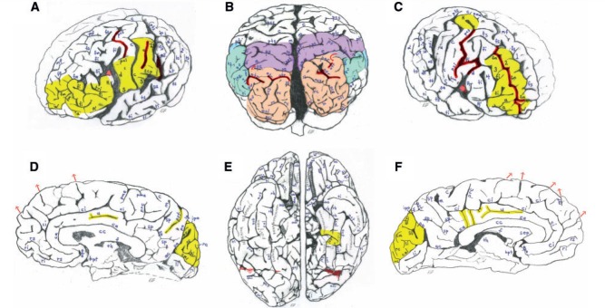
Figure 10.
Highlights of Einstein’s brain. (A) Figure 2 of the left lateral surface of Einstein’s brain highlighted to summarize interesting features, which have been darkened. These include a connected precentral superior and inferior sulcus, a long unnamed sulcus in the inferior primary somatosensory cortex, and a posterior ascending limb of the Sylvian fissure that, contrary to the literature, is not confluent with the postcentral inferior sulcus. Unusually expanded primary somatosensory (posterior to the central sulcus) and primary motor cortices (rectangular region below the precentral inferior sulcus) are highlighted in yellow, as are the unusually convoluted surface of the pars triangularis (part of Broca’s speech area) and the frontal polar region. (B) Figure 7 of an occipital view of Einstein’s brain coloured to indicate the approximate boundaries of the superior parietal lobule (purple), inferior parietal lobule (aqua/blue) and occipital lobes (salmon). Presence of four transverse occipital sulci (darkened) is extremely rare, if not unique. Parts of the posterior temporal lobes are uncoloured below the inferior parietal lobules and rostral to the occipital lobes. Although the small striped patch between the superior and inferior parietal lobules on the right belongs with the superior parietal lobule rather than the angular gyrus of the inferior parietal lobule, its relationship with the bordering intraparietal sulcus is usually associated with a location in the angular gyrus. It would therefore be interesting to study the cytoarchitecture of this enigmatic patch of cortex. Notice that the inferior parietal lobule is favoured on the left (and see Fig. 4), while the superior parietal lobule is relatively greater on the right. There is also an asymmetry that favours the right posterior temporal region, and the right occipital lobe is shifted forward relative to the left. (C) Figure 2 of the right lateral surface of Einstein’s brain highlighted to summarize interesting features, including sulci that are darkened. Unusual sulcal patterns include a connected precentral superior and inferior sulcus, a caudal segment of the inferior frontal sulcus that is connected with both the diagonal and precentral inferior sulci, and a long midfrontal sulcus that terminates in the fronto-marginal sulcus of Wernicke. The midfrontal sulcus divides the middle frontal region into two distinct gyri (highlighted in yellow), which causes Einstein’s right frontal lobe to have four rather than the typical three gyri. The enlarged ‘knob’ that probably represents motor cortex for the left hand and the highly convoluted frontal polar region are also highlighted in yellow. (D) Figure 8 of the right medial surface of Einstein’s brain with unusual features highlighted in yellow. The cingulate gyrus has a long unnamed sulcus, the transverse parietal sulcus seems relatively elongated and the cuneus appears to be unusually convoluted. (E) Figure 6 of the basal surface of Einstein’s brain highlighted to show that the left collateral sulcus is divided into two segments, and that part of the fusiform gyrus bridges between these segments to merge with the parahippocampal gyrus. (F) Figure 8 of the left medial surface of Einstein’s brain with unusual features highlighted in yellow. The cingulate gyrus has a long unnamed sulcus, and the cingulate sulcus gives off four inferiorly directed branches (two of which are tiny), which suggest that the cingulate gyrus may be relatively convoluted. The cuneus appears to be unusually convoluted. The figures are reproduced with permission from the National Museum of Health and Medicine.

