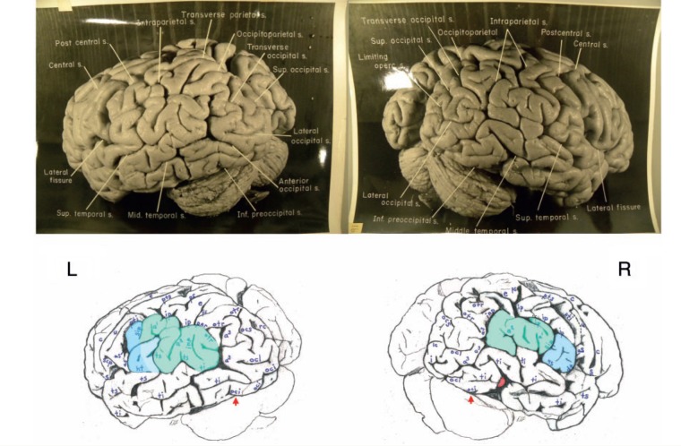
Figure 4.
Top: Photographs of the left (L) and right (R) lateral surfaces of Einstein’s brain taken with the back of the brain rotated towards the viewer, with original labels. Bottom: Our identifications. The arrows indicate the pre-occipital notch at the inferolateral border of each hemisphere, which indicate the approximate inferior boundary between the lateral surfaces of the temporal and occipital lobes; on the right, an apparent artificial cut severed the rostral tip (shaded red) of a gyrus in the posterior part of the inferior temporal lobe. This cut appears to be a lateral extension of that observed on the right side of the base of the brain (Fig. 6). Typically, the supramarginal gyrus surrounds the posterior ascending limb of the Sylvian, and the angular gyrus surrounds the upturned end(s) of superior temporal sulcus. These gyri are separated approximately at the level of the intermedius primus sulcus of Jensen and together form the inferior parietal lobule. The supramarginal gyri are shaded blue; the angular gyri are aqua. In the left hemisphere, part of the cortical region above posterior terminal horizontal branch of the Sylvian is shaded an inbetween colour because it could arguably belong to either gyrus. Einstein’s inferior parietal lobules have different shapes in the two hemispheres, and appear to be relatively larger on the left side. Sulci: a1 = ascending branch of the superior temporal sulcus; a2 = angular; a3 = anterior occipital; aS = posterior ascending limb of the Sylvian; c = central; dt = descending terminal branch of the Sylvian; e = processus acuminis; ht = posterior terminal horizontal branch of the Sylvian; i = inferior polar; inp = intermediate posterior parietal; ip = intraparietal; lc = lateral calcarine; oci = inferior occipital; ocl = lateral occipital; ocs = superior occipital; otr = transverse occipital; par = paroccipital; ps = superior parietal; pti = postcentral inferior; pts = postcentral superior; rc = retrocalcarine; S = Sylvian fissure; scp = subcentral posterior; sip = intermedius primus of Jensen; ti = inferior temporal; ts = superior temporal; u = unnamed. The figure is reproduced with permission from the National Museum of Health and Medicine.

