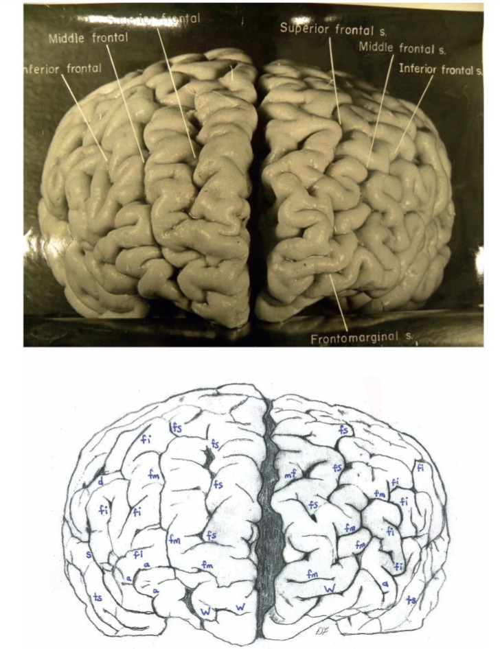
Figure 5.
Top: Photograph of a frontal view of Einstein’s brain in an unconventional orientation, with original labels. Bottom: Our identifications of sulci. a = additional inferior frontal; fi = inferior frontal; fm = midfrontal; fs = superior frontal; mf = medial frontal; S = Sylvian fissure; ts = superior temporal; W = fronto-marginal of Wernicke. The figure is reproduced with permission from the National Museum of Health and Medicine.

