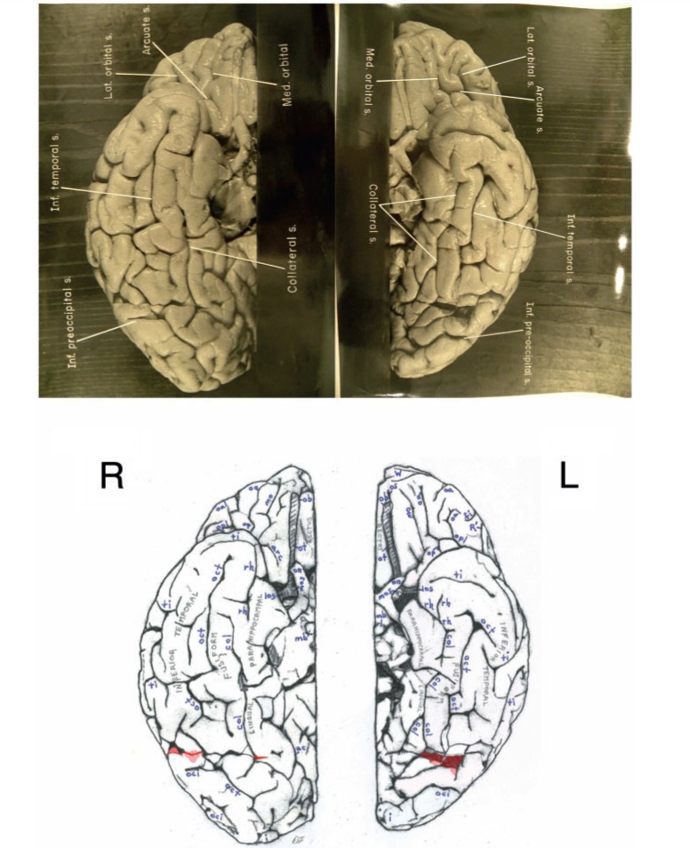
Figure 6.
Top: Separate photographs of the right (R) and left (L) basal views of Einstein’s bisected brain with cerebellum removed and original labels. Bottom: Our identifications. The two photographs are not to the same scale and the right hemisphere is rotated slightly laterally compared with the left, as suggested by a published basal photograph of the entire brain with its cerebellum attached (Witelson et al., 1999b). The base of Einstein’s brain appears to have been accidentally cut, perhaps with a scalpel, as indicated in red shading. This may have occurred during removal of the dura mater (tentorium cerebelli) that separates the dorsum of the cerebellum from the inferior surface of the occipital lobes. Magnifying the photographs on a computer screen should facilitate observation of these cuts. See Fig. 4 for an extension of this cut that reached the right lateral surface of the temporal lobe where it severed the tip of a gyrus (shaded in red). Sulci: arc = arcuate orbital; col = collateral; fi = inferior frontal; i = inferior polar; mo = medial orbital; oa = anterior orbital; oal = lateral anterior orbital; oci = inferior occipital; oct = occipito-temporal; op = posterior orbital; opl = lateral posterior orbital; os = olfactory; R’ = horizontal ramus of anterior Sylvian fissure; rh = rhinal; ti = inferior temporal. Abbreviations of other features: los = lateral olfactory stria; mb = mammillary body; mos = medial olfactory stria; ob = olfactory bulb; on = optic nerve; ot = olfactory tract. The figure is reproduced with permission from the National Museum of Health and Medicine.

