Abstract
Objective
To investigate the bioecological relationship between Chagas disease peridomestic vectors and reptiles as source of feeding.
Methods
In a three-story building, triatomines were captured by direct search and electric vacuum cleaner search in and outside the building. Then, age structure of the captured Triatoma maculata (T. maculata) were identified and recorded. Reptiles living in sympatric with the triatomines were also searched.
Results
T. maculata were found living sympatric with geckos (Thecadactylus rapicauda) and they bit residents of the apartment building in study. A total of 1 448 individuals of T. maculata were captured within three days, of which 74.2% (1 074 eggs) were eggs, 21.5% were nymphs at different stages, and 4.3% were adults.
Conclusions
The association of T. maculata and T. rapicauda is an effective strategy of colonizing dwellings located in the vicinity of the habitat where both species are present; and therefore, could have implications of high importance in the intradomiciliary transmission of Chagas disease.
Keywords: Chagas disease, Gekkonidae, Reduviidae, Thecadactylus rapicauda, Triatoma maculate, Trypanosoma cruzi, Vector domiciliation
1. Introduction
It is estimated that in Central and South America there exist approximately 12 to 16 million people infected with Trypanosoma cruzi (T. cruzi) in endemic areas[1]. In Venezuela, the areas at higher risk of T. cruzi infection have traditionally been Anzoátegui, Barinas, Cojedes, Carabobo, Trujillo, Aragua, Yaracuy and Portuguesa states, which represent 66.8% confirmed cases of Chagas disease[2]. During the 1960s, the Chagas disease endemic areas in the country possessed triatomine vector infestation indexes of 70% in small villages. This alarming infestation prompted chemical control programs and improvement in the construction of rural housing. As a consequence of the anti-triatomine battle, the percentage of humans testing positive for Chagas antibodies decreased significantly to 6.2% in 1995 and even lower (4.3%) in 1996[3].
At the first half of the 20th century, the following order of importance for the three main vector species of T. cruzi in Venezuela was established: Rhodnius prolixus (Stal 1859) (R. prolixus), Triatoma maculata (Erichson 1848) (T. maculata) and Panstrongylus geniculatus (Latreille 1811) (P. geniculatus). This classification was based on the epidemic relevancies for the Chagas disease such as hematophagic habits, times of defecation, rates of natural infection with T. cruzi and the biological attributes that made them compatible with domiciliary environments.
In Latin America, within the last two decades, different studies have emphasized the significant changes in the diversity of triatomine species, which were originally wild; however, now have intruded residential homes and have subsequently been domiciled[4]. This was the case for P. geniculatus, where populations of peri and intradomiciliary have been reported in Brazil and Venezuela[4]–[6]. Concomitantly, it has been suggested that it could be playing an important role in the maintenance of the domestic cycle of T. cruzi inside certain social-environmental conditions[5],[6].
In November 2007, P. geniculatus was responsible for T. cruzi oral transmission of 123 patients in Chacao (Reyes-Lugo personal communication) by guava juice contaminated with feces of this triatomine, and it was also suspected of infecting 50 individuals in Chichiriviche de la Costa in March 2009 (Rodriguez-Acosta personal communication) both locations within the state of Miranda, Venezuela.
T. maculata can be found in Aruba, Bonaire, Brazil, Colombia, Curazao, Guyana, Suriname and Venezuela[7], it is an ornitophilic specie, considered opportunistic because on occasion it has been found to colonize near henhouses in Brazil and Venezuela[8],[9]. At the same time, T. maculata is considered to be responsible for the Chagas diseases peridomestic cycle in some of the countries mentioned above[10],[14]. This work describes for the first time a population of domiciliated T. maculata strongly associated with the gecko Thecadactylus rapicauda (T. rapicauda) (Houttuyn 1782) (Reptilia: Squamata: Saurian: Gekkonidae), which is distributed in Central and South America[10].
2. Materials and methods
2.1. Ethical statement
The human study (blood samples test) was performed in accordance with the principles of the Declaration of Helsinki, and the norms of the Tropical Medicine Institute of the Universidad Central de Venezuela Bioethics Committee.
All the experimental events concerning the use of live animals were done by specialized personnel. The Venezuelan pertinent regulations as well as institutional guidelines, according to protocols approved by the Tropical Medicine Institute of the Universidad Central de Venezuela Bioethics Committee.
2.2. Study area
The study area was in a three-story residential building (Car-arte) in the town of Pitahaya State of Miranda, Venezuela, which locates 45 km south of Caracas. It is 293 m above sea level near a dry tropical forest. Pitahaya has a total annual precipitation of 1 350 mm and an average annual temperature of 26 °C. The plants that characterize the surrounding ecosystem belong to the families of Loganiaceae, Malpighiaceae, Malvaceae, Meliaceae, Moraceae, Nyctaginaceae, Orchidaceae, Passifloraceae, Phytolacaceae, Poaceae, Polygonaceae, Portulacaeae, Rubiaceae, Sapindaceae, Smilacaeae, Ulmaceae, Vitaceae, Zygophyllaceae and Mimosacea[11]. Currently, this woodland is under heavy construction. Charallave, the nearest town is essentially a mosaic of suburban areas, including a small relict forest, which at an earlier time covered a large area, and secondary savannas of different dimensions. Charallave held 14 329 homes in 1990[19] and 129 213 inhabitants in 2005[12].
2.3. Triatomines search
Triatomines were detected in an exhaustive, manually revised building, exteriorly and interiorly. The roof, walls and floors were inspected externally. Internally, all furniture, objects and equipment were revised. The direct search included a total of 10 hours, nine participants, for a capture effort of 1.11 hours/man.
An electric vacuum cleaner connected to a 6 m-hose with a diameter of 4 cm was used (Genie® brand, PB15 Model / 110 - 120 V, 60 Hz, 7.8 amp, 1.5 Peak HP Wet-Dry 10 gallons[4] for areas difficult to access (e.g. cracks and holes in floors, walls and roofs, cumulative objects, switches and lamps, conduit of the building's electric system, etc). The vacuum cleaner samples were analyzed in the laboratory with the help of a magnifying stereoscopic (Wild, Germany). Entomological pincers and a fine paintbrush were used to separate the live and dead triatomines, as well as other parts of their anatomy, exuviae and collected eggs. The identification of the insects was carried out according to methods of Ramiréz-Pérez and Carcavallo et al[13],[14].
The triatomines (live, dead as well as exhuvia and eggs), collected by both methods, were separated and stored in properly identified 600 mL, plastic containers. The animals were transferred in a 20 L, polycarbonate container with ice and a humid atmosphere in order to maintain the insects at a constant environment. The live T. maculata were maintained in our Insectary to start a new colony. During the search for triatomines, information was gathered in regards to characteristics of the inspected areas. For instance, the size of the residential area, living conditions and description of surrounding areas. The temperature and the environmental relative humidity in situ were also measured.
The geckos, living in sympatric with the triatomines, were also captured manually using entomology nets and transferred to the Tropical Medicine Institute Serpentarium for their later identification with the Donoso-Barros (1968) keys[15].
2.4. Detection of T. cruzi
Parasitological conventional techniques (Giemsa staining and agar-blood cultures) were applied to determine the presence of promastigots of T. cruzi in the captured triatomines. The intestinal contents and fresh feces of some T. maculata specimens were used in these techniques.
A morphological comparison between intact red blood cells in the digestive tract of some triatomines and the geckos' red blood cells was carried out with the purpose of establishing the blood ingest origin in the triatomines. However, opossum (Didelphis marsupialis) feces were observed in a nearby area. The colony of T. maculata studied in Pitahaya was feeding off the geckos (T. rapicauda Houttuyn 1782), which was evidenced by the presence of nucleated red blood cells (characteristic of reptiles) found in the colony when analyzed in the laboratory. In addition to feeding off the geckos, at least ten individuals were feeding off humans. The colony of T. maculata moved from the roof to the bedrooms via the conduit of the building's electric system.
The human inhabitants bitten by T. maculata were subjected to clinical and parasitological studies for T. cruzi infection at the Tropical Medicine Institute of the Universidad Central de Venezuela.
3. Results
3.1. Description of the homes colonized by T. maculata
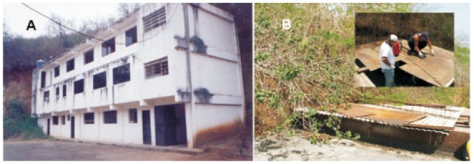
The homes occupied by T. maculata were in a three-story building with a total of three apartments (200 m2 each). Each apartment consists of a sitting and dining room, a kitchen with a laundry area, two bedrooms and two bathrooms, which were typical modern 21st century homes. The walls and ceilings were built with clay blocks and had a nice paint finish; the floor of the first-story apartment was made of cement and was sealed with an epoxy coat, while the floors of the rest of the apartments were made of ceramic. The residence in study had a water pipe system and the electric power ran through internal wired ducts. However, these services were functional only for the first floor. At the moment of this study, in this floor, some rooms and a bathroom were being used as a depot for material, and bats and geckos were observed living in and around these areas (Figure 1). All floors contained many light switches (some without switch plates), uncovered lamps, and 15 functional incandescent light bulbs were distributed throughout the external part of the building and in the stairways (Figure 1).
Figure 1. The homes colonized by T. maculata.

A: Apartment complex located in Pitahaya, Miranda state, Venezuela 10°12′ 2339 N and 66°51′ 1326 W, where T. maculata was associated with the gecko T. rapicauda. B: Location of T. maculata colony. Trash accumulation, due to the falling leaves from the neighboring trees, can be seen. The improvised roof with metal sheets, acerolit® and wooden pieces placed in the exit of the stairway which leads to the roof was dismantled, and a vacuum cleaner was used to collect samples.
Five people occupied the building, which included the watchman living on the first floor and the family in study, composed of four members (3 years old boy, 6 years old girl, 36 years old mother and 42 years old father), on the second floor. The third floor was uninhabited without windows or doors. The toilet and hygiene conditions of the inhabited areas were deemed appropriate.
The largest colony of T. maculata next to a small population of the geckos T. rapicauda (20 individuals) was discovered on the building's roof, which was an area containing wooden, plastic and metallic sheets in a scattered fashion. Between these sheets, an accumulation of dirt, dried leaves (4 kg) and rust (8 kg) was found.
The mean temperature and relative humidity registered in the three consecutive days (April 19-21, 2007; 14:00 h) inside the apartment, exactly below the improvised roof of sheets where the T. maculata colony, was (29.65±3.46) °C and (54.21±12.58)%, respectively.
3.2. Capture and age structure of the T. maculata domiciliary populations
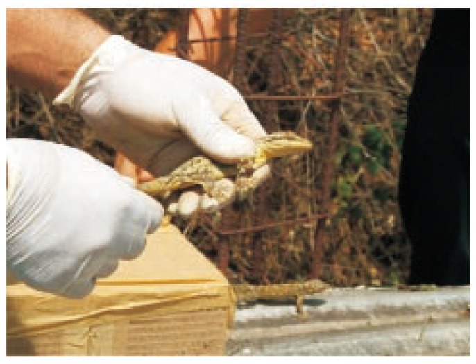
The results of the captured triatomines are presented in Table 1. The total number of T. maculata was 1 448 and eggs collected inside the apartment complex acount for 74.2% (1 074 eggs), Nymphs were 21.5%, and adults were 4.3%. All the captured triatomines were engorged with blood, with the exception of two newly emerged adults (1 female and 1 male) evidenced by the nymph V exhuvia found next to the captured adults. During the inspection of the T. maculata nest, a gecko burrow was discovered with 20 individuals (another gecko was seen escaping), varying in ages and sizes, and also including some eggs (Figure 2).
Table 1. The number of T. maculata specimens captured by developmental stages in the studied building.
| Stage of development | Method of capture |
||||||
| Direct searchinga |
Vacuum cleanerb,c |
||||||
| Live | Deaths | Complete exhuvias | Live | Complete exhuvias | Total | % | |
| Eggs | 10 | 35 | 300d | 0 | 729d | 1074 | 74.2 |
| Nymphs I | 7 | 4 | 3 | 3 | 1 | 18 | 1.2 |
| Nymphs II | 15 | 5 | 18 | 1 | 1 | 40 | 2.8 |
| Nymphs III | 8 | 16 | 30 | 4 | 2 | 60 | 4.1 |
| Nymphs IV | 24 | 14 | 8 | 2 | 1 | 49 | 3.4 |
| Nymphs V | 76 | 37 | 26 | 1 | 5 | 145 | 10.0 |
| Females | 15 | 4 | 20 | 1 | 2 | 42 | 2.9 |
| Males | 8 | 3 | 6 | 0 | 3 | 20 | 1.4 |
| Total | 163 | 118 | 411 | 12 | 744 | 1448 | 100.0 |
a Time used during direct roof search: nine participants /10 hours = 1.11 hour/person
b Time used with an electric vacuum cleaner roof search: two participants = 1 h 6 min.
c Collected composition: triatomines, 522 grams of trash and 538 grams of paper; Odontomachus spp (Formicidae; Hymenoptera), Ligaeidae (Hemiptera), Piralidae (Lepidoptera) were the associated fauna found.
d Corium
Figure 2. A gecko T. rapicauda captured in a T. maculata nest, in an apartment complex in Pitahaya, Miranda state. To the bottom right of the photo, another gecko can be seen escaping.

Thirteen nymphs were manually captured by the inhabitants in the apartment complex and brought to the Tropical Medicine Institute where the entomological inspection was carried out. Ten triatomines were located in the space between the two mattresses occupied by the children, and the mother captured three at the moment that the triatomines were ingurgitating on one of the children.
4. Discussion
Domiciliation of triatomines can be visualized as an expansion of the evolutionary plan from predator to a particular area within a habitat occupied by a hematophagous organism, where the domestic habitat merely symbolizes a specific type of “bed” prepared by vertebrates. The course of action is a specialized one and given that it appears to engage generalizations such as genetics in addition to morphological characters, it is possible to accept the process to be permanent[16].
Chagas disease mainly occurs in the poor, rural areas of Mexico, Central and South America and in very rare occasion in the Southern United States[17]. In Central and South America, these blood sucking bugs are consequences of human activities intruding on the original habitats of these insects, which lead to the formation of mosaic areas of vegetation coverage next to homes. These environmental alterations affect the populations of wild triatomines when reducing their living areas and their food sources, so that the ultimate effect is the establishment of triatomine populations in peridomicile and intradomicile[18]. In addition, the distribution and dispersion of the triatomines has also been associated with the change in climatic conditions, biogeographical aspects, and the nature of the shrubbery environment[19].
The following characteristics play key roles in developing domicile triatomine populations: (1) similar microclimatic conditions of homes to the natural ecotopes, particularly the temperature and relative humidity, (2) scarcity of wild animals for blood feeding, and (3) drastic environmental modifications to their natural habitats forcing them to seek shelter next to homes. The persistence of these stimuli will allow triatomine populations to better adaptation to that habitat once the colonizing process has been initiated[18].
Some of the original wild triatomines that have adapted fairly well to recent domiciliation are: P. geniculatus and Eratyrus mucronatus in Venezuela; Panstrongylus rufotuberculatus in Colombia; Triatoma dimidiata in Costa Rica; Triatoma infestans in Paraguay and Brazil; Triatoma rubovaria in Uruguay; Triatoma sordida and Triatoma guasayana in Argentina; Triatoma maculata in Brazil and Colombia[5],[19]–[24].
Many of the locations where T. maculata is currently present have different levels of human interventions, such as housing construction and farming areas, the main factors of the original habitat modifications for this triatomine. In Venezuela, in particular, they are found in the states of Anzoátegui, Bolívar, Lara, Miranda, Nueva Esparta (Antolín del Campo, Marcano, Díaz and Mariño municipalities) and Sucre. These triatomines, contrarily to past observations, have increased their presence in domiciles and peridomiciles in different towns throughout these Venezuelan states. For instance, in the Venezuelan Guayana, T. maculata was found naturally infected with T. cruzi and Triatoma rangeli[25]. The disappearance of their natural habitats has occurred in parallel with the disappearance of their natural feeding sources. Alternatively, it is considered that this condition could favor its biotic potential because it increases the contact frequency with the blood of mammals in peri and intradomicile[10], which possesses epidemiological importance in Chagas disease transmission. In Talaigua Nuevo (Bolívar, Colombia), it was demonstrated that this species verified the life cycle inside the domicile, feeding on mammals, reaching a 58.3% index of T. cruzi natural infection, thus taking on the responsibility for the intradomicile transmission of this protozoan. In Venezuela immature stages of triatomines have been observed feeding on mammals inside homes; however, resulting negative to T. cruzi infection[25],[26].
Other indicatives of the T. maculata biotic potential could be the fact that it can multiply in conditions of low humidity and relatively high temperature[27] and their proven ability to feed on lizards and snakes[28]. This would explain the findings in this current work of 1 448 T. maculata specimens at different development stages co-inhabiting with 20 specimens of geckos (T. rapicauda).
Furthermore, the distribution of these individuals collected in different development stages is in agreement with the population levels described for this species found in henhouses in Brazil and with a population equivalent to that reported for a typical domiciliary species such as Rhodnius prolixus[8],[9]. In Roraima (Brazil), T. maculata was the most numerous species found in henhouses, with a peridomiciliary infestation index of 16.7% (number of triatomines captured/number of captured × 100) and a crowding index of 12 500% (number of triatomines captured/number of houses × 100)[17].
T. maculata is considered to be responsible for maintaining the domestic cycle of Chagas disease in the peridomicile environment in the towns where it has been detected[8]–[29], with their T. cruzi natural infection indexes varying from 0% to 58.3%[23],[29]. This peridomiciliation is of particular interest in Chagas disease transmission. The adaptation of vector species to human dwellings in deforested areas is thought to be linked to recent findings of T. cruzi in human populations in the Amazon[30],[31] where the low frequency of human autochthonous cases of Chagas disease contrasts with the high infection index found in reservoirs and vectors[32].
The triatomines' colonization strategy to the contemporary peri-urban domicile appears novel if taken into consideration with the existing knowledge, which points out that the process of colonization of the domestic environment by these insects normally begins in the peridomiciliary surroundings. These surrounding include the housings of domestic animals or in the burrows of small synanthropics mammals such as pigs, rodents(Rattus rattus) and marsupials (Didelphis marsupialis). The latter two are those most frequently cited, which are also considered as T. cruzi reservoirs in the urban environment[33].
Today, triatomines have been observed to be associated with wild or domestic birds, and in rare opportunities with mammals [21],[23], validating the discovery in this report as the first description, to our knowledge, of co-habitation between the triatomine T. maculata and the gecko T. rapicauda.
The gecko, T. rapicauda, is a depredatory opportunist species that feeds mainly on insects. The possibility to feed on triatomines comes about through their reported consumption of other invertebrates such as terrestrial mollusks and arachnids[34],[35].
T. rapicauda has a wide geographical distribution. Today, it is common to observe in Center and South America reptiles of the Gekkonidae family colonizing homes located in areas occupied by tropical forests (dry and rainy). This colonization could be considered as a mechanism that facilitates the domiciliation of triatomines.
T. rapicauda is usually associated to debris and corrugated metallic roofs in abandoned constructions[36], which corresponds with the current work. These geckos have been described in rainy, humid and dry primary and secondary tropical forests, from 0 to 650 m altitude. They are a nocturnal species usually found around house lights, where they can be found feeding on insects. This situation can possess particular relevance for the indirect transmission of Chagas diseases in those communities that are located near these ecosystems; for they have been known to travel freely to and from these communities.
Nowadays, at least in Venezuela, it is relatively more common to observe populations of peri or intradomiciliary geckos than peridomiciliary pigs and hens in the area occupied by T. maculata. The association triatomine-gecko in the domestic environment could have implications of high importance for intradomiciliary transmission of Chagas disease due to its eclectic alimentary habits on birds, reptiles and mammals. It has been reported that trypanosomes are able to infect reptiles[37],[38]. These investigators successfully infected reptiles with parasites from patients with trypanosomiasis, verifying that reptiles could be reservoirs for human trypanosomes. Furthermore, Njagu et al[39] found pathogenic mammalian trypanosomes in monitor lizards in an endemic area for human trypanosomiasis, substantiating that reptiles are reservoirs for these parasites.
The matter becomes more prominence, recalling the work done by Medina[40], in which he found that T. rapicauda was easily infected with Leishmania parasites (a Trypanosomatid protozoa-related) add to this that parasites can invade the bloodstream in reptiles, we can understand the interest of deepening the study on lizards and clarify their status as potential reservoirs of human trypanosomiasis.
Although, in this particular study, the triatomines found feeding on the geckos were negative for T. cruzi, the fact that geckos provide a source of food for these triatomines in these domesticated areas could indirectly pose problematic in the spread of Chagas disease. All of these findings may have significant repercussions on potential policies to control Chagas disease, and could also explain earlier outbreaks of human trypanosomiasis in Venezuela.
Acknowledgments
We would like to thank the family of Pitahaya Town who kindly allowed us to conduct this study in the building.
Footnotes
Foundation Project: Supported by Proyecto Control de Vectores IMT-UCV.
Conflict of interest statement: We declare that we have no conflict of interest.
References
- 1.Dias J, Schofield C. The evolution of Chagas disease (American trypanosomiasis) control after 90 years since Carlos Chagas discovery. Mem Inst Oswaldo Cruz. 1999;94(Suppl 1):103–121. doi: 10.1590/S0074-02761999000700011. [DOI] [PubMed] [Google Scholar]
- 2.Ramirez N, Silva LC, Kiriakos D, Rodríguez-Morales AJ. Enfermedad de Chagas en Venezuela: Un bosquejo de su impacto sobre la salud publica. Acta Cient Estud. 2004;2:148–156. [Google Scholar]
- 3.PAHO . Washington: Oficina Sanitaria Panamericana; 1998. La Salud en las Américas. PAHO. Publicación Científica No. 569 Volumen II. Organización Panamericana de la Salud; pp. 1–592. [Google Scholar]
- 4.Reyes-Lugo M, Rodríguez-Acosta A. Domiciliation of selvatic Chagas disease vector Panstrongylus geniculatus Latreille 1811 (Triatominae: Reduviidae) in Venezuela. Trans Roy Soc Trop Med Hyg. 2000;94:508. doi: 10.1016/s0035-9203(00)90068-3. [DOI] [PubMed] [Google Scholar]
- 5.Reyes-Lugo M. ¿Realmente el chipo Panstrongylus geniculatus tiene poca importancia como transmisor de la Enfermedad de Chagas en Venezuela? Arch Ven Med Trop. 1999;3:3–4. [Google Scholar]
- 6.Carrasco HJ, Torrellas A, García C, Segovia M, Feliciangeli MD. Risk of Trypanosoma cruzi I Kinetoplastida: Trypanosomatidae transmission by Panstrongylus geniculatus Hemiptera: Reduviidae in Caracas Metropolitan District and neighboring States, Venezuela. Int J Parasitol. 2005;35:1379–1384. doi: 10.1016/j.ijpara.2005.05.003. [DOI] [PubMed] [Google Scholar]
- 7.Schofield CJ. Triatominae. Biologíay control. UK: Euro-conmmunica Publications; 1994. p. 74. [Google Scholar]
- 8.Luitgards-Moura JF, Borges-Pereira J, Costa J, Lago ZP, Rosa-Freitas MG. On the possibility of autochthonous Chagas disease in Roraima (Amazon region, Brazil) (2000-2001) Rev Inst Med Trop Sao Paulo. 2005a;47:45–54. doi: 10.1590/s0036-46652005000100008. [DOI] [PubMed] [Google Scholar]
- 9.Luitgards-Moura JF, Vargas AB, Almeida CE, Magno-Esperanca G, Agapito-Souza R, Folly-Ramos E, et al. A Triatoma maculata (Hemiptera Reduviidae Triatominae) population from Roraima (Amazon Region, Brazil) has some bionomic characteristics of a potential Chagas disease vector. Rev Inst Med Trop Sao Paulo. 2005b;47:131–137. doi: 10.1590/s0036-46652005000300003. [DOI] [PubMed] [Google Scholar]
- 10.Kronauer DJC, Bergmann PJ, Mercer JM, Russell AP. A phylogeographically distinct and deep divergence in the widespread Neotropical turnip-tailed gecko, Thecadactylus rapicauda. Mol Phylogenet Evol. 2005;34:431–437. doi: 10.1016/j.ympev.2004.10.009. [DOI] [PubMed] [Google Scholar]
- 11.Fajardo L, González V, Nassar JM, Lacabana P, Portillo CA, Carrasquel F, et al. Tropical dry forests of venezuela: characterization and current conservation status. Biotropica. 2005;37:531–546. [Google Scholar]
- 12.OCEI . Anuario estadístico de Venezuela. Caracas: Oficina Central de Estadística e Información (OCEI); 2005. pp. 1–440. [Google Scholar]
- 13.Ramírez Pérez J. Revisión de los triatominos (Hemiptera: Reduviidae) en Venezuela. Bol Dir Malariol San Amb. 1987;27:118–146. [Google Scholar]
- 14.Carcavallo RU, Galíndez-Girón I, Jurberg J, Galvao C, Lent H. Pictorial keys for tribes genera and species of the subfamily Triatominae. In: Carcavallo RU, Galindez-Girón I, Jurberg J, Lent H, editors. Atlas of Chaga's disease vectors in the Americas. Rio de Janeiro: Fiocruz; 1998a. pp. 127–131. [Google Scholar]
- 15.Donoso-Barros R. The lizards of venezuela (checklist and key) Caribb J Sci. 1968;8:105–122. [Google Scholar]
- 16.Schofield CJ, Diotaiuti L, Dujardin JP. The process of domestication in triatominae. Mem Inst Oswaldo Cruz. 1999;94(Suppl 1):375–378. doi: 10.1590/s0074-02761999000700073. [DOI] [PubMed] [Google Scholar]
- 17.WHO . Control of Chagas disease. Geneva: WHO; 2002. p. 109. [Google Scholar]
- 18.Forattini OP. Biogeografia, origem e distribuicao da domiciliacao de Triatomineos no Brasil. Rev Saude Pub. 1980;14:265–299. doi: 10.1590/s0034-89101980000300002. [DOI] [PubMed] [Google Scholar]
- 19.Zeledón R, Ugalde JA, Paniagua LA. Entomological and ecological aspects of six sylvatic species of Triatomines (Hemiptera: Reduviidae) from the collection on the National Biodiversity Institute of Costa Rica, Central America. Mem Inst Oswaldo Cruz. 2001;96:757–764. doi: 10.1590/s0074-02762001000600002. [DOI] [PubMed] [Google Scholar]
- 20.Ruas-Neto AL, Krug L. Epidemiologia da doença de Chagas no Rio Grande do Sul: A distribução vetorial. Bol Soc Zool Uruguay. 1995;9:20–32. [Google Scholar]
- 21.Rojas de Arias A. Chagas disease in Paraguay. PAHO/HCP/HCT/72/96. 1996 [Google Scholar]
- 22.Soto-Vivas A, Barazarte H, Molina de Fernández D. Primer registro de Eratyrus mucronatus Stål 1959 (Hemiptera: Reduviidae) en el ambiente domiciliario en Venezuela. Entomotropica. 2001;16:215–217. [Google Scholar]
- 23.Cortés L, Suárez H. Triatominos (Reduviidae): Triatominae en un foco de enfermedad de Chagas en Talaigua Nuevo Bolívar, Colombia. Biomédica Bogotá. 2004;25:568–574. [PubMed] [Google Scholar]
- 24.Guhl F, Aguilera G, Pinto N, Vergara D. Actualización de la distribución geográfica y ecoepidemiología de la fauna de triatominos (Reduviidae: Triatominae) en Colombia. Biomédica. 2007;27(suppl 1):143–162. [PubMed] [Google Scholar]
- 25.Dias E. Doença de Chagas nas Américas. IV. Colômbia, Venezuela e Guianas. Rev Bras Malar. 1952;4:255–280. [PubMed] [Google Scholar]
- 26.Pifano CF. La dinámica epidemiológica de la Enfermedad de Chagas en el Valle de los Naranjos, estado de Carabobo, Venezuela. Arch Ven Med Trop Parasitol Med. 1973;5:31–45. [Google Scholar]
- 27.Feliciangeli MD, Rabinovich JE. Vital statistics of triatominae hemiptera: reduviidae under laboratory conditions II Triatoma maculata. J Med Entomol. 1985;22:43–48. [Google Scholar]
- 28.Urdaneta-Morales S, McLure I. Observations upon hematophagy in Venezuela triatomines fed upon poikilotherms. Acta Cient Ven. 1972;23:161–164. [PubMed] [Google Scholar]
- 29.Pifano CF. Algunos aspectos de la Enfermedad de Chagas en Venezuela. Arch Ven Med Trop Parasitol Med. 1960;3:73–99. [PubMed] [Google Scholar]
- 30.Coura JR. Doença de Chagas como endemia na Amazônia Brasileira: risco ou hipótese? Rev Soc Bras Med Trop. 1990;23:67–70. doi: 10.1590/s0037-86821990000200001. [DOI] [PubMed] [Google Scholar]
- 31.Walsh JF, Molyneux DH, Birley MH. Deforestation: effects on vector-borne diseases. Parasitology. 1993;106:S55–S75. doi: 10.1017/s0031182000086121. [DOI] [PubMed] [Google Scholar]
- 32.Valente SAS, Valente VC. Situação atual da doença de Chagas na Amazônia. Rev Soc Brasil Med Trop. 1993;26(Suppl 2):68–70. [Google Scholar]
- 33.Herrera L, Urdaneta-Morales S, Carrasco H. Trypanosoma cruzi: comportamiento de metatripomastigotes obtenidos de Didelphis marsupialis y Panstrongylus geniculatus. Rev Cient FCV-LUZ. 2003;4:307–311. [Google Scholar]
- 34.Duellman WE. The biology of an equatorial herpetofauna in Amazonian Ecuador. Lawrence: University Press of Kansas; 1978. pp. 1–352. [Google Scholar]
- 35.Hoogmoed MS. Notes on the herpetofauna of Suriname IV: the lizards and amphisbaenians of Suriname. Biogeographica. 1973;4:1–419. [Google Scholar]
- 36.Avila-Peres TCS. The lizards of Brazilian Amazônia. Zool Verh Leiden. 1995;299:1–706. [Google Scholar]
- 37.Woo P, Soltys MA. The experimental infection of reptiles with Trypanosoma brucei. Ann Trop Med Parasitol. 1969;63:35–38. doi: 10.1080/00034983.1969.11686597. [DOI] [PubMed] [Google Scholar]
- 38.Molyneux DH. Animal reservoirs and Gambian trypanosomiasis. Ann Soc Belge Med Trop. 1973;53:605–618. [PubMed] [Google Scholar]
- 39.Njagu Z, Mihok S, Kokwaro E, Verloo D. Isolation of Trypanosoma brucei from the monitor lizard Varanus niloticus, in an endemic focus of Rhodesian sleeping sickness in Kenya. Acta Trop. 1999;72:137–148. doi: 10.1016/s0001-706x(98)00092-8. [DOI] [PubMed] [Google Scholar]
- 40.Medina R. Leishmaniasis experimental en animales silvestres. Rev Dermatol Venez. 1966;7:91–119. [Google Scholar]


