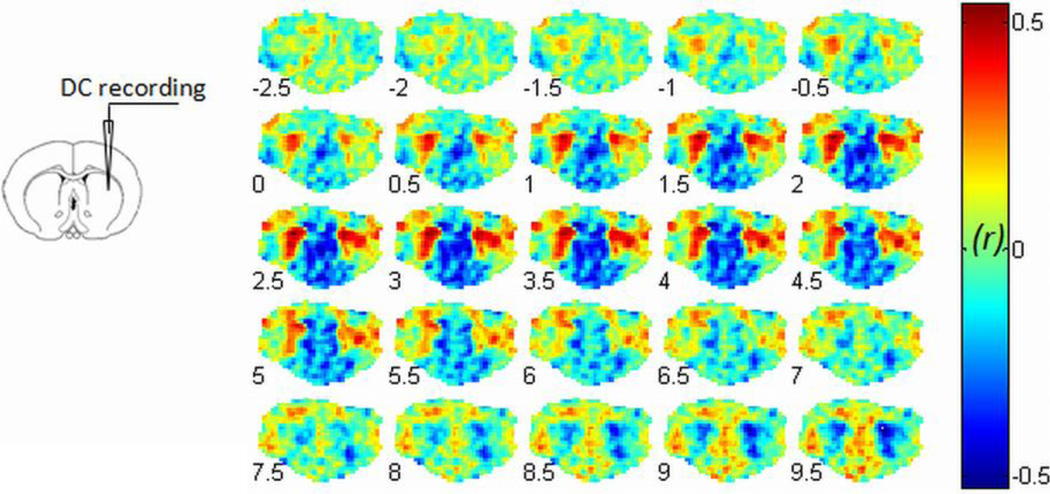
Fig. 2.
Simultaneous DC recording from CP during fMRI. The recording site in the caudate putamen (CP) is illustrated at left panel. The BOLD/infraslow LFP correlation results with varied BOLD lags (−2.5 sec to 9.5 sec) are shown in the right panel. The maximum correlation is observed in bilateral CP when the BOLD signal is delayed by 2.5 sec relative to infraslow LFP under dexmedetomidine. The left side of the brain is on the left side of the image.

