Case notes
We developed, in portable document format (PDF), a detailed 3-dimensional (3D) interactive anatomic model of 23 pelvic structures that include the muscles, ligaments, and fascia of the pelvic floor and the organs it supports. Bones, blood vessels, and the perineum are illustrated as well. To produce this tool, 3D volumetric models were created from serial 5-mm–thick images that were obtained with a 3-Tesla magnetic resonance scanner. The subject was a healthy, 45-year-old, multiparous woman who was at the 50th percentile for height. Magnetic resonance images were then imported into 3D Slicer software (version 3.4.1; Brigham and Women’s Hospital, Boston, MA). Each structure was traced with the use of the most clearly visible axial and/or coronal plane images and lofted into a 3D virtual model that was based on our previous anatomic work.1-5 Models were validated against the original scans and tracings.
Next, the 3D models that had been generated by 3D Slicer were imported into Adobe 3D Reviewer (version 9.0; Adobe Systems Inc, San Jose, CA), exported as a universal 3D file, and, with the aid of the program’s 3D tool, inserted into Adobe Acrobat 9 Pro (Adobe Systems Inc) and saved as a PDF file.
Conclusion
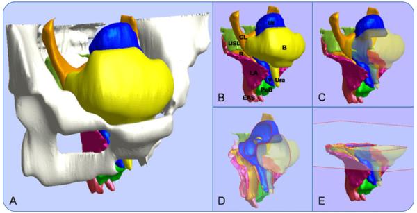
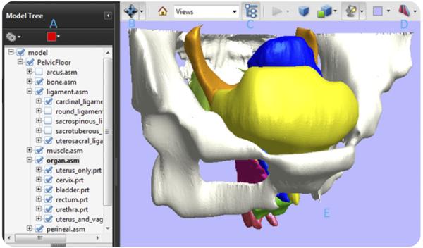
The 3D PDF file allows the user to view and manipulate detailed 3D models of pelvic anatomy without any specialized visualization software. For example, the model can be rotated and magnified; and structures can be hidden or rendered transparent, which makes it easier to understand complex anatomic relationships (Figures 1 and 2; an interactive model is available online at www.AJOG.org). All that is needed is the readily available (and free) Adobe Reader; cross-section capability is available with Adobe Acrobat. Availability of this anatomically accurate model is critical to avoiding errors in comprehension that can arise from conceptually based anatomic representations.
FIGURE 1.
The user can manipulate the 3-dimensional model of pelvic structures. A, A three-quarter right anterolateral view. B, Hiding the bones reveals selected features. C, Making the bladder and urethra transparent reveals the underlying structures. D, Sample sagittal cross-section of the remaining structures. E, Sample axial cross-section.
B, bladder; CL, cardinal ligament; EAS, external anal sphincter; LA, levator ani; PeB, perineal body; R, rectum; Ura, urethra; USL, uterosacral ligament; Ut, uterus; V, vagina.
Luo. A model patient: female pelvic anatomy viewed in diverse 3-dimensional images. Am J Obstet Gynecol 2011.
FIGURE 2.
The manipulation interface includes a model tree and toolbar. A, The model tree allows users to hide, isolate, or render transparent individual anatomic structures by right clicking on the label. B, Using this button in the 3-dimensional toolbar at the top, it is possible to zoom in or out, rotate the model, spin it, and pan over it. C, The model tree can be toggled on and off with this button. D, Cross-sections can be cut at a given location and orientation. E, The 3-dimensional model is activated by clicking on the portable document format file.
Luo. A model patient: female pelvic anatomy viewed in diverse 3-dimensional images. Am J Obstet Gynecol 2011.
Supplementary Material
Acknowledgments
Supported by the Eunice Kennedy Shriver National Institute of Child Health and Human Development, Grant R01 HD038665, and the Office for Research on Women’s Health, Specialized Centers of Research: Sex and Gender Factors Affecting Women’s Health, Grant P50 HD044406.
J.O.L.D. and J.A.A-M. receive research support from American Medical Systems and Kimberly Clark Corporation. J.O.L.D. has been a consultant for Johnson and Johnson and American Medical Systems. J.A.A-M. has been a consultant for American Medical Systems. J.L.’s doctoral studies are funded partially by American Medical Systems and Kimberly Clark Corporation.
Footnotes
Presented as a short oral presentation at the 31st Annual Scientific Meeting of the American Urogynecologic Society, Long Beach, CA, Sept. 30-Oct. 2, 2010.
Click Supplementary Content under the article title in the online Table of Contents
Contributor Information
Jiajia Luo, Biomechanics Research Laboratory, Department of Mechanical Engineering, College of Engineering, Medical School, University of Michigan, Ann Arbor, MI.
James A. Ashton-Miller, Biomechanics Research Laboratory, Department of Mechanical Engineering, College of Engineering, Medical School, University of Michigan, Ann Arbor, MI.
John O.L. DeLancey, Department of Obstetrics and Gynecology, Medical School, University of Michigan, Ann Arbor, MI.
REFERENCES
- 1.Hsu Y, Fenner DE, Weadock WJ, DeLancey JO. Magnetic resonance imaging and 3-dimensional analysis of external anal sphincter anatomy. Obstet Gynecol. 2005;106:1259–65. doi: 10.1097/01.AOG.0000189084.82449.fc. [DOI] [PMC free article] [PubMed] [Google Scholar]
- 2.Margulies RU, Hsu Y, Kearney R, Stein T, Umek WH, DeLancey JO. Appearance of the levator ani muscle subdivisions in magnetic resonance images. Obstet Gynecol. 2006;107:1064–9. doi: 10.1097/01.AOG.0000214952.28605.e8. [DOI] [PMC free article] [PubMed] [Google Scholar]
- 3.Hsu Y, Lewicky-Gaupp C, DeLancey JO. Posterior compartment anatomy as seen in magnetic resonance imaging and 3-dimensional reconstruction from asymptomatic nulliparas. Am J Obstet Gynecol. 2008;198:651.e1–7. doi: 10.1016/j.ajog.2007.11.032. [DOI] [PMC free article] [PubMed] [Google Scholar]
- 4.Brandon CJ, Lewicky-Gaupp C, Larson KA, DeLancey JO. Anatomy of the perineal membrane as seen in magnetic resonance images of nulliparous women. Am J Obstet Gynecol. 2009;200:583.e1–6. doi: 10.1016/j.ajog.2009.03.004. [DOI] [PMC free article] [PubMed] [Google Scholar]
- 5.Larson KA, Yousuf A, Lewicky-Gaupp C, Fenner DE, DeLancey JO. Perineal body anatomy in living women: 3-dimensional analysis using thin-slice magnetic resonance imaging. Am J Obstet Gynecol. 2010;203:494.e15–21. doi: 10.1016/j.ajog.2010.06.008. [DOI] [PMC free article] [PubMed] [Google Scholar]
Associated Data
This section collects any data citations, data availability statements, or supplementary materials included in this article.


