Abstract
Methanobactin (mb) is a small copper-binding peptide produced by methanotrophic bacteria and is intimately involved in both their copper metabolism and their role in the global carbon cycle. The structure for methanobactin comprises seven amino acids plus two chromophoric residues that appear unique to methanobactin. In a previously published structure, both chromophoric residues contain a thiocarbonyl attached to a hydroxyimidazolate ring. In addition, one is attached to a pyrrolidine ring, while the other to an isopropyl ester. A published X-ray determined structure for methanobactin shows these two chromophoric groups forming an N2S2 binding site for a single Cu(I) ion with distorted tetrahedral geometry. In this report we show that NMR, mass spectrometry, and chemical data, reveal a chemical structure that is significantly different than the previously published one. Specifically, the 1H and 13C NMR assignments are inconsistent with an N-terminal isopropyl ester and point instead to a 3-methylbutanoyl group. Our data also indicate that oxazolone rings instead of hydroxyimidazolate rings form the core of the two chromophoric residues. Because these rings are directly involved in the binding of Cu(I) and other metals by methanobactin, and are likely involved in the many chemical activities displayed by methanobactin, their correct identity is central to developing an accurate and detailed understanding of methanobactin’s many chemical and biological roles. For example, the oxazolone rings make methanobactin structurally more similar to other bacterially produced bactins and siderophores and suggest pathways for its biosynthesis.
Methanobactin (mb) is a small, copper binding peptide produced by methanotrophic bacteria, which use methane as their primary source of energy and play an integral role in the global carbon cycle. Mb can be isolated from the growth media of methanotrophs1,2,3 and is also found within the bacterial cells and associated with the copper and iron containing particulate methane mono-oxygenase (pMMO) enzyme.4,5 pMMO, along with its soluble form (sMMO) produced under limiting copper levels, is responsible for catalyzing the oxidation of methane to methanol. Thus, this enzyme has received much attention for its role in removing a potent greenhouse gas from the atmosphere, and for the development of new hydroxylation catalysts that could make more efficient use of natural gas.6
Mb is involved in many biological processes, including scavenging copper from the environment,1,7,8 serving as a copper chaperone for pMMO,1,4,7 complexing reactive oxygen species,4,9 and mediating both electron flow to pMMO3,4,9 and the genetic expression of pMMO.1,10 Copper-free mb can be isolated under low copper levels.3 It binds Cu(II) with subnanomolar affinity,8 reducing it to Cu(I).3,11,12 In addition, mb can bind Cd(II), Co(II), Fe(III), Mn(II), Ni(II), Zn(II), and Pb(II), and can bind and reduce Ag(I), Au(III), and possible Hg(II).14
The best characterized mb peptide is isolated from Methylosinus trichosporium OB3b.2,11 It contains seven amino acids and two unusual chromophoric residues, each containing a thiocarbonyl/enethiol attached to a conjugated ring system. Through a combination of mass spectrometry and X-ray crystallography, these ring systems were assigned as hydroxyimidazolates (Figure S1), which were ligated to Cu(I) in an N2S2 distorted tetrahedral geometry.2,11,13 However, it has been difficult to reconcile the chemical composition of these residues with possible biosynthetic pathways that bacteria use to produce mb. Since these residues directly participate in metal ion binding and are likely to be involved in many other chemical activities displayed by mb,9 it is crucial to establish their correct chemical structures.
In this communication, we report a revised structure for mb, which reconciles chemical analysis, NMR, X-ray, and mass spectrometry data with possible biosynthetic pathways for expression of mb in bacteria. The revised structure also clears up confusion in the literature2,7,11,12 on reporting both the chemical structure and the chemical formula for mb.
We isolated mb as described by Choi et al.3 and incubated it with between 0.5 and 1.0 Cu(II) ions per mb to obtain the Cu(I)-bound form. We purified the Cu(I)-bound mb by HPLC, and after lyophilization, re-dissolved the peptide in either 100% D2O or 90% H2O/10% D2O containing 9 mM sodium phosphate (pH 6.5) for NMR analysis (see Supporting Information). We collected the NMR spectra at 400 MHz or 600 MHz at either 5 °C or 25 °C. We used a combination of [1H,1H]-COSY, [1H,1H]-TOCSY, [1H,1H]-ROESY, [1H,15N]-HSQC, [1H,13C]-HSQC, and [1H,13C]-HMBC experiments to assign all of the non-hydroxyl 1H and 15N resonances, along with all of the 13C resonances except for the ones belonging to the conjugated rings (Table S1). While the sequential 1H and 13C assignments are consistent with the seven amino acid residues in the published structure (Figure S1), those for the N-terminal chromophoric residue are not. As shown in Figure 2, the spin system observed in [1H,1H]-TOCSY spectra for the N-terminal alkyl group is consistent with an isobutyl group rather than the previously reported isopropyl group.11 The [1H,13C]-HMBC experiment confirmed that this isobutyl moiety is attached to a carbonyl to form a 3-methylbutanoyl group.
Figure 2.
600 MHz spectra of methanobactin in 9 mM phosphate/10% D2O, pH6.5, 25°C. The sample was exposed to Cu(II) at 0.55 Cu:mb prior to isolation by HPLC. Selected regions of NMR spectra are shown indicating the presence of a 3-methylbutanoyl group. Connectivities and chemical shifts from A) [1H,1H]-TOCSY and B) [1H,13C]-HMBC spectra indicate an A3B3MXY type spin system (ie, CH3(CH3)CHCH2−). Additional multiple bond connectivity from [1H,13C]-HMBC spectra in C) and D) confirm attachment of a carbonyl group to this moiety.
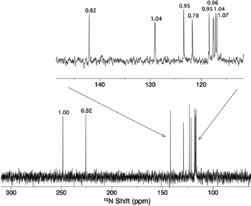
For a full characterization of the nitrogen content of the peptide, we used 15N direct detection on a uniformly 15N-labeled sample of Cu(I)-bound mb. Figure 3 shows the 1D proton decoupled 15N spectrum. We observed only ten 15N resonances with nearly equal intensities. This confutes the previously published structure with hydroxyimidazolates rings, which should display a total of twelve nitrogen resonances. Eight nitrogen resonances are attached to protons and were assigned using an [1H,15N]-HSQC experiment (Figure S2). The remaining two nitrogen resonances at 226 and 249 ppm show no proton coupling and require long cycle times (>10 s) to be observed, suggesting long longitudinal spin relaxation times (T1). Guided by the X-ray structure, we assigned these resonances to nitrogens belonging to two oxazolone rings and not hydroxyimidazolate rings.
Figure 3.
The proton-decoupled, 15N spectrum of U-15N Cu(I)-bound methanobactin at 25°C. A total of ten resonances are observed with the relative integrated intensities for each indicated.
To confirm the mass of the peptide, we carried out ESI-TOF mass spectrometry on Cu(I)-bound mb. In agreement with previously published data,2,11,12 we observe an m/z of 1215 for the (M-2H+63Cu)1− species. The presence of a 3-methylbutanoyl group instead of an isopropyl ester should result in a theoretical m/z of 1213. However, the inclusion of the two alkylidene oxazolone rings (instead of the originally reported hydroxyimidazolate rings) results in an expected m/z of 1215, which agrees well with our measurements. We repeated the mass spectrometry analysis multiple times, obtaining values for m/z that are within ±1.2 ppm of that expected for our revised structure (1215.1781), but lying 8 to 10 ppm below that expected for the previously reported structure11 (1215.1893) (Figure S3a).
The presence of oxazolone rings was also substantiated by chemical degradation of uncomplexed mb using methanolysis. Oxazolone rings contain a lactone that is susceptible to methanolysis15 and the concomitant loss of the conjugated ring systems can be followed by UV/Vis spectrophotometry.8 Interestingly, the oxazolone B ring is more susceptible to methanolysis than the A ring (Figure S4).8 As expected for the methanolysis of oxazolone rings, the mass of the product formed upon methanolysis of the oxazolone B ring corresponds to an increase of one equivalent of methanol (Figure S5). The opening of the oxazolone A ring requires higher concentrations of methanolic-HCl (0.01 vs. 0.001 saturated methanolic-HCl) and results in an additional increase in the mass by a second equivalent of methanol.
In summary, NMR, mass spectrometry and chemical degradation all point to a primary structure for mb containing two alkylidene oxazolone rings rather than the previously reported hydroxyimidazolate rings. This new revised mb structure also suggests possible pathways for the biosynthesis of mb from common amino acids. A scheme illustrating how mb could be synthesized by modifications of a peptide (LSGSCYPSSCM) is reported in the Supporting Information (Scheme S1). There are two proposed pathways. One is consistent with the fact that oxazolones have long been used as intermediates in the chemical syntheses of amino acids.15 The other is consistent with numerous examples of bacterially produced bactin and siderophore peptides containing the related oxazoline ring.16
Supplementary Material
Figure 1.
The revised structure for methanobactin; 1-(N-[mercapto-{5-oxo-2-(3-methylbutanoyl)oxazol-(Z)-4-ylidene}methyl]-Gly1-l-Ser2-l-Cys3-l-Tyr4)-pyrrolidin-2-yl-(mercapto-[5-oxo-oxazol-(Z)-4-ylidene]methyl)-l-Ser5-l-Cys6-l-Met7. The model shown is the (M-2H+63Cu)1− charged species, with the backbone traced in blue. The published X-ray crystal structure11 shows the chiral amino acids are all the l-enantiomers, the configurations of the exocyclic double bonds are both Z, and the chiral carbon 2 of the pyrrolidine ring is S.
Acknowledgement
This work was supported by an RSEC grant from the Chemistry Department, University of Minnesota (L.A.B and W.H.G), and by internal grants from ORSP, University of Wisconsin-Eau Claire (L.A.B and W.H.G). The 400 MHz Bruker Avance II NMR spectrometer and the Agilent 6210 ESI-TOF LC/MS used in these studies were funded with NSF grants (CHE-0521019 and CHE-0619296) to the UW-Eau Claire. NSF funding (BIR-961477) was also provided to the University of Minnesota NMR Facility.
Footnotes
Supporting Information Available: Experimental procedures,1H, 13C and 15N resonance assignments; additional 1H, 15N, 13C NMR and mass spectra; the methanolysis data, proposed schemes for the biosynthesis of mb; complete references for citations 8, 9 and 14. This material is available free of charge via the Internet at http://pubs.acs.org.
References
- 1.DiSpirito AA, Zahn JA, Graham DW, Kim HJ, Larive CK, Derrick TS, Cox CD, Taylor A. J Bacteriol. 1998;180:3606–3613. doi: 10.1128/jb.180.14.3606-3613.1998. [DOI] [PMC free article] [PubMed] [Google Scholar]
- 2.Kim HJ, Galeva N, Larive CK, Alterman M, Graham DW. Biochemistry. 2005;44:5140–5148. doi: 10.1021/bi047367r. [DOI] [PubMed] [Google Scholar]
- 3.Choi DW, Antholine WE, Do YS, Semrau JD, Kisting CJ, Kunz RC, Campbell D, Rao V, Hartsel SC, DiSpirito AA. Microbiology. 2005;151:3417–3426. doi: 10.1099/mic.0.28169-0. [DOI] [PubMed] [Google Scholar]
- 4.Choi DW, Kunz RC, Boyd ES, Semrau JD, Antholine WE, Han JI, Zahn JA, Boyd JM, de la Mora AM, DiSpirito AA. J Bacteriol. 2003;185:5755–5764. doi: 10.1128/JB.185.19.5755-5764.2003. [DOI] [PMC free article] [PubMed] [Google Scholar]
- 5.Martinho M, Choi DW, Dispirito AA, Antholine WE, Semrau JD, Munck E. J Am Chem Soc. 2007;129:15783–15785. doi: 10.1021/ja077682b. [DOI] [PMC free article] [PubMed] [Google Scholar]
- 6.Lieberman RL, Rosenzweig AC. Nature. 2005;434:177–182. doi: 10.1038/nature03311. [DOI] [PubMed] [Google Scholar]
- 7.Balasubramanian R, Rosenzweig AC. Curr Opin Chem Biol. 2008;12:245–249. doi: 10.1016/j.cbpa.2008.01.043. [DOI] [PMC free article] [PubMed] [Google Scholar]
- 8.Choi DW, et al. Biochemistry. 2006;45:1442–1453. doi: 10.1021/bi051815t. [DOI] [PubMed] [Google Scholar]
- 9.Choi DW, et al. J Inorg Biochem. 2008;102:1571–1580. doi: 10.1016/j.jinorgbio.2008.02.003. [DOI] [PubMed] [Google Scholar]
- 10.Knapp CW, Fowle DA, Kulczycki E, Roberts JA, Graham DW. Proc Natl Acad Sci U S A. 2007;104:12040–12045. doi: 10.1073/pnas.0702879104. [DOI] [PMC free article] [PubMed] [Google Scholar]
- 11.Kim HJ, Graham DW, DiSpirito AA, Alterman MA, Galeva N, Larive CK, Asunskis D, Sherwood PM. Science. 2004;305:1612–1615. doi: 10.1126/science.1098322. [DOI] [PubMed] [Google Scholar]
- 12.Hakemian AS, Tinberg CE, Kondapalli KC, Telser J, Hoffman BM, Stemmler TL, Rosenzweig AC. J Am Chem Soc. 2005;127:17142–17143. doi: 10.1021/ja0558140. [DOI] [PMC free article] [PubMed] [Google Scholar]
- 13.Kim HJ. Ph.D. Thesis. University of Kansas; 2003. [Google Scholar]
- 14.Choi D, et al. J Inorg Biochem. 2006;100:2150–2161. doi: 10.1016/j.jinorgbio.2006.08.017. [DOI] [PubMed] [Google Scholar]
- 15.Fisk JS, Mosey RA, Tepe J. J. Chem Soc Rev. 2007;36:1432–1440. doi: 10.1039/b511113g. [DOI] [PubMed] [Google Scholar]
- 16.Roy RS, Gehring AM, Milne JC, Belshaw PJ, Walsh CT. Nat Prod Rep. 1999;16:249–263. doi: 10.1039/a806930a. [DOI] [PubMed] [Google Scholar]
Associated Data
This section collects any data citations, data availability statements, or supplementary materials included in this article.


