

Abstract
Pigmented epithelioid melanocytoma (PEM) is a recently proposed term which encompasses those melanocytic tumours previously designated as ‘animal-type melanoma’ or ‘pigment-synthesising melanoma’ and ‘epithelioid blue nevus’, the latter known to be associated with Carney's complex. We report a case of PEM in a previously well 26-year-old Caucasian woman who presented with a dark pigmented nodule on the anterior chest wall.
Background
In 2004, Zembowitz et al1 proposed the term ‘pigmented epithelioid melanocytoma’ (PEM) for a group of lesions including cases previously diagnosed as human animal-type melanoma and epithelioid blue nevus.
Case presentation
A 26-year-old Caucasian woman, with an unremarkable medical history, consulted for a blue–black nodule situated on the anterior chest wall. The lesion had appeared de novo and had slowly progressed. There was no personal or family history of skin cancer.
Physical examination revealed 1 cm blue–black, oval-shaped firm and quite mobile nodule, with smooth surface. Its clinical characteristics were compatible with those of a blue nevus. There was no palpable lymphadenopathy. A complete excision was performed and the lesion was sent for histological examination. This revealed PEM (figures 1–3). Further treatment including sentinel lymph node biopsy (SLNB) was negative for metastases. A complementary wide surgical excision was performed to include a 2 cm margin of normal skin.
Figure 1.
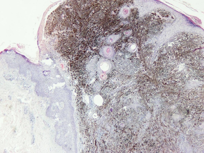
Pigmented epithelioid melanocytoma (H&E ×40).
Figure 2.
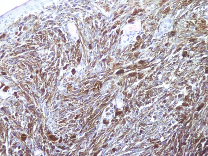
Tumour cells containing melanin granules that obscure nuclear details (H&E ×100).
Figure 3.
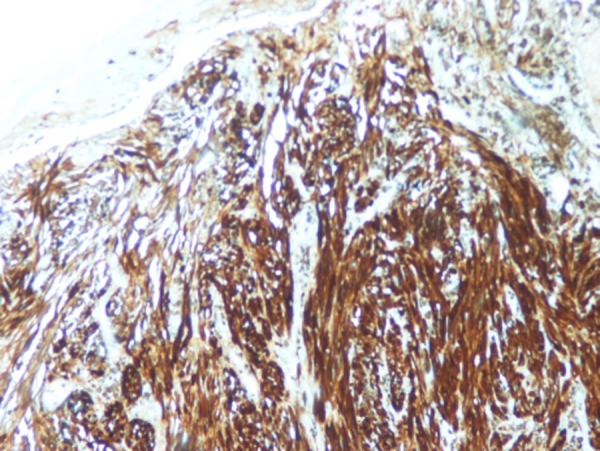
Immunohistochemical staining with S-100 confirms the melanocytic nature of the neoplastic cells (S-100 ×100).
Investigations
Haematological, biochemical and tumour marker (lactate dehydrogenase, LDH) profiles were within normal limits. Further investigation including positron emission tomography scan showed no abnormalities and was negative for secondary localisations.
Differential diagnosis
Clinical differential diagnosis often includes blue nevus, combined nevus or melanoma.2 3
Treatment
The nodule was completely excised with 2 cm margin of normal skin. SLNB was negative for metastases.
Outcome and follow-up
She was followed up every 3 months during the first-year postsurgery, every 6 months the second, and yearly thereafter. She is doing well 4 years after surgery.
Discussion
Animal-type melanoma (ATM) is a rare melanocytic lesion that was described in grey horses for the first time in 1832 by Dick.4 A case series of 14 ATMs reported in the literature showed occurrence over a broad age range with predilection for children and young adults.5 Although uncommon, aggressive behaviour has been documented, with metastases to regional lymph nodes as well as distant sites (such as liver, spleen, bone marrow and parotid gland).4 6 To date three deaths have been reported.6 7
Epithelioid blue nevus (EBN) was first reported in association with Carney's complex in 1996. However, it can also occur sporadically.8 Carney's complex is a familial syndrome characterised by lentigines, blue nevi, myxomas and rare tumours such as calcifying sertoli cell tumour of the testis and psammomatous melanotic schwanoma.8 Clinical and histological features of ATMs and EBNs overlap.
In 2004, the term ‘Pigmented Epithelioid Melanocytoma’ was coined by Zembowicz et al following clinicopathological study of 40 patients with tumours previously diagnosed as ATM and EBN.1
PEM is a distinct clinicopathological entity with unique clinical presentation and histological features. It presents as a slowly growing or recently changing pigmented lesion or dermal tumour with blue or blue/grey colour. PEM occurs in patients over a broad age range but mainly in children, adolescents and young adults.1 Most PEMs arise de novo, but occasionally they arise in association with a common compound, dermal or congenital nevus. It is reported that PEM has no ethnic predilection and suggested that exposure to sun is unlikely to be considered a major factor in pathogenesis.1 Cutaneous ulceration has rarely been observed.1 From an immunohistochemical point of view PEMs express melanoma antigen recognised by T-cells (MART)-1, S-100 (figure 3) and human melanoma black (HMB)-45. Although sentinel lymph node metastases were found to be frequent (43%), there was no spread beyond regional lymph nodes.1 Only one patient in the initial cohort experienced distant metastasis of the liver. Remarkably, this patient is doing well more than 3.5 years after surgical excision of the metastasis.1 Despite the limited follow-up (median 24 months) the patients appeared to have favourable prognosis.1 However, the current follow-up is still too short to make definitive statement about prognosis. No histological criteria were found to be predictive of metastatic behaviour.1
Current experience indicates that PEM is best classified as a low-grade melanoma or borderline melanocytic tumour with metastatic potential.
Learning points.
Pathologists and surgeons should recognise a new concept of pigmented epithelioid melanocytoma (PEM).
When a diagnosis of PEM is made, sentinel lymph node sampling should be recommended.
PEM has an indolent clinical course, so it requires long follow-up.
Footnotes
Competing interests: None.
Patient consent: Obtained.
Provenance and peer review: Not commissioned; externally peer reviewed.
References
- 1.Zembowicz A, Carney JA, Mihm MC. Pigmented epithelioid melanocytoma, a low grade melanoma indistinguishable from animal type melanoma and epithelioid nevus. Am J Surg Pathol 2004;28:31–40 [DOI] [PubMed] [Google Scholar]
- 2.McCarthy SW, Scolyer RA. Melanocytic lesions of the face: diagnostic pitfalls. Ann Acad Med Singapore 2004;33:3–14 [PubMed] [Google Scholar]
- 3.Ito K, Mihm MC. Pigmented epithelioid melanocytoma: report of first Japanese cases previously diagnosed as cellular blue nevus. J Cutan Pathol 2009;36:439–43 [DOI] [PubMed] [Google Scholar]
- 4.Dick W. Melanosis in men and horses (letter). Lancet 1832;192 [Google Scholar]
- 5.Anthony FC, Sanclemente G, Shaikh H, et al. Pigment synthesizing melanoma (so-called animal type melanoma): a clinicopathological study of 14 cases of a poorly known distinctive variant of melanoma. Histopathology 2006;48:754–62 [DOI] [PubMed] [Google Scholar]
- 6.Pathy AL, Helm TN, Elston D, et al. Malignant melanoma arising in a blue nevus with features of pillar neurocristic hamartoma. J Cutan Pathol 1993;20:459–64 [DOI] [PubMed] [Google Scholar]
- 7.Butolo D, Lentini M. Human equine type melanoma clinicopathologic study of 4 cases. Pathologica 2004;96:18–22 [PubMed] [Google Scholar]
- 8.Carney JA, Ferreiro JA. The epithelioid blue nevus: a multicentric familial tumour with associations, including cardiac myxomas and psammomatous melanotic schwanoma. Am J Surg Pathol 1996;20:259–72 [DOI] [PubMed] [Google Scholar]