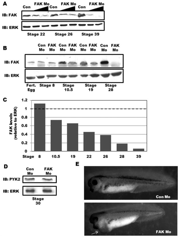
FIG. 1.
Depletion of FAK in Xenopus laevis leads to pericardial edema. A: FAK or Control morpholinos (20 and 40 ng) were injected into fertilized oocytes and embryonic FAK protein levels were assessed at the indicated stages by Western blotting. Levels of ERK are shown as a control for loading. B: Western blot analysis for FAK in Con Mo- and FAK Mo-injected embryos (40 ng/embryo) at the indicated stages of development. Levels of ERK are shown as a control for loading. C: Densitometric analysis of Western blots comparing FAK band intensity relative to ERK. Data are presented as FAK levels in FAK Mo-embryos relative to Con Mo-embryos (set to 1) at each developmental stage analyzed. D: Western blot analysis for PYK2 (and ERK) in stage 30 Con Mo- and FAK Mo-injected embryos. E: Gross morphology of Control and FAK morphant tadpoles at Stage 37. FAK morphants exhibit a slightly shortened anteroposterior axis and pericardial edema (arrow).

