FIG. 7.
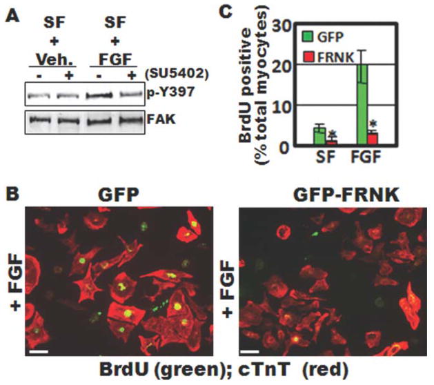
FAK is activated by FGF and is necessary for FGF-dependent myocyte proliferation. A: Western blot analysis of cell lysates isolated from primary embryonic rat cardiomyocytes. Cells were maintained in serum free (SF) media and treated with bFGF (100 ng/mL) or vehicle (veh.) for 30 min with or without 10 min pretreatment with the specific FGFR-inhibitor, SU5402 (10 μM). Lysates were immunoblotted with antibodies directed towards phospho-specific Y397-FAK or total FAK. B: BrdU incorporation in isolated embryonic cardiomyocytes infected with GFP- or GFP-FRNK adenovirus (10 m.o.i). Cells were maintained in serum-free medium and treated with vehicle (not shown) or bFGF (100 ng/mL) for 24 h. Costaining with anticardiac troponin T (cTnT) was performed to identify cardiomyocytes. C: Quantification of BrdU- positive cardiomyocytes (means ± SEM; N = 3; minimum of 300 cells/condition). Scale bar is 20 μm.

