Those of us that had been hammering away in the field for 40 years or more cannot deny the excitement in learning that Brian Kobilka, a former postdoctoral fellow of Robert Lefkowitz, in collaboration with Roger Sunahara, had determined the structure of the agonist-bound β2-adrenergic receptor-Gs protein complex (1). To borrow a phrase from Henry Bourne, this is “the Big Enchilada” for the G protein-coupled receptor (GPCR)/G protein field. This work followed just five years after Kobilka and Schertler’s determination of the crystal structure of the β2-adrenergic receptor (β2AR) bound to the inverse agonist carazolol (2), and over two and a half decades after the reports by Robert Lefkowitz’s group of the purification, cloning, and sequencing of the mammalian β2AR (3, 4). These remarkable achievements and others discussed below led to the award of the 2012 Nobel Prize in Chemistry to Kobilka and Lefkowitz. This was another big win for GPCRs, G proteins, and second messengers, a field that has already been richly rewarded, and justifiably so, for the role they play in hormone and neurotransmitter signaling and pharmacology.
Brian K. Kobilka. Image Copyright © Nobel Foundation 2012/Ulla Montain.
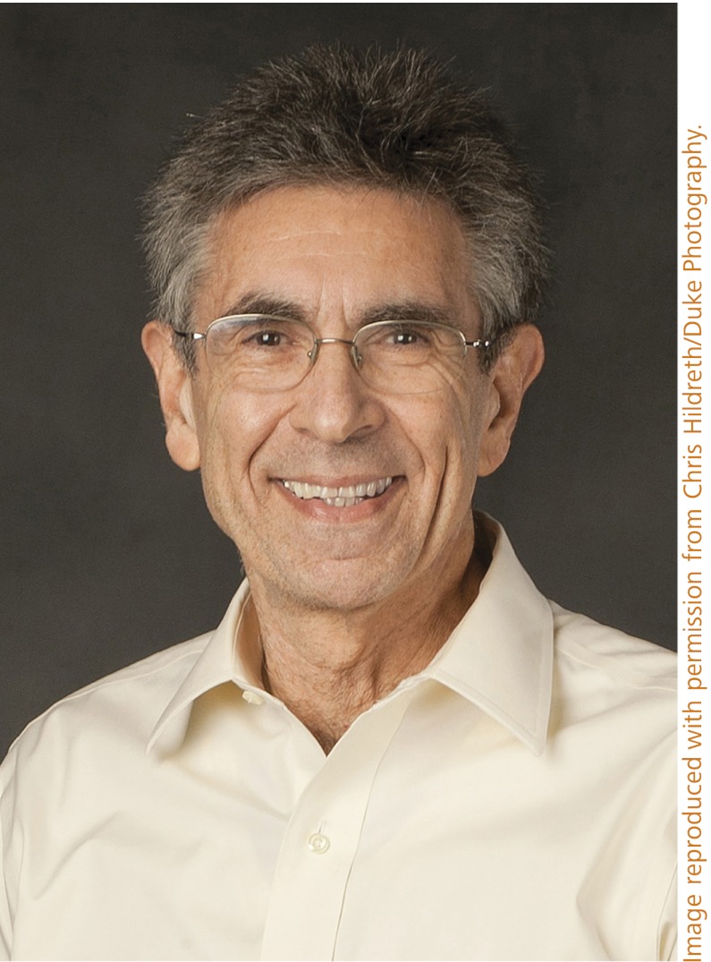
Robert J. Lefkowitz. Image reproduced with permission from Chris Hildreth/Duke Photography.
This story began over three decades ago when Bob tackled the extremely difficult job of identifying, purifying, and cloning the β2AR, an intrinsic membrane protein present at extremely low levels. Success evolved from the groups’ development of radiolabled antagonists (after realizing the pitfalls of using the rapidly oxidized catecholamines), the alprenolol affinity column, and identification of batches of digitonin that would preserve activity over the 100,000-fold or so purification required. Another key to Bob’s success was his ability to attract a long list of talented postdoctoral fellows (too many to list, but notably Kobilka, Limbird, Strader, Benovic, Cerione, Lohse, Bouvier, Dohlman, Luttrell, and his longtime colleague Marc Caron). Once the task of purifying the β2AR was accomplished, it was delivered to Richard Dixon, Catherine Strader, and Irving Sigal at Merck, who determined a partial peptide sequence and were able to find, remarkably and luckily enough, a genomic intronless clone (3). The sequence of the mammalian β2AR, along with that of the turkey β2AR reported by Elliot Ross’s group and the muscarinic acetylcholine receptor by Numa and colleague’s group (5), revealed the stunning similarity of the overall seven transmembrane helices topology to rhodopsin previously sequenced by Paul Hargrave et al. (6). These results linked the parallel studies of rhodopsin pioneered by Hermann Kuhn and colleagues (7), Jeremy Nathans and D. S. Hogness (8), Lubert Stryer, Krzysztof Palczewski, and many others, and suggested all GPCRs might share this homology.
Beyond the β2AR structure, the Lefkowitz group played a major role in establishing β2AR regulation as the paradigm for the GPCR field. Important areas pioneered by the group were in establishing the ternary complex model (9), and characterization of the desensitization of the β2AR, first demonstrating its phosphorylation in response to agonist stimulation. Interestingly, these findings were greatly aided by use of the S49 lymphoma somatic cell mutants, the cyc−-lacking Gs, and the kin−-lacking PKA, isolated by Henry Bourne’s group. These cell lines were used to demonstrate that desensitization of the β2AR proceeded through G protein-dependent and independent pathways (10–12). Building on this finding led to another landmark achievement of Bob’s group, the purification of one of the β2AR GPCR kinases (termed βARK or β-adrenergic receptor kinase originally and later GRK2) from the kin− cell line (13). Later, the group was able to show that arrestin was required for desensitization of the GRK-phosphorylated β2AR by binding to and completely uncoupling β2AR stimulation of Gs. After the discovery of the role of the G-protein βγ subunits in augmenting receptor phosphorylation by GRKs (14, 15), Bob Lefkowitz and John Tesmer reported the crystal structure of GRK2 in a complex with the G-protein βγ subunit (16). More recently, the Lefkowitz group was first to establish the novel concept that arrestins not only work through uncoupling and desensitizing the receptor, but also work as positive signaling proteins, scaffolding proteins such as MAP kinases (17), Src (18), and many other important regulatory proteins (19).
Brian Kobilka, after leaving his postdoctoral fellowship with Lefkowitz, quickly established his own reputation in the field by achieving the first animal knockouts of the β2AR and other adrenergic receptors, even as he was quietly and doggedly pursuing his real love of getting to the structure and molecular dynamics of the β2AR. Brian admitted this to me during a visit at Stanford some 20 years or so back, although it was readily apparent because a good bit of our conversing was done as Brian was running in and out of his laboratory purifying a fluorescently labeled receptor. It is important to understand that at the time, Brian’s goal of getting crystals and the structure of the β2AR was one shared by innumerable groups throughout the world working on their favorite receptors. The field had the rhodopsin crystal structure as the prototype, but the crystal structure of a hormone- or neurotransmitter-regulated GPCR was elusive for the very reasons given above. Through novel and creative approaches involving a long, exhausting series of dead ends, Brian obtained workable crystals that led to the first structure of an inverse agonist-bound β2AR with Schertler and colleagues (2). Soon after, Kobilka and colleagues and Stevens and colleagues reported a higher resolution structure based on using a fusion protein of T4-lysozyme inserted into the third intracellular loop (20, 21). These breakthroughs were trumped by Brian’s determination of the structure of the agonist-bound β2AR in complex with Gs. This required even more ingenious engineering, notably use of llama antibodies (coined nanobodies) to stabilize the activated receptor first (22) and later the β2AR/Gs complex (1). This work established the receptor/Gs contact sites, demonstrated the dramatic movements of the fifth and sixth transmembrane segments relative to the inactive structure, and revealed an unexpected major conformational movement of the non-Ras helical component of Gs. Brian’s work has been followed by a streak of GPCR crystal structures and it is clear that these collective achievements have far reaching applicability to development of new drugs and our understanding of how agonists (strong, weak, inverse, and biased), and potentially allosterically acting ligands differentially regulate the huge family of GPCRs. The likelihood for getting structures of GPCRs with arrestin and GRKs and determining the mechanism of biased ligands signaling looks far more promising at this juncture. As is often the case, many individuals are needed to advance a scientific field resulting in an award, as can be seen from this Profile; nevertheless Bob and Brian will be great ambassadors to represent all of this work to the world.
References
- 1.Rasmussen SG, et al. Crystal structure of the β2 adrenergic receptor-Gs protein complex. Nature. 2011;477(7366):549–555. doi: 10.1038/nature10361. [DOI] [PMC free article] [PubMed] [Google Scholar]
- 2.Rasmussen SG, et al. Crystal structure of the human beta2 adrenergic G-protein-coupled receptor. Nature. 2007;450(7168):383–387. doi: 10.1038/nature06325. [DOI] [PubMed] [Google Scholar]
- 3.Dixon RA, et al. Cloning of the gene and cDNA for mammalian beta-adrenergic receptor and homology with rhodopsin. Nature. 1986;321(6065):75–79. doi: 10.1038/321075a0. [DOI] [PubMed] [Google Scholar]
- 4.Benovic JL, Shorr RG, Caron MG, Lefkowitz RJ. The mammalian beta 2-adrenergic receptor: purification and characterization. Biochemistry. 1984;23(20):4510–4518. doi: 10.1021/bi00315a002. [DOI] [PubMed] [Google Scholar]
- 5.Kubo T, et al. Cloning, sequencing, and expression of complementary DNA encoding the muscarinic acetylcholine receptor. Nature. 1986;323:411–416. doi: 10.1038/323411a0. [DOI] [PubMed] [Google Scholar]
- 6.Hargrave PA, et al. The structure of bovine rhodopsin. Biophys Struct Mech. 1983;9(4):235–244. doi: 10.1007/BF00535659. [DOI] [PubMed] [Google Scholar]
- 7.Wilden U, Hall SW, Kühn H. Phosphodiesterase activation by photoexcited rhodopsin is quenched when rhodopsin is phosphorylated and binds the intrinsic 48-kDa protein of rod outer segments. Proc Natl Acad Sci USA. 1986;83(5):1174–1178. doi: 10.1073/pnas.83.5.1174. [DOI] [PMC free article] [PubMed] [Google Scholar]
- 8.Nathans J, Hogness DS. Isolation and nucleotide sequence of the gene encoding human rhodopsin. Proc Natl Acad Sci USA. 1984;81(15):4851–4855. doi: 10.1073/pnas.81.15.4851. [DOI] [PMC free article] [PubMed] [Google Scholar]
- 9.De Lean A, Stadel JM, Lefkowitz RJ. A ternary complex model explains the agonist-specific binding properties of the adenylate cyclase-coupled beta-adrenergic receptor. J Biol Chem. 1980;255(15):7108–7117. [PubMed] [Google Scholar]
- 10.Strasser RH, Sibley DR, Lefkowitz RJ. A novel catecholamine-activated adenosine cyclic 3′,5′-phosphate independent pathway for beta-adrenergic receptor phosphorylation in wild-type and mutant S49 lymphoma cells: Mechanism of homologous desensitization of adenylate cyclase. Biochemistry. 1986;25(6):1371–1377. doi: 10.1021/bi00354a027. [DOI] [PubMed] [Google Scholar]
- 11.Green DA, Clark RB. Adenylate cyclase coupling proteins are not essential for agonist-specific desensitization of lymphoma cells. J Biol Chem. 1981;256(5):2105–2108. [PubMed] [Google Scholar]
- 12.Clark RB, Friedman J, Dixon RA, Strader CD. Identification of a specific site required for rapid heterologous desensitization of the beta-adrenergic receptor by cAMP-dependent protein kinase. Mol Pharmacol. 1989;36(3):343–348. [PubMed] [Google Scholar]
- 13.Benovic JL, Strasser RH, Caron MG, Lefkowitz RJ. Beta-adrenergic receptor kinase: Identification of a novel protein kinase that phosphorylates the agonist-occupied form of the receptor. Proc Natl Acad Sci USA. 1986;83(9):2797–2801. doi: 10.1073/pnas.83.9.2797. [DOI] [PMC free article] [PubMed] [Google Scholar]
- 14.Haga K, Haga T. Activation by G protein beta gamma subunits of agonist- or light-dependent phosphorylation of muscarinic acetylcholine receptors and rhodopsin. J Biol Chem. 1992;267(4):2222–2227. [PubMed] [Google Scholar]
- 15.Pitcher JA, et al. Role of beta gamma subunits of G proteins in targeting the beta-adrenergic receptor kinase to membrane-bound receptors. Science. 1992;257(5074):1264–1267. doi: 10.1126/science.1325672. [DOI] [PubMed] [Google Scholar]
- 16.Lodowski DT, Pitcher JA, Capel WD, Lefkowitz RJ, Tesmer JJ. Keeping G proteins at bay: A complex between G protein-coupled receptor kinase 2 and Gbetagamma. Science. 2003;300(5623):1256–1262. doi: 10.1126/science.1082348. [DOI] [PubMed] [Google Scholar]
- 17.McDonald PH, et al. Beta-arrestin 2: A receptor-regulated MAPK scaffold for the activation of JNK3. Science. 2000;290(5496):1574–1577. doi: 10.1126/science.290.5496.1574. [DOI] [PubMed] [Google Scholar]
- 18.Luttrell LM, et al. Beta-arrestin-dependent formation of beta2 adrenergic receptor-Src protein kinase complexes. Science. 1999;283(5402):655–661. doi: 10.1126/science.283.5402.655. [DOI] [PubMed] [Google Scholar]
- 19.Reiter E, Lefkowitz RJ. GRKs and beta-arrestins: Roles in receptor silencing, trafficking and signaling. Trends Endocrinol Metab. 2006;17(4):159–165. doi: 10.1016/j.tem.2006.03.008. [DOI] [PubMed] [Google Scholar]
- 20.Cherezov V, et al. High-resolution crystal structure of an engineered human beta2-adrenergic G protein-coupled receptor. Science. 2007;318(5854):1258–1265. doi: 10.1126/science.1150577. [DOI] [PMC free article] [PubMed] [Google Scholar]
- 21.Rosenbaum DM, et al. GPCR engineering yields high-resolution structural insights into beta2-adrenergic receptor function. Science. 2007;318(5854):1266–1273. doi: 10.1126/science.1150609. [DOI] [PubMed] [Google Scholar]
- 22.Rasmussen SG, et al. Structure of a nanobody-stabilized active state of the β(2) adrenoceptor. Nature. 2011;469(7329):175–180. doi: 10.1038/nature09648. [DOI] [PMC free article] [PubMed] [Google Scholar]


