Abstract
Although the hair shaft is derived from the progeny of keratinocyte stem cells in the follicular epithelium, the growth and differentiation of follicular keratinocytes is guided by a specialized mesenchymal population, the dermal papilla (DP), that is embedded in the hair bulb. Here we show that the number of DP cells in the follicle correlates with the size and shape of the hair produced in the mouse pelage. The same stem cell pool gives rise to hairs of different sizes or types in successive hair cycles, and this shift is accompanied by a corresponding change in DP cell number. Using a mouse model that allows selective ablation of DP cells in vivo, we show that DP cell number dictates the size and shape of the hair. Furthermore, we confirm the hypothesis that the DP plays a crucial role in activating stem cells to initiate the formation of a new hair shaft. When DP cell number falls below a critical threshold, hair follicles with a normal keratinocyte compartment fail to generate new hairs. However, neighbouring follicles with a few more DP cells can re-enter the growth phase, and those that do exploit an intrinsic mechanism to restore both DP cell number and normal hair growth. These results demonstrate that the mesenchymal niche directs stem and progenitor cell behaviour to initiate regeneration and specify hair morphology. Degeneration of the DP population in mice leads to the types of hair thinning and loss observed during human aging, and the results reported here suggest novel approaches to reversing hair loss.
Keywords: Hair, Dermal papilla, Stem cell niche
INTRODUCTION
The hair shaft is formed from keratinocytes derived from a progenitor population at the base of the hair follicle during the anagen or growth phase of the hair cycle. The dermal papilla (DP) serves as a physical niche for these matrix progenitors (Legué and Nicolas, 2005) and provides signals that contribute to specifying the size, shape and pigmentation of the hair shaft (Enshell-Seijffers et al., 2010a; Enshell-Seijffers et al., 2008; Enshell-Seijffers et al., 2010b). At the end of the growth phase, these progenitors stop dividing and either differentiate or degenerate (supplementary material Fig. S1). The majority of cells in the epithelial layers of the lower follicle encircling the hair shaft degenerate during the catagen phase. The base of the hair shaft rises through the degenerating outer root sheath of the lower follicle and becomes anchored in the ‘permanent’ region of the follicular epithelium in proximity to the keratinocyte stem cells in the follicular bulge. The DP is also drawn to a position at the base of the permanent follicle, where it persists through the quiescent or telogen phase. At the onset of a new growth phase, the keratinocytes at the base of the follicle engulf the DP and regenerate a new progenitor population as the follicular epithelium extends into the sub-dermis, ultimately reconstituting the hair bulb and generating a new hair shaft that is extruded through the surrounding outer root sheath to project from the skin surface.
A correlation between hair size and DP cell number has been noted in human hair follicles. This is true both in the context of variation between follicles in individuals and in the context of follicular decline in progressive alopecia where the size of the follicle and hair shaft is reduced in successive hair cycles until the cycling terminal hair follicle is ultimately reduced to a miniaturized vellus hair follicle (Alcaraz et al., 1993; Elliott et al., 1999; Miranda et al., 2010; Van Scott and Eckel, 1958). A similar correlation was observed in rodent vibrissae in an injury-induced regeneration model (Ibrahim and Wright, 1982). Despite this general correlation, the issue remains of whether changes in DP cell number, particularly the reduction in DP cells associated with follicular miniaturization, is the cause or an effect of changes in follicle and hair size. To address this experimentally, DP cell number was analysed and manipulated in hair follicles of the mouse.
MATERIALS AND METHODS
Mice
Strains were constructed using the previously described alleles. Corin-cre (Enshell-Seijffers et al., 2010a), ROSA:LNL: rtTA-IRES-EGFP (Belteki et al., 2005), ROSA:LNL:tTA (Wang et al., 2008), tetODTA/+ (Lee et al., 1998) and rYFP (Srinivas et al., 2001). DPTetOn was made using Corin-cre/+; ROSA:LNL: rtTA-IRES-EGFP/ROSA:LNL rtTA-IRES-EGFP; tetODTA/+. Controls were Corin-cre/+; ROSA:LNL: rtTA-IRES-EGFP/ROSA:LNL rtTA-IRES-EGFP and ROSA:LNL: rtTA-IRES-EGFP/ROSA:LNL: rtTA-IRES-EGFP; tetODTA/+, which have similar phenotypes. Control data in Fig. 5 are from Corin-cre/+; ROSA:LNL: rtTA-IRES-EGFP/ROSA:LNL rtTA-IRES-EGFP. DPTetOff was made using Corin-cre/+; ROSA:LNL:tTA/ROSA:LNL:tTA; tetODTA/+. Controls were Corin-cre/+; ROSA:LNL:tTA/ROSA:LNL:tTA and ROSA:LNL:tTA/ROSA:LNL:tTA; tetODTA/+, which have similar phenotypes. Control data in Fig. 3 are from ROSA:LNL:tTA/ROSA:LNL:tTA; tetODTA/+.
Fig. 5.
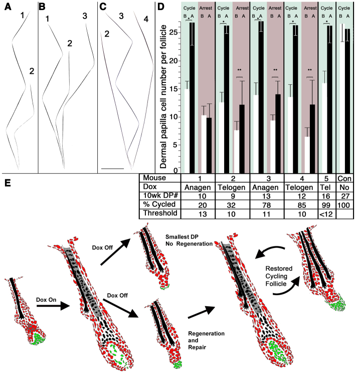
DP number is restored in follicles that enter the anagen phase. (A-C) Follicles from DPtetOn mice treated with doxycyline during the second growth phase have reduced second cycle hairs (2) compared with first cycle hairs (1). Many follicles fail to generate a third hair (A), whereas follicles that generate a slightly larger second hair go on to make a more normal third hair (B). (C) A follicle that produced a reduced awl hair in the second cycle (2) made a normal-sized zigzag in the third cycle (3) and progressed to a normal auchene in the fourth (4). Scale bar: 1 mm. (D) Average DP cell number for follicles that made zigzag hairs in the first and second cycles are shown at 10 weeks (B, before) and 20 weeks (A, after) for five treated mice and one control. For each mouse, the time of doxycycline treatment (Anagen; Tel, telogen; No, None) and average number of DP cells per follicle among the entire zizgzag population at 10 weeks (10wk DP#) is shown below. The data have been separated into the follicles that cycled (green) and those that did not (grey). The fraction that had cycled at 20 weeks is listed below the graph for each mouse (20-100%). Black bars represent the average DP cell number scored at 20 weeks for the fraction that did (green shaded area, ‘Cycle’) or did not (grey shaded area, ‘Arrest’) produce a third hair. The white bar (B) shows the average DP number after doxycycline treatment, before the skin re-enters anagen, inferred for the fraction of the sample that will (Cycle, green) or will not (Arrest, grey) make a third hair (see text). The inferred threshold number of DP cells required to re-enter the anagen phase for each mouse is shown below (Threshold). Mean and s.d., *P<3×10⁻⁶, **P<0.001 (n=25, 37, 40, 38 and 20 follicles at 10 weeks, 37, 27, 23, 22 and 20 follicles at 20 weeks, for mice 1-5, respectively). (E) Schematic representation of this experiment in which follicles with the fewest DP cells (green) arrest in the telogen phase, while follicles with a few more DP cells cycle and restore DP cell number, hair size and cycling activity.
Fig. 3.
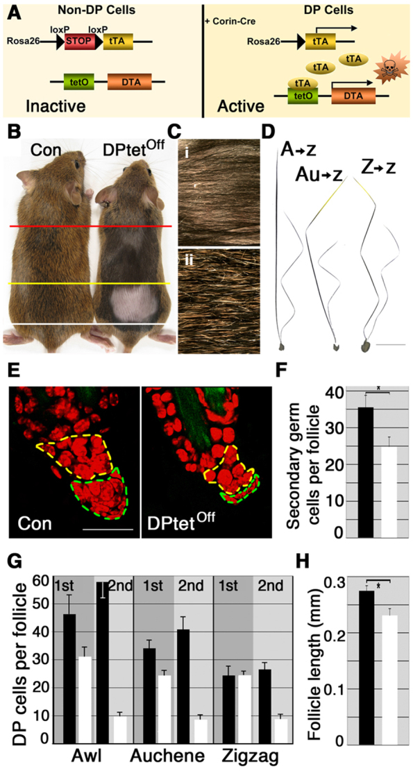
DP cell depletion results in smaller hairs and failure to re-enter the anagen phase of the hair cycle. (A) All cells in DPtetOff mice start with inactive r26tTA and tetO-DTA alleles. Corin-cre expresses cre-recombinase in DP cells, which removes the stop sequence from r26tTA and leads to the production of the tet transactivator protein (tTA). This binds to the tetO sequence and initiates transcription of the DTA transgene. DTA kills the cell. (B) Control (Con) and DPtetOff are shown. Animals were shaved between the red and white lines after the first hair cycle (P21), and between the yellow and white lines after the second hair cycle (P63). Above the red line the pelage consists of the first, second and, in control, third hair coats. Between the red and yellow lines, the shorter, thinner, darker hairs produced in the second cycle by DPtetOff contrast with the normal second and third hairs of the control. Between the yellow and white lines, the lack of hair in DPtetOff reveals the failure to re-enter the anagen phase, while the control has completed the production of a third hair coat. (C) Close-up view of the thin second hair coat that does not completely cover the skin in DPtetOff (i) and corresponding region of the control (ii). (D) Dissected follicles from a DPtetOff animal reveal largely normal first hairs. However, many follicles that produced awl or auchene hairs in the first cycle produce zigzag hairs in the second cycle (A→z and Au→z, respectively), and all second cycle hairs are shorter and thinner than normal. Scale bar: 1 mm. (E) Optical sections of follicles harvested at 10 weeks of age that had produced zigzag hairs in control (Con) and DPtetOff mice. The DP is marked with a broken green line, the secondary germ region with a broken yellow line. Scale bar: 25 μm. (F) The number of cells in the secondary germ region differs significantly between control and DPtetOff follicles that produced a zigzag hair in both hair cycles (mean and s.d., n=20 follicles from two mice each,*P=1.0×10⁻¹²). (G) DP cell number per follicle in control (black bars) and DPtetOff (white bars) after the first and second hair cycles in follicles that made awl, auchene or zigzag hairs in the first hair cycle. DP cell number and hair morphology are only slightly affected after the first cycle, but dramatically reduced after the second cycle (mean and s.d., n=20/type second cycle, n=10/type first cycle). The difference between DP number in control and DPtetOff after the second cycle is significant for all three hair types shown (P<6×10-16). (H) Follicle length during second telogen, measured from the skin surface to the base of the DP differs significantly between control and DPtetOff follicles that produced a zigzag hair in both hair cycles (mean and s.d., n=44 follicles con, n=53 follicles DPtetOff from 3 mice each, * P=6.8×10-36).
Whole-mount follicle preparation
A 5×3 mm full-thickness skin biopsy was fixed overnight in 4% paraformaldehyde (4°C), rinsed in PBS, incubated in methanol overnight (4°C) and stained with To-Pro3 (Molecular Probes) 1:1000 in PBS for 4 hours (22°C). Follicles were dissected and mounted in 50% glycerol. Serial optical sections of the dermal papilla were collected on a Nikon ECLIPSE Ti microscope. Images of complete hairs were assembled from overlapping photomicrographs to achieve the resolution required to reveal hair structure.
DP cell counting
Data in Fig. 2 from anagen follicles was collected from unpigmented follicles (tyrc/c) to prevent melanin from obscuring cells in the upper DP. Two independent methods to genetically mark DP cells, Cor-cre/+; rYFP/+ (Enshell-Seijffers et al., 2010a) and Sox2GFP (Biernaskie et al., 2009), yielded similar results. All data on anagen DP cell numbers relied on fluorescent reporter expression. Data from Fig. 2 were collected from male mice on an FVB background. Follicles from pigmented mice (DPTetOn, DPTetOff) were scored in telogen, when the follicle is unpigmented. Wild-type mice expressing fluorescent reporters were used to confirm that the boundary of the DP in telogen could be reliably distinguished by morphological criteria. Although DPTetOn mice express GFP in most DP cells and this assisted in scoring, morphological criteria were used as any unlabelled DP cells would be selectively enriched by the experimental treatment. DP cells in DPTetOff mice do not express fluorescent markers and were identified by morphological criteria. Individual nuclei within the boundaries of the DP were counted in z-stacks. In all cases where comparisons between genetically manipulated and control mice are made, the comparisons are between follicles that produced the same hair type in each of the hair cycles (e.g. Z>Z versus Z>Z but not A>Z). Much of the analysis reported here emphasizes follicles that produced zigzag hairs in both or all three hair cycles, because the underlying difference in DP number between follicles producing different hair types in control and experimental mice adds variation that would tend to obscure the changes caused by experimental treatment within a discrete follicle class. However, qualitatively similar results are observed in matched groups of hair follicles producing other hair types. Cells in the secondary germ area were identified by their position in the follicle, defined as keratinocytes lying more than one cell layer below the base of the lowest club hair to the base of the follicular epithelium.
Fig. 2.
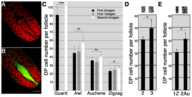
Follicles producing different hair types have different numbers of DP cells. (A,B) Optical sections showing hair bulb from a Sox2GFP/+ mouse (nuclei indicated in red) (A) and overlay of the same section with DP cells that express GFP (green) (B). Scale bar: 25 μm. (C) DP cell numbers were counted in optical sections of isolated intact hair follicles producing each hair type during first (morphogenetic) anagen (P11, black bars), first telogen (P20, grey bars) and second anagen (P30, white bars). Mean and s.d. are shown. Differences between hair types are significant (P<0.001) at all time points. Differences within hair type between the first and second cycle are significant (*P<0.01; **P<0.001; ***P<0.03). All follicles producing auchene hairs in the second hair cycle produced zigzag hairs in the first cycle. Guard hairs were not analysed during telogen. (Minimum n=20 hairs from two mice per type/time point, except guards, n=6/time point.) (D) Examples of two and three medulla cell thick awls are shown above. There is a significant difference (*P=0.002) between the number of DP cells in follicles that produce thin (2) versus thick (3) awl hairs in the same hair cycle (mean and s.d., n=5 and n=9, respectively). (E) The average DP cell number and s.d. for the 20% of zigzag follicles with the most DP during the first hair cycle (1Z, n=7), and that for follicles producing an auchene in the second hair cycle (2Au, n=20) are shown. All follicles that produced an auchene in the second cycle produced a zigzag hair in the first cycle. The significant difference (*P=1.5×10⁻⁸) between these two populations demonstrates that DP cell number has increased as these follicles shift from making zigzag to making auchene hairs.
Doxycyline administration and subsequent analysis
Animals were provided with chow containing 6 g/kg doxycycline ad libitum and 0.5 mg/ml in the drinking water (Bio-Serve, Laurel, MD). Females, which have a slightly longer first telogen phase were used for these experiments. To evaluate the effects of doxycycline on DP cell number, animals were anesthetized at 10 weeks, after cessation of doxycycline treatment and a 3×5 mm full thickness skin biopsy was taken from the dorsal midline. Semi-permanent hair dye was applied to a patch on the back of the animal. A small patch on the posterior was shaved to monitor hair cycling. At 20 weeks, a skin biopsy taken from the dyed area for analysis. Dye experiments confirmed that club hairs formed in successive hair cycles anchor progressively lower in the follicle and this criteria was used to distinguish the order of hairs when more than one dyed hair was present. Experiments involving mice were approved by the IACUC of Massachusetts General Hospital (Boston, MA, USA). The probability of significant difference was calculated using Student’s t-test.
RESULTS
A single follicle produces different hair types in successive hair cycles
The pelage of the mouse is composed of four hair types; guard, awl, auchene and zigzag (Fig. 1). Of the two straight hair types, guard hairs are longer and smoothly tapered, while awls are thicker and blunter. Auchenes are thinner than awls and have a single oblique angle bend in the middle. Zigzags are the thinnest hair type and have alternating oblique angle bends. It is often assumed that the follicles that produce different hair types are distinct both in embryological origin and epigenetic identity. The follicles that make guard hairs form first in embryogenesis and normally continue to make guard hairs throughout the life of the animal. However, the follicles that make awls, auchenes and zigzags can make different hair types in successive hair cycles (Fig. 1). In the mouse, the hairs produced in the previous cycle, referred to as club hairs, remain anchored in the permanent region of the follicle. Many follicles retain a zigzag club hair while generating an awl or auchene hair in the second cycle. Virtually all of the follicles with an auchene club hair from the first cycle generate an awl hair in the second hair cycle. Follicles with an awl club hair continue to produce awls in successive cycles, but the later-generated hair is usually thicker than that produced in the prior hair cycle. Unlike the other hair types, the club hair in a guard follicle from the previous cycle is shed in the ensuing cycle and a single club hair is retained at telogen. However, dissection of follicles in anagen while the club hair is retained confirms that guard hair follicles continue to produce guard hairs (not shown). The shift in representation of the different hair types within the hair coat predicted by this analysis of dissected follicles is observed by scoring the frequency of each hair type in the first and second hair coats. Guards are unchanged, whereas zigzags decrease and awls increase from the first to second hair coats. The representation of auchenes is little changed between the first and second coats, as the shift from auchene to awl production is roughly balanced by the shift from zigzag to auchene production (Fig. 1B). Although not every follicle produces more than one hair type; those that do, progress up the morphocline that ranges from zigzag to auchene to thin awl to thick awl. Conversion between hair types in the other direction is not observed in healthy young animals.
Fig. 1.
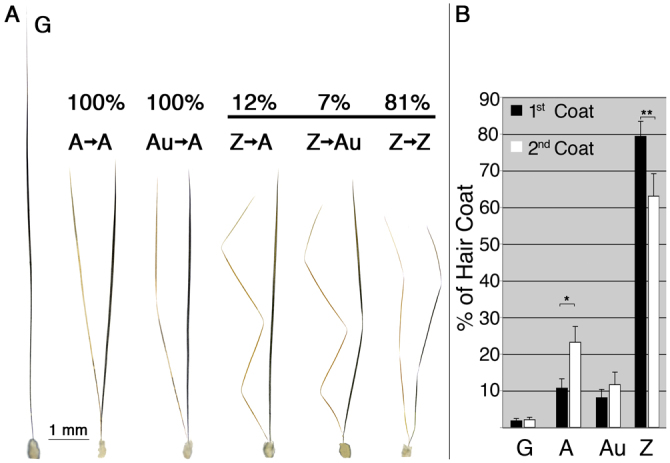
Follicles produce different hair types in successive cycles. (A) Hair was dyed at the end of the first hair cycle and follicles were dissected at the end of the second hair cycle. Examples of each type observed are shown: G, guard; A→A, awl to awl; Au→A, auchene to awl; Z→A, zigzag to awl; Z→Au, zigzag to auchene; Z→Z, zigzag-zigzag. Within follicles that made each hair class in the first cycle, the percentage making different hair classes in the second cycle is shown above. (G, n=51; A, n=160; Au, n=128; Z, n=1889 follicles from five mice). (B) The average frequency and s.d. of each hair type in the first (black) and second (white) hair coats is shown (n=21 mice, minimum of 300 hairs scored in each hair coat). G, guard; A, awl; Au, auchene; Z, zigzag. *P<7×10-12, **P<8×10-13.
DP cell number correlates with hair type and size
Guard hair follicles have more DP cells than other types (Sharov et al., 2006) but a difference in DP cell number between other follicle types has not been reported. Optical sectioning of dissected but intact hair follicles reveals that follicles producing guard, awl, auchene and zigzag hairs have significantly different numbers of DP cells when scored during the growth phase at postnatal day (P) 11 (Fig. 2A-C, P<0.001 for all comparisons). The thickness of awl hairs can be conveniently scored by the number of medulla cells in the thickest region. Within this hair class, thickness also correlates with DP cell number (Fig. 2D). During the first telogen phase, DP cell number is unchanged from that observed at P11 (Fig. 2C). However, during the early anagen phase of the second hair coat, recruitment of new cells to the DP occurs (Chi et al., 2010). When scored at P30, when the morphogenesis of the hair shaft is at a similar stage to that observed at P11, the average DP cell number per follicle has increased for each hair type (Fig. 2C).
When a follicle switches from the production of one hair type to a larger hair type, a corresponding shift in the number of DP cells is observed. Approximately 20% of follicles containing a zigzag club hair make an awl or auchene hair in the second hair cycle. Even if we assume that only those follicles with the most DP cells in the first cycle convert to the production of larger hair types in the second cycle, there is a significant increase (36%, P=1.5×10⁻⁸) in the DP cell number per follicle in the follicles that convert to the production of auchene hairs (Fig. 2E). The larger difference between the minimum number of DP cells observed in awl hairs in the second cycle (41 DP cells at P30) and the largest number of DP cells observed in first cycle zigzag hairs (34 DP cells at P20) also demonstrates this fact. Thus, the correlation between hair size and DP cell number extends across the three alternative morphotypes that can be generated by a single hair follicle in successive cycles. The same keratinocyte stem cell pool produces hairs of different sizes and types under the direction of different numbers of DP cells.
DP cell number specifies hair size and shape
A mouse model that allows selective ablation of DP cells in vivo was developed to test the role of DP cell number in specifying hair morphology. Corin-cre was used to initiate expression of a cre-dependent tet transactivator allele (r26tTA) specifically in DP cells of mice that also harbour an allele that encodes the diptheria toxin A chain (DTA) under the control of tet-Operator sequences (tetO-DTA) in Corin-cre/+; r26tTA/r26tTA; tetO-DTA/+ mice (hereafter, DPtetOff) (Enshell-Seijffers et al., 2010b; Lee et al., 1998; Wang et al., 2008) (Fig. 3A). The tet transactivator protein (tTA) expressed from the recombined r26tTA allele binds to the tet-Operator and initiates expression of DTA, which kills the cell. To distinguish hairs produced in successive cycles, a region of the coat was clipped at P21, and a subset of this region was clipped again at P63 (Fig. 3B). Fur plucked from the region that had been clipped in the preceding telogen phase was used to characterize overall coat structure produced in a given cycle, while intact follicles dissected from a biopsy taken from the unclipped region at each telogen phase were used to analyse the structures and hair production histories of individual follicles.
Although most DP cells recombine the r26tTA allele during the first growth cycle (supplementary material Fig. S2), the relatively low efficiency of this tetOff/tetO-DTA system results in a progressive reduction in DP cell numbers, primarily during the second hair cycle (Fig. 3). As a result, there is little effect on the first hair coat, although there is a reduction in the thickness of awl hairs that reflects the preferential reduction of DP cell number in this population during the first cycle (Fig. 3G and data not shown). However, the more effective depletion of DP cell numbers in the second hair cycle has dramatic effects. There is a marked reduction in the length and thickness of the hairs generated in the second hair cycle (Fig. 3B-D). In addition, the switch from production of smaller hair types to larger ones normally observed in many follicles is blocked. Instead, the opposite is frequently observed as diminutive zigzag hairs from the second hair cycle are found in follicles that had produced auchenes or awls in the first cycle (Fig. 3D). This conversion to production of smaller hair types is reflected in the representation of the different hair types in the first and second coats. Although the frequency of awl and auchenes increases in the second hair coat at the expense of zigzags in controls, the frequency of zigzags rises from 78% to 94% in the DPtetOff animals from the first to the second coat, while the frequency of awls and auchenes together drops from 20% in the first to 4% in the second coat (n=3 animals, minimum of 300 hairs per animal per time point). Follicles in these mice assume a normal telogen morphology at the end of the second hair cycle, but they are smaller than controls (Fig. 3E). This reduction is observed not only in the DP, which has less than half the normal number of cells (Fig. 3E,G), but also in the epithelial follicle where the secondary germ region is notably reduced in size (Fig. 3E,F,H).
The DP regulates hair cycling
These experiments also document the role of signalling from the DP to initiate regeneration of the follicle. Follicles in DPtetOff mice, which average fewer than 10 DP cells after the second hair cycle, remain arrested in the telogen phase long after follicles in control mice have entered and completed a third hair cycle (n=12/12 mice, Fig. 3B; supplementary material Fig. S3). By contrast, follicles in DPtetOff mice with a single dose of the r26tTA allele (Corin-cre/+; r26tTA/+; tetODTA/+) show a more modest reduction in DP cell number, re-enter the hair cycle and generate a third hair coat (supplementary material Fig. S3).
Temporally controlled DP cell ablation during the anagen phase of the hair cycle
Although the DPtetOff mice show ongoing stochastic DP cell death that serves as a good model of hair thinning and loss, the ongoing damage to DP cells hampers more-detailed analysis. To gain further insight, ablation of DP cells was restricted to a discrete period using an analogous DPtetOn system. In this model [Corin-cre/+; r26rtTA-IRES-EGFP/r26rtTA-IRES-EGFP; tetODTA/+ (hereafter DPtetOn)], a cre-dependent reverse tet-transactivator allele (r26rtTA-IRES-EGFP) (Belteki et al., 2005) replaces r26tTA. The reverse tet-transactivator (rtTA) only activates transcription of the tetODTA transgene while doxycycline is administered (Fig. 4A). This allowed us to alter DP cell numbers during a discrete time period and then track the consequences in the absence of ongoing DP cell death.
Fig. 4.
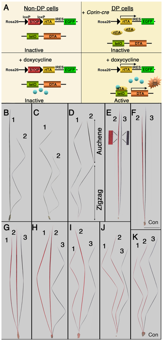
A fixed period of DP damage alters hair morphology. (A) Schematic of the DPtetOn system. All cells in DPtetOn mice start with inactive r26rtTA and tetO-DTA alleles. Corin-cre expresses cre-recombinase in DP cells, which removes the stop sequence from r26rtTA and leads to the production of the reverse tet transactivator protein (rtTA) and EGFP. rtTA is inactive until bound by doxycycline. In the presence of doxycycline it binds to the tetO sequence and initiates transcription of the DTA transgene. DTA kills the cell. (B-K) Dissected follicles from DPtetOn mice treated with doxycycline during the anagen phase (P30-56, B-D), second telogen phase (P42-63, E,G-J) or control (F,K). The hair cycle in which the hair was generated is indicated (1, 2, 3). For animals treated in telogen and control, the first two hairs were dyed red at 9 weeks, prior to formation of the third hair (black). (B,C) Treatment during anagen results in either a shift to production of a smaller hair type (B, awl to zigzag) or production of a smaller hair of the same class (C, zigzag to smaller zigzag). (D) Example of a chimeric hair produced with treatment during anagen. The distal half of the second hair has the morphology of an auchene, whereas the proximal region has the morphology of a zigzag. In follicles that produced awl hairs in the second hair cycle, DP deletion during the second telogen phase causes production of smaller hairs or hair types during third anagen. (E) This follicle produced a normal awl in the second cycle and a smaller awl after DP cell ablation. A small segment is magnified for comparison of thickness and structure. (F) A control awl follicle produces a slightly larger hair in the third cycle. Scale bar: 1 mm. (G,H) A follicle that produced a normal auchene hair in the first cycle (1), an awl hair in the second (2) and made an auchene (G) or reduced zigzag hair (H) in the third cycle after DP cell depletion (3). (I) Follicles that produced zigzag hairs in the first cycle and auchenes in the second produce zigzag hairs after DP cell deletion during second telogen. (J) In contrast to follicles producing larger hair types, the follicles that produced zigzag hairs in the second hair cycle and re-entered the third cycle produce a zigzag hair that is only slightly smaller than its second cycle counterpart. (K) Control follicle that produced zigzag hairs of increasing size in all three cycles.
Although the defects observed in the DPtetOff model are ultimately caused by expression of DTA in DP cells, initiation of this expression during morphogenesis of the follicle could somehow interfere with normal development of the epithelial compartment of the follicle and contribute to the phenotypes observed in the second hair coat. DP cell number was reduced in structurally normal follicles by doxycycline administration, starting during the early anagen phase of the second hair cycle (P30 females). Results were similar to those observed with the DPtetOff system, confirming that these phenotypes also result from DP cell depletion during the second hair cycle after a normal morphogenetic (first) cycle. The conversion to production of larger hair types between the first and second hair coats was inhibited or reversed, and shorter thinner hairs were produced (Fig. 4B,C). For example, 78% of awl hairs in the second coat had two or fewer rows of medulla cells, whereas only 1% of control awls were this thin (DPtetOn, n=115 from three mice; control, n=123 from two mice). In addition, hairs with chimeric morphologies where distal structures represent larger hair types and proximal structures represent smaller hair types were also observed (Fig. 4D). Thus, hair type can be affected by DP cell depletion within the course of an anagen phase. Follicles in these mice assumed a normal telogen morphology at the end of the hair cycle, but exhibited reduced numbers of DP cells. Many hair follicles failed to enter a third hair cycle (see below).
DP cell ablation in the telogen follicle
To further isolate the immediate effects of DP cell deletion from any persisting effects on the keratinocyte pool from the prior cycle, DPtetOn animals were treated with doxycycline from P42-63 during the extended second telogen phase after a normal resting follicle structure was established. They were maintained in the absence of doxycycline for several weeks prior to the onset of the next anagen phase to preclude any potential lingering effects of sub-lethal expression of DTA in DP cells that were not killed. A full thickness skin biopsy was taken at 10 weeks and a region of the coat was dyed. Individual follicles were dissected from the biopsy and the distribution of DP cell number per follicle and structure of the epithelial follicle was evaluated in follicles bearing different hair types. The structure of the epithelial follicle was unchanged after doxycyline treatment. The number of keratinocytes lying between the DP and the bulge (secondary germ region) in these treated DPtetOn mice (35.6±3.8) was indistinguishable from that of control follicles (35.6±3.6, n=10 follicles with two zigzag hairs per mouse, two mice per treatment; supplementary material Fig. S4). DP cell number in these follicles was variably reduced. Despite the normal follicular epithelium structure and the absence of ongoing DTA expression, both aspects of the DP cell depletion phenotype were observed in a biopsy taken at 20 weeks, after the third hair coat had been completed in control animals. Many follicles failed to undergo a third hair cycle (Fig. 5). Smaller hairs were generated in many of the follicles that made a third hair (Fig. 4E,G-J). Among follicles that had produced either awls or auchenes in the second cycle, both production of hairs with reduced size within a hair class (20%, n=13 of 64 follicles from two mice) and a switch to the production of smaller hair types (80%; n=51 of 64 follicles from two mice) were observed (Fig. 4E-I). The conversion to production of smaller hair types was not observed in control animals. Many follicles that had produced zigzag hairs in the second cycle failed to enter a third cycle. However, it is noteworthy that those that did produced a zigzag hair that was only slightly reduced in size from controls hairs and comparable with that produced during the first hair cycle (Fig. 4J,K). The proportion of follicles that completed a third hair cycle was determined by scoring the presence or absence of a third undyed hair in dissected follicles from the 20-week skin biopsy. This proportion correlated with the severity of DP cell depletion observed at 10 weeks during the telogen phase. In mice with average DP cell numbers of 9.3±3.0, 12.3±3.4 and 16.1±2.2 at 10 weeks among follicles that had produced two zigzag hairs prior to doxycycline treatment, 32%, 85% and 99% of these follicles generated a third hair (Fig. 5D, Telogen). These results confirm that many of the effects on hair morphology and cycling can be directly attributed to effects on DP cell number.
Regulative restoration of DP cell number in cycling follicles
The relatively normal structure of zigzag hairs produced during the third cycle in the follicles of DPtetOn animals after doxycycline treatment had been stopped (Fig. 4J; Fig. 5B,C) contrasts with the reduced size observed in hairs generated while doxycycline was administered during the anagen phase (Fig. 4C; Fig. 5A,B). This might suggest that DTA expression during anagen has additional effects on signalling from the DP beyond the reduction of DP cell numbers, or that active cell killing has other indirect effects. However, a comparison between DP cell numbers prior to and after the third hair cycle revealed a different explanation, regardless of whether the animal was treated with doxycycline during anagen or telogen. DP cell number increased dramatically during the anagen phase in those follicles that generated a third hair in the absence of ongoing DTA expression (Fig. 5D, green). At 20 weeks, a comparison of the number of DP cells in the follicles that produced a third hair to those that failed to cycle within an individual mouse revealed a significant difference between these two populations in all of the mice (P<1.2×10⁻⁸). DP cell number in follicles that produced a zigzag hair in the third cycle was indistinguishable from that of control mice at 20 weeks (Fig. 5D, green). By contrast, DP cell number per follicle among the follicles that failed to generate a third hair by 20 weeks remained significantly lower in all experimental animals than that observed in control animals (P<4.0×10⁻¹³, n=4 mice; Fig. 5D, grey).
The normal structure of these third hairs might suggest that only a subset of follicles that had escaped damage went on to cycle. This is not the case. The presence of a miniaturized second hair in the follicles of mice treated with doxycyline during the preceding anagen phase provides direct confirmation that regulative regeneration has occurred in individual follicles (Fig. 5B,C). Analysis of DP cell numbers confirms that this is also the case for mice treated during the telogen phase. In each mouse, the range of DP cell numbers in the follicles that completed a third cycle (20 weeks, Fig. 5D, green) does not overlap that of the starting population (10 weeks) as a whole. As the most stringent test, we assume that only the follicles with the largest number of DP cells at 10 weeks produced a third hair. In this scenario, the subset of the largest of the rank-ordered 10-week DP cell numbers corresponding to the fraction of follicles that cycled is the starting distribution of DP cell numbers for follicles that went on to generate a third hair. This can be compared with the actual range of DP cell number per follicle observed at 20 weeks in the follicles that had cycled, which were identified by the presence of an undyed hair. The significant difference between these two distributions confirms that DP cell number was restored in the follicles that cycled (maximum P<3.0×10⁻⁶, n=5 mice). These follicles continue to cycle, and in some cases progress to the production of larger hair types (Fig. 5C). These results demonstrate that the follicle is capable of regulative restoration of DP cell number. However, this is a follicle-autonomous effect that depends on successful re-entry into the anagen phase of the hair cycle (Fig. 5E).
A threshold number of DP cells required for regeneration
The comparison between DPtetOff mice with one or two copies of the r26tTA allele suggested the existence of a crucial threshold between 10 and 20 DP cells that is required to initiate follicular regeneration (supplementary material Fig. S3). In both treatments of DPtetOn animals, DP cell ablation initiated during anagen or telogen, mice with different average numbers of DP cells in the second telogen phase (10 weeks) were generated (Fig. 5D, 10 wk DP#). In four of these animals, only a subset of follicles within a contiguous region generated a third hair (Fig. 5A,B,D, mouse 1-4). The proportion of follicles that generated a third hair in different animals differs with the severity of DP cell depletion within a treatment group (Anagen or Telogen, Fig. 5D, % Cycled). Nevertheless, if we assume that the follicles with more DP cells cycle and apply the observed cycling frequency to the rank ordered DP cell number data, a consistent threshold is revealed. This ranges from 10 (15th ascending percentile in mouse 4) to 13 (80th percentile in mouse 1) DP cells (Fig. 5D, Threshold). This analysis indicates that follicles in which the DP cell number was at or below this threshold usually failed to cycle, whereas follicles with a few more DP cells re-entered the hair cycle and generated a third hair (Fig. 5D and data not shown).
DISCUSSION
In summary, these experiments demonstrate that the DP specifies hair morphology and regulates re-entry into the growth phase of the hair cycle. Our previous work had documented one mechanism by which DP cells regulate hair follicle morphology and cycling. Blocking the capacity of DP cells to respond to Wnt signals caused a change in the production per DP cell of secreted factors that act on adjacent keratinocytes to regulate hair morphogenesis and anagen re-entry (Enshell-Seijffers et al., 2010a; Enshell-Seijffers et al., 2010b). In the work reported here, we reveal a different mechanism whereby changes in the number of DP cells specify changes in hair size, shape and cycling frequency.
Follicles can produce larger hairs within a hair class or switch to production of a larger hair type as the animal grows. This is accompanied by an increase in DP cell number, which can be achieved in part by the recruitment of new cells to the DP (Chi et al., 2010). It has been suggested that there is some intrinsic difference between the DP cells of different hair follicle types, perhaps reflecting alternative differentiated states of dermal fibroblasts when the follicles first form, or even distinct cell lineages (Driskell et al., 2009; Driskell et al., 2012). The shift between hair types produced by a single follicle is inconsistent with these models. The fact that this shift is normally uni-directional is formally consistent with a developmental ratchet model in which successive DP identities are mutually exclusive and perhaps epigenetically distinct. However, the observations that reducing DP cell number can reverse this switch between hair types and that the same follicle can shift up, down and back up this morphocline in the experimentally manipulated DPtetOn mice undermine a ratchet model. Instead they support the idea that these distinctive morphologies are not driven by functionally distinct DP cell populations. Instead, either qualitative or quantitative changes in the aggregate signalling behaviour of different numbers of functionally equivalent DP cells guides these different morphogenetic programs.
The precise sequence of events that initiates the activation of the telogen hair follicle remains obscure, but signals from the DP are thought to play a crucial role (Enshell-Seijffers et al., 2010a; Greco et al., 2009; Sun et al., 1991). Recent work had shown that complete ablation of the DP prevents anagen initiation, at least temporarily (Rompolas et al., 2012). Our findings extend that work by showing that when DP cell number falls below a critical threshold, a follicle fails to enter anagen and remains in telogen for months or more, while adjacent follicles with a few more DP cells re-enter the growth cycle. Thus, while tissue-level signals coordinate anagen re-entry among competent follicles (Plikus et al., 2011; Plikus et al., 2008), follicle-intrinsic signalling that is dependent on a critical DP cell number is required to initiate follicular regeneration, even in the context of a permissive or activating macro-environment.
DP cell depletion in mice causes the changes in hair follicle structure and cycling that characterize progressive alopecia in humans, including reduction in both hair and follicle size, a prolonged telogen phase and an ultimate failure to produce new terminal hairs (Kligman, 1988). The selective reduction of the secondary germ region observed in these mice as an indirect consequence of DP cell depletion is also observed in androgenetic alopecia in humans (Garza et al., 2011). In this context, it is noteworthy that when the underlying cause of DP cell depletion is removed in our mouse model, the follicle exhibits an intrinsic capacity for regulative regeneration to produce normal hairs, but only when DP cell number is sufficient to stimulate anagen re-entry. Treatments to arrest follicular decline are available for some forms of human hair thinning, but effective repair of diminished follicles has remained elusive. In our mouse model, the difference of a few DP cells distinguishes whether a follicle will arrest and fail to make a new hair or will re-enter the hair cycle and undergo regenerative repair. This suggests that therapeutic interventions in humans that cause only a minor direct improvement in DP cell number may be sufficient to restore hair cycling and harness the natural regenerative ability of the follicle for further progress towards robust hair growth.
In addition to the anagen-dependent and follicle intrinsic regenerative response these experiments reveal, our data suggest a weaker and potentially non-autonomous regenerative effect in the follicles that fail to cycle. If the assumption that the follicles with the fewest DP cells fail to cycle is correct, then a small but significant increase in DP cell number was observed in the follicles that did not cycle in three out of the four mice in which a subset of follicles did not cycle (Fig. 5D, grey; P<5.8×10⁻⁵, mouse 2; P<1.1×10⁻⁶, mouse 3; P<0.00036, mouse 4). This effect was not observed in the animal where the fewest follicles cycled (Fig. 5D, mouse 1, grey). This suggests a mechanism that is not dependent on anagen re-entry by the follicle, but may depend on adjacent cycling follicles. Although modest in magnitude compared with the full restoration of DP number in cycling follicles, the apparent threshold of DP cell number required for anagen re-entry suggests even this small effect, if confirmed by subsequent work, would be biologically significant if it poised the follicle for successful regeneration in the next hair cycle.
The hair follicle serves as a model to understand stem cell biology and tissue regeneration. Although much research has focused on the attributes of keratinocyte stem cells in this system, the work reported here emphasizes the important role of the DP in guiding organ regeneration. The natural capacity to alter DP cell number in a reproducible manner during growth of the animal and the intrinsic ability to restore DP cell number to normal levels after damage suggest an active mechanism to regulate DP size. This is likely to entail dynamic communication between the epithelial and mesenchymal components of the follicle, as is observed during morphogenesis of this mini-organ in the embryo. The ability to perturb this system and follow its response over time in living tissue holds promise for further dissection of the mechanisms by which appropriate organ size and shape are achieved by feedback between epithelium and mesenchyme.
Supplementary Material
Acknowledgments
The authors thank R.Czyzewski for technical assistance.
Footnotes
Funding
This work was supported by a grant from the National Institutes of Health, National Institute of Arthritis, Musculoskeletal and Skin Diseases [R01AR055256 to B.A.M.]. Deposited in PMC for release after 12 months.
Competing interests statement
The authors declare no competing financial interests.
Supplementary material
Supplementary material available online at http://dev.biologists.org/lookup/suppl/doi:10.1242/dev.090662/-/DC1
References
- Alcaraz M. V., Villena A., Pérez de Vargas I. (1993). Quantitative study of the human hair follicle in normal scalp and androgenetic alopecia. J. Cutan. Pathol. 20, 344–349 [DOI] [PubMed] [Google Scholar]
- Belteki G., Haigh J., Kabacs N., Haigh K., Sison K., Costantini F., Whitsett J., Quaggin S. E., Nagy A. (2005). Conditional and inducible transgene expression in mice through the combinatorial use of Cre-mediated recombination and tetracycline induction. Nucleic Acids Res. 33, e51 [DOI] [PMC free article] [PubMed] [Google Scholar]
- Biernaskie J., Paris M., Morozova O., Fagan B. M., Marra M., Pevny L., Miller F. D. (2009). SKPs derive from hair follicle precursors and exhibit properties of adult dermal stem cells. Cell Stem Cell 5, 610–623 [DOI] [PMC free article] [PubMed] [Google Scholar]
- Chi W. Y., Enshell-Seijffers D., Morgan B. A. (2010). De novo production of dermal papilla cells during the anagen phase of the hair cycle. J. Invest. Dermatol. 130, 2664–2666 [DOI] [PMC free article] [PubMed] [Google Scholar]
- Driskell R. R., Giangreco A., Jensen K. B., Mulder K. W., Watt F. M. (2009). Sox2-positive dermal papilla cells specify hair follicle type in mammalian epidermis. Development 136, 2815–2823 [DOI] [PMC free article] [PubMed] [Google Scholar]
- Driskell R. R., Juneja V. R., Connelly J. T., Kretzschmar K., Tan D. W., Watt F. M. (2012). Clonal growth of dermal papilla cells in hydrogels reveals intrinsic differences between Sox2-positive and -negative cells in vitro and in vivo. J. Invest. Dermatol. 132, 1084–1093 [DOI] [PMC free article] [PubMed] [Google Scholar]
- Elliott K., Stephenson T. J., Messenger A. G. (1999). Differences in hair follicle dermal papilla volume are due to extracellular matrix volume and cell number: implications for the control of hair follicle size and androgen responses. J. Invest. Dermatol. 113, 873–877 [DOI] [PubMed] [Google Scholar]
- Enshell-Seijffers D., Lindon C., Morgan B. A. (2008). The serine protease Corin is a novel modifier of the Agouti pathway. Development 135, 217–225 [DOI] [PMC free article] [PubMed] [Google Scholar]
- Enshell-Seijffers D., Lindon C., Kashiwagi M., Morgan B. A. (2010a). Beta-catenin activity in the dermal papilla regulates morphogenesis and regeneration of hair. Dev. Cell 18, 633–642 [DOI] [PMC free article] [PubMed] [Google Scholar]
- Enshell-Seijffers D., Lindon C., Wu E., Taketo M. M., Morgan B. A. (2010b). Beta-catenin activity in the dermal papilla of the hair follicle regulates pigment-type switching. Proc. Natl. Acad. Sci. USA 107, 21564–21569 [DOI] [PMC free article] [PubMed] [Google Scholar]
- Garza L. A., Yang C. C., Zhao T., Blatt H. B., Lee M., He H., Stanton D. C., Carrasco L., Spiegel J. H., Tobias J. W., et al. (2011). Bald scalp in men with androgenetic alopecia retains hair follicle stem cells but lacks CD200-rich and CD34-positive hair follicle progenitor cells. J. Clin. Invest. 121, 613–622 [DOI] [PMC free article] [PubMed] [Google Scholar]
- Greco V., Chen T., Rendl M., Schober M., Pasolli H. A., Stokes N., Dela Cruz-Racelis J., Fuchs E. (2009). A two-step mechanism for stem cell activation during hair regeneration. Cell Stem Cell 4, 155–169 [DOI] [PMC free article] [PubMed] [Google Scholar]
- Ibrahim L., Wright E. A. (1982). A quantitative study of hair growth using mouse and rat vibrissal follicles. I. Dermal papilla volume determines hair volume. J. Embryol. Exp. Morphol. 72, 209–224 [PubMed] [Google Scholar]
- Kligman A. M. (1988). The comparative histopathology of male-pattern baldness and senescent baldness. Clin. Dermatol. 6, 108–118 [DOI] [PubMed] [Google Scholar]
- Lee P., Morley G., Huang Q., Fischer A., Seiler S., Horner J. W., Factor S., Vaidya D., Jalife J., Fishman G. I. (1998). Conditional lineage ablation to model human diseases. Proc. Natl. Acad. Sci. USA 95, 11371–11376 [DOI] [PMC free article] [PubMed] [Google Scholar]
- Legué E., Nicolas J. F. (2005). Hair follicle renewal: organization of stem cells in the matrix and the role of stereotyped lineages and behaviors. Development 132, 4143–4154 [DOI] [PubMed] [Google Scholar]
- Miranda B. H., Tobin D. J., Sharpe D. T., Randall V. A. (2010). Intermediate hair follicles: a new more clinically relevant model for hair growth investigations. Br. J. Dermatol. 163, 287–295 [DOI] [PubMed] [Google Scholar]
- Plikus M. V., Mayer J. A., de la Cruz D., Baker R. E., Maini P. K., Maxson R., Chuong C. M. (2008). Cyclic dermal BMP signalling regulates stem cell activation during hair regeneration. Nature 451, 340–344 [DOI] [PMC free article] [PubMed] [Google Scholar]
- Plikus M. V., Baker R. E., Chen C. C., Fare C., de la Cruz D., Andl T., Maini P. K., Millar S. E., Widelitz R., Chuong C. M. (2011). Self-organizing and stochastic behaviors during the regeneration of hair stem cells. Science 332, 586–589 [DOI] [PMC free article] [PubMed] [Google Scholar]
- Rompolas P., Deschene E. R., Zito G., Gonzalez D. G., Saotome I., Haberman A. M., Greco V. (2012). Live imaging of stem cell and progeny behaviour in physiological hair-follicle regeneration. Nature 487, 496–499 [DOI] [PMC free article] [PubMed] [Google Scholar]
- Sharov A. A., Sharova T. Y., Mardaryev A. N., Tommasi di Vignano A., Atoyan R., Weiner L., Yang S., Brissette J. L., Dotto G. P., Botchkarev V. A. (2006). Bone morphogenetic protein signaling regulates the size of hair follicles and modulates the expression of cell cycle-associated genes. Proc. Natl. Acad. Sci. USA 103, 18166–18171 [DOI] [PMC free article] [PubMed] [Google Scholar]
- Srinivas S., Watanabe T., Lin C. S., William C. M., Tanabe Y., Jessell T. M., Costantini F. (2001). Cre reporter strains produced by targeted insertion of EYFP and ECFP into the ROSA26 locus. BMC Dev. Biol. 1, 4 [DOI] [PMC free article] [PubMed] [Google Scholar]
- Sun T. T., Cotsarelis G., Lavker R. M. (1991). Hair follicular stem cells: the bulge-activation hypothesis. J. Invest. Dermatol. 96, 77S–78S [DOI] [PubMed] [Google Scholar]
- Van Scott E. J., Ekel T. M. (1958). Geometric relationships between the matrix of the hair bulb and its dermal papilla in normal and alopecic scalp. J. Invest. Dermatol. 31, 281–287 [DOI] [PubMed] [Google Scholar]
- Wang L., Sharma K., Deng H. X., Siddique T., Grisotti G., Liu E., Roos R. P. (2008). Restricted expression of mutant SOD1 in spinal motor neurons and interneurons induces motor neuron pathology. Neurobiol. Dis. 29, 400–408 [DOI] [PubMed] [Google Scholar]
Associated Data
This section collects any data citations, data availability statements, or supplementary materials included in this article.


