INTRODUCTION
The extraction of genomic DNA from Western clawed frog (Xenopus tropicalis) tissue samples is an essential step for genotyping known mutations, identifying live animals carrying nonfluorescent transgenes, and reverse genetic screening techniques. We describe here a method for sampling tail tissue from X. tropicalis tadpoles at stage 48 and later. This method of tissue sampling allows a significant amount of genomic DNA to be obtained from tadpoles without killing them. In both Xenopus laevis and X. tropicalis, the tip of the tail is able to regenerate following surgery and its removal does not have a significant effect on the survival of the tadpole when performed carefully. Genomic DNA purification can be carried out in “deep-well” 96-well plates, making it amenable to high-throughput applications. Typically, the DNA yield is sufficient for more than 100 standard 50-μL polymerase chain reactions (PCRs) (using 1–5 μL of DNA per reaction), so it can be used for genetic screening and mapping studies.
RELATED INFORMATION
This method for genomic DNA purification was modified from that described by Wienholds et al. (2003) for zebrafish. See The Western Clawed Frog (Xenopus tropicalis): An Emerging Vertebrate Model for Developmental Genetics and Environmental Toxicology (Showell and Conlon 2009) for an introduction to X. tropicalis as a model organism.
MATERIALS
CAUTIONS AND RECIPES: Please see Appendices for appropriate handling of materials marked with <!>, and recipes for reagents marked with <R>.
Reagents
Ethanol (70%, v/v)
<!>Isopropanol
Nuclease-free H2O, sterile
<R>Tissue lysis buffer, prewarmed to 55°C
-
<!>Tricaine (ethyl 3-aminobenzoate methanesulfonate salt)
Store as a 2.5% (w/v) stock solution in sterile H2O at 4°C. X. tropicalis tadpole at stage 48 or later (Nieuwkoop and Faber 1967)
Equipment
Incubator or water bath preset to 55°C (see Step 4)
-
Microcentrifuge
For processing 96-well plates, use a centrifuge with a microplate rotor for 96-well plates. -
Microcentrifuge tubes (1.5 mL)
Alternatively, use deep-well 96-well plates (see Step 2). Net, small (e.g., brine shrimp net)
Petri dishes (15 cm)
Scalpel with a curved blade
Sealing film for PCR plates (for processing 96-well plates only; see Step 4)
-
Single-channel pipette with 1000-μL capacity and disposable filter tips
For processing 96-well plates, a multichannel pipette with ≥500-μL capacity may be used instead. Tank containing chlorine-free aquatic system water (pH 6.5–7.0, conductivity 600–800 μS)
Teaspoon
Transfer pipettes (plastic, 3.5-mL)
Vortex mixer
METHOD
-
Using a net, transfer a X. tropicalis tadpole at stage 48 or later (Nieuwkoop and Faber 1967) to a Petri dish containing a 0.025% (w/v) solution of tricaine. Wait ~30 sec for the anesthetic to take effect.
The duration of anesthesia should be kept to the minimum required to sufficiently immobilize the tadpole for tissue sampling. Excessive anesthesia results in death. When taking tissue samples from larger numbers of animals, tadpoles can be anesthetized safely in groups of five. -
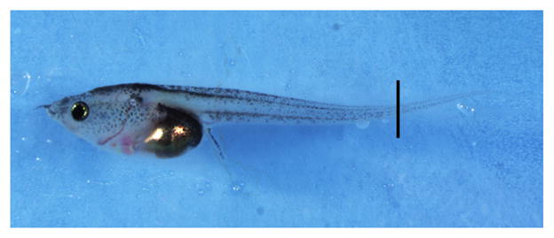
Cleanly sever the tail at a point 3–5 mm from its tip using a scalpel (Fig. 1), and transfer the tail tissue to a 1.5-mL microcentrifuge tube using a plastic transfer pipette.
Do not remove too much tissue, because cuts made too far anterior to the tail tip are likely to sever the major blood vessels running along the tail, leading to excessive bleeding and death. When taking tissue samples from larger numbers of animals, deep-well 96-well plates can be used instead of 1.5-mL microcentrifuge tubes. -
Using a teaspoon, transfer the tadpole to a tank that contains aquatic system water at a depth of 1 in. for recovery.
Tadpoles should recover from anesthesia within ~5 min, after which they should be returned to normal housing conditions.See Troubleshooting. -
Add 200 μL of prewarmed tissue lysis buffer to the tail tissue. Incubate for 12–16 h at 55°C (in a suitable incubator or water bath), and vortex periodically to aid effective lysis of the tissue.
If processing in 96-well format, seal the plate with sealing film. After incubation, centrifuge the sealed plates for 1 min at 6000g to prevent cross-contamination of the wells when removing the film. Add 150 μL of isopropanol, vortex gently, and then centrifuge at 6000g for 15 min at room temperature.
Aspirate the supernatant using a single-channel pipette. Change pipette tips between samples to avoid cross-contamination.
Wash the DNA pellet by adding 300 μL of 70% ethanol, briefly vortexing, and then centrifuging at 6000g for 10 min at room temperature.
-
Remove the 70% ethanol by aspiration and allow the DNA pellet to air dry.
When dry, the DNA pellet will appear opaque and white. -
Resuspend the DNA pellet in 0.5 mL of sterile nuclease-free H2O.
Resuspension is aided by allowing the pellet to sit at room temperature for 10 min after adding the H2O. -
Store the DNA at 4°C for up to 6 mo or at −20°C indefinitely.
Routine PCR amplification of genomic sequences can be carried out using a 1:50 dilution of the resulting DNA with typical PCR reaction components.See Troubleshooting.
FIGURE 1.

Tail tissue sampling. The position at which the tip of the tail should be cut is indicated by a black line on a X. tropicalis tadpole at stage 48.
TROUBLESHOOTING
Problem: The tadpoles do not recover from anesthesia.
[Step 3]
Solution: Reduce the length of time for which the tadpoles are kept in the anesthetic solution in Step 1.
Problem: PCR-amplifiable genomic DNA is not recovered.
[Step 10]
Solution: Consider the following:
Ensure that the pellet containing the genomic DNA is not discarded along with the 70% ethanol in Step 8.
Ensure that there is no carry-over of ethanol between Steps 8 and 9.
Ensure that the DNA is thoroughly resuspended in Step 9.
Decrease the volume of sterile nuclease-free H2O used to resuspend the DNA in Step 9.
References
- Nieuwkoop PD, Faber J, editors. Normal table of Xenopus laevis (Daudin): A systematical and chronological survey of the development from the fertilized egg till the end of metamorphosis. 2. North-Holland; Amsterdam: 1967. [Google Scholar]
- Showell C, Conlon FL. The Western clawed frog (Xenopus tropicalis): An emerging vertebrate model for developmental genetics and environmental toxicology. Cold Spring Harb Protoc. 2009 doi: 10.1101/pdb.emo131. (this issue) [DOI] [PMC free article] [PubMed] [Google Scholar]
- Wienholds E, van Eeden F, Kosters M, Mudde J, Plasterk RH, Cuppen E. Efficient target-selected mutagenesis in zebrafish. Genome Res. 2003;13:2700–2707. doi: 10.1101/gr.1725103. [DOI] [PMC free article] [PubMed] [Google Scholar]


