Summary
The master transcription factor, Pparγ regulates the general differentiation program of both brown and white adipocytes. However, it has been unclear whether Pparγ also controls fat lineage-specific characteristics. Here, we show that Early B-Cell Factor-2 (Ebf2) regulates Pparγ binding activity to determine brown versus white adipocyte identity. The Ebf DNA-binding motif was highly enriched within brown adipose-specific Pparγ binding sites that we identified by genome-wide ChIP-Seq. Of the Ebf isoforms, Ebf2 was selectively expressed in brown relative to white adipocytes and was bound at brown adipose-specific Pparγ target genes. When expressed in myoblasts or white pre-adipose cells, Ebf2 recruited Pparγ to its brown-selective binding sites and reprogrammed cells to a brown fat fate. Brown adipose cells and tissue from Ebf2-deficient mice displayed a loss of brown-specific characteristics and thermogenic capacity. Together, these results identify Ebf2 as a key transcriptional regulator of brown fat cell fate and function.
Keywords: Ebf2, Pparγ, Prdm16, brown adipose tissue, obesity, Ucp1
Introduction
Adipose tissues play an integral role in regulating systemic metabolism and energy homeostasis. White adipocytes store excess energy in the form of triglycerides for future need. By contrast, brown adipocytes metabolize lipid and glucose to produce heat in a process known as nonshivering thermogenesis (Cannon and Nedergaard, 2004). Brown adipocytes are packed with specialized mitochondria that contain Uncoupling Protein-1 (Ucp1) in their inner membrane. When activated, Ucp1 dissipates the electrochemical gradient used for ATP synthesis, resulting in increased heat production.
In rodents, brown adipose tissue (BAT) is a major site for cold-induced thermogenesis, which is essential for protection against hypothermia. BAT is also activated by overfeeding in mice as a physiological countermeasure to limit weight gain (Rothwell and Stock, 1979). Indeed, mouse strains that have increased BAT activity and/or increased numbers of brown-like (beige) adipocytes within their WAT are lean and healthy (Bostrom et al., 2012; Cederberg et al., 2001; Chiang et al., 2009; Guerra et al., 1998; Kopecky et al., 1996; Scime et al., 2005; Seale et al., 2011). By contrast, mice lacking brown and/or beige fat activity are susceptible to obesity (Feldmann et al., 2009; Lowell et al., 1993).
A natural role for BAT in adult humans was doubted until recently. Through the use of PET imaging, significant deposits of Ucp1-expressing adipocytes that take up large amounts of glucose and lipid were identified in most, if not all, adult humans (Cypess et al., 2009; Nedergaard et al., 2007; Ouellet et al., 2012; Saito et al., 2009; van Marken Lichtenbelt et al., 2009; Virtanen et al., 2009). Moreover, human BAT activity is inversely correlated with body mass, suggesting that natural variations in BAT function may contribute to the regulation of body weight in humans. Thus, there is great interest in identifying factors and pathways that control brown fat cell development and/or function.
Many transcriptional factors have been shown to regulate the brown adipocyte phenotype. Notable among these are Peroxisome proliferator-activated receptor gamma coactivator 1-alpha (Pgc1α) and PR-domain containing protein-16 (Prdm16). Pgc1α is a transcriptional co-activator that is especially involved in the acute thermogenic response of brown fat cells to β-adrenergic stimuli (Kleiner et al., 2012; Lin et al., 2004; Puigserver et al., 1998; Uldry et al., 2006). However, Pgc1α is not required for determination of brown adipocytes, nor is it necessary for the differentiation-linked induction of Ucp1 and other brown fat-specific genes (Lin et al., 2004; Uldry et al., 2006). Prdm16 is a large zinc finger-containing protein that binds and co-regulates Pparγ, Pparα, c/EBPβ and Pgc1α to induce brown fat-specific genes (Hondares et al., 2011; Kajimura et al., 2009; Seale et al., 2008; Seale et al., 2007). However, Prdm16 expression is induced quite late during the adipogenesis process (Seale et al., 2007), and thus probably does not regulate lineage commitment per se.
Brown and white adipocytes both express a core set of transcriptional factors that are believed to regulate common aspects of the differentiation process. A central component of this pathway is Peroxisome proliferator-activated receptor-γ (Pparγ), which is required for the development of all types of fat cells (Barak et al., 1999; Nedergaard et al., 2005; Rosen et al., 1999; Tontonoz et al., 1994). However, whether Pparγ also plays a fundamental role in controlling the brown or white fate of adipocytes had not been assessed.
Here, we used chromatin immunoprecipitiation analysis combined with massively parallel sequencing (ChIP-seq) to identify Pparγ binding sites in BAT and WAT. We found a surprisingly large number of binding sites that were fat depot-selective. Motif analyses identified the consensus binding site for Early B-cell factor (Ebf) as being highly enriched in BAT-specific Pparγ binding regions. Ebfs have been shown to regulate the initiation of adipogenesis in white fat cells (Akerblad et al., 2002; Jimenez et al., 2007) but their role in brown versus white fat cell determination had not been evaluated. We found that Ebf2 was expressed at much higher levels in brown relative to white adipose cells and tissue. Cell culture and genetic studies in mice revealed that Ebf2 recruits Pparγ to brown fat-selective gene targets, including Prdm16 to determine brown identity.
Results
The DNA motif for Early B cell factor (Ebf) is enriched at BAT-specific Pparγ binding sites
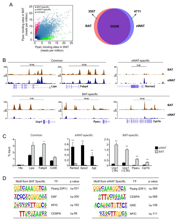
Pparγ regulates the adipogenic process in all fat cells and binds to thousands of loci during white adipocyte differentiation (Lefterova et al., 2008; Mikkelsen et al., 2010; Nielsen et al., 2008). We asked whether Pparγ has unique transcriptional targets in BAT and WAT. To do this, we performed genome-wide ChIP-seq analysis of Pparγ binding in interscapular BAT and epididymal WAT (eWAT) from 8-week- old 129Sv mice. Although 80% of binding sites were common to BAT and eWAT, there were ~3500 and ~4700 sites that were highly specific to BAT and eWAT, respectively (Figure 1A). ChIP-seq tracks show representative examples of target genes bound by Pparγ in: (1) both depots (e.g. Lipe, Fabp4); (2) eWAT only (Rarres2); and BAT only (Ucp1, Pparα, Cpt1b and Prdm16) (Figure 1B, 5A). We also used ChIP-qPCR in independent tissue samples to confirm the differential binding of Pparγ at select sites (Figure 1C). These results reveal that ~10% of Pparγ binding sites in BAT and eWAT are depot-selective.
Figure 1. The Ebf motif is highly enriched at BAT-specific Pparγ binding sites.
(A) ChIP for Pparγ followed by massively parallel sequencing and genome alignment was used to profile binding sites in mouse eWAT and BAT. (B) Representative Pparγ ChIP-sequencing tracks in BAT (brown) and eWAT (blue) at common, BAT-specific and WAT-specific sites. (C) Independent ChIP-qPCR validation of Pparγ binding in BAT and eWAT at select sites (mean ± SD; n=3; *p<0.05, **p<0.01). (D) Motif analyses of BAT- and eWAT-specific Pparγ binding regions.
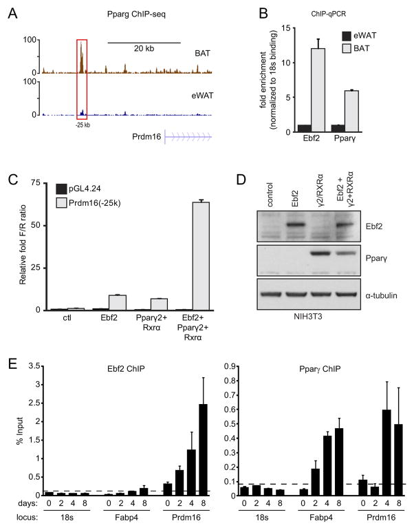
Figure 5. Ebf2 and Pparγ cooperatively activate transcription.
(A) Pparγ ChIP-seq tracks from BAT (brown) and WAT (blue) at the Prdm16 locus. (B) ChIP-qPCR analysis of Ebf2 and Pparγ binding to the −25 kb Prdm16 region (red box in (A)) in eWAT and BAT. (C) Transcriptional activity of the −25 kb region of Prdm16 or control reporter construct (pGL4.24) in response to expression of: Ebf2, Pparγ/Rxrα, or the combination of Ebf2 and Pparγ/Rxrα in NIH-3T3 cells (n=4; mean ± SD). (D) Western blot analysis of Ebf2 and Pparγ levels in NIH-3T3 lysates that were used for transcription assays in (C). (E) ChIP-qPCR analysis of Ebf2 and Pparγ binding to the −25 kb Prdm16 region and Fabp4 during a time-course of brown adipocyte differentiation.
We then performed motif analysis to determine which, if any, transcription factor binding sites were associated with BAT- or WAT-specific Pparγ binding regions. As anticipated, the DR-1 motif, which is bound directly by Pparγ, was the highest scoring motif in both tissue types. The next most enriched motif in BAT-selective Pparγ sites was the binding sequence for Early B-cell factor (Ebf) (Figure 1D), which prompted us to examine the role of Ebfs in brown adipocyte differentiation.
Ebf2 is selectively expressed in brown relative to white and beige adipocytes
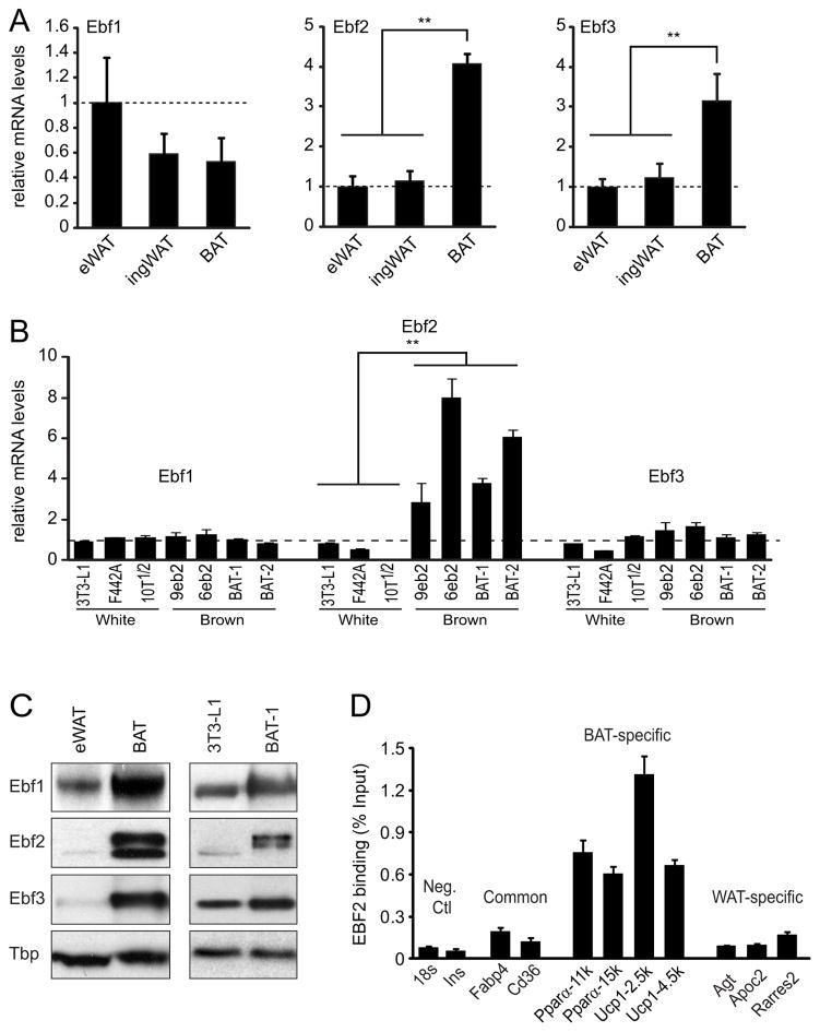
There are four members of the Ebf protein family (Ebf1-4), which could potentially bind the “Ebf” motif. By qPCR, we found that Ebf2 and Ebf3 mRNAs were expressed at higher levels in BAT relative to both epididymal and inguinal WAT (Figure 2A). Ebf4 was either expressed at very low levels or absent in the fat depots we examined. In established cell lines, only Ebf2 transcripts were dramatically enriched (~6-fold) in brown (4 independent lines) relative to white adipocytes (3T3-L1, 3T3-F442A, 10T1/2) (Figure 2B). Interestingly, the brown-selective expression of Ebf2 was apparent at the preadipocyte stage, though its expression also rose during the differentiation process (Figure S1A, B).
Figure 2. Ebf2 is selectively expressed in brown relative to white adipose cells and tissue.
(A) mRNA levels of Ebf1, Ebf2 and Ebf3 in eWAT, ingWAT (inguinal) and BAT (mean ± SD; n=3; ** p <0.01). (B) mRNA levels of Ebf1, Ebf2 and Ebf3 in a panel of white and brown adipose cell lines (mean ± SD; n=3; ** p <0.01). (C) Western blot analysis of Ebf1, Ebf2 and Ebf3 protein expression in representative samples from eWAT, BAT (left panel) and adipocytes (day 7) from white (3T3-L1) and brown (BAT 1) preadipocytes. Tbp was used for a loading control. (D) ChIP-qPCR analysis of Ebf2 binding in BAT at sites bound by Pparγ in: BAT and WAT (common), BAT-only and WAT-only (mean ± SD; n=3–5).
Thermogenic adipocytes, known as beige or brite [brown-in-white] cells develop within WAT in response to various stimuli, including β-adrenergic agonists. These beige adipocytes have many of the characteristics of brown fat cells, but are derived from a separate cellular lineage (Seale et al., 2008). Wu et al. recently cloned out several beige and white preadipocyte cell lines from the inguinal WAT of mice (Wu et al., 2012). Interestingly, we found that Ebf2 was expressed at similar levels in beige and white adipocytes lines; much lower than its levels in brown adipocytes, cloned by the same procedures (Figure S1C). These results suggest that Ebf2 is preferentially expressed in “classic” brown adipocytes relative to other types of adipocytes.
We also examined Ebf protein levels in adipose cells and tissue by western blotting using commercially available antibodies. First, we assayed the Ebf-isoform specificity by probing lysates from C2C12 cells that had been transiently transfected with Flag tagged versions of Ebf1, Ebf2 or Ebf3 (Figure S1D). The Ebf2 antibody was highly specific, whereas Ebf1 and Ebf3 antibodies cross-reacted to a small degree with the other Ebf isoforms. In agreement with mRNA analysis, Ebf2 and Ebf3 protein levels were dramatically higher in BAT relative to eWAT but Ebf2 protein levels were also very highly enriched in brown relative to white (3T3-L1) adipocytes (Figure 2C).
The next logical question was whether Ebf2 binds at/near brown-specific genomic targets of Pparγ in BAT. Using ChIP-qPCR, we found that Ebf2 was enriched by ~6–15-fold at several BAT-specific Pparγ binding sites relative to control regions (Figure 2D). There was much less binding of Ebf2 with Pparγ binding sites that are common between WAT and BAT (Fabp4, Cd36), and no detectable enrichment of Ebf2 at WAT-specific Pparγ binding sites (Agt, Apoc2, Rarres2). These results show that Ebf2 is expressed selectively in BAT and binds to BAT-specific Pparγ gene targets.
Ebf2 expression stimulates a brown adipose-specific differentiation program
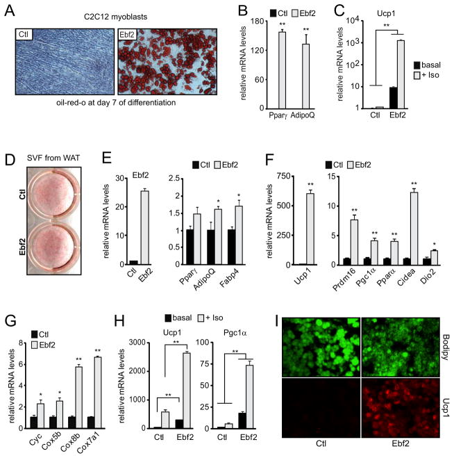
Brown fat cells originate from Myf5- and Pax7-expressing precursors that also give rise to skeletal muscle. To examine whether Ebf2 is sufficient to drive brown adipocyte differentiation, we first used retrovirus to express Ebf2 or control vector in C2C12 cells, a committed muscle precursor cell line. Ebf2- and control-expressing cultures were grown to confluence and then induced to undergo adipocyte differentiation. Strikingly, the Ebf2-expressing cultures underwent very efficient conversion into lipid-containing adipocytes, whereas control cultures were devoid of adipocytes (Figure 3A). Ebf2 expression stimulated adipogenesis in C2C12 cells to almost the same extent as retroviral Pparγ2 expression (not shown). Consistent with its effects on cell morphology, Ebf2 increased the expression levels of general adipocyte-selective genes, such as Pparγ and AdipoQ (Figure 3B). But, unlike Pparγ2, Ebf2 also activated the brown fat-specific gene program, including high levels of Ucp1 and Cidea (Figure 3C, S2). Finally, Ebf2 expression also enabled adipocytes to acutely increase their expression levels of Pgc1α and Ucp1 in response to the pan β-adrenergic agonist, isoproterenol (Figure 3C, S2).
Figure 3. Ebf2 expression drives a brown fat-specific differentiation program.
(A) Ctl- and Ebf2-expressing C2C12 myoblasts were induced to differentiate into adipocytes followed by staining of triglycerides with oil-red-o. (B, C) Above cultures were analyzed for their expression levels of: Pparγ and AdipoQ (B); and Ucp1 levels ± Isoproterenol treatment (C) (n=3). (D–I) Ctl- and Ebf2-expressing white preadipocytes were induced to differentiate into adipocytes followed by staining with oil-red-o (D). (E–H) mRNA levels of: Ebf2 and general adipocyte markers (E); brown-selective genes (F); mitochondrial genes (G); Ucp1 and Pgc1α ± Isoproterenol treatment (H) (n=3–5 pools/construct). (I) Bodipy staining of triglycerides (green) and Ucp1 immunostaining (red). All expression data are: mean ± SD; * p <0.05; ** p <0.01.
The powerful adipogenic action of Ebf2 in C2C12 cells made it difficult to isolate the effects of Ebf2 on the induction of brown-specific genes. We thus wanted to investigate the function of Ebf2 in preadipose cells that are naturally competent to undergo adipocyte differentiation. The stromal vascular fraction (SVF) of WAT contains bona fide preadipocytes that differentiate into white adipocytes. We expressed Ebf2 or vector control in primary SVF cultures isolated from inguinal WAT of 8–12 week old male mice. Cells transduced with Ebf2 virus expressed ~4 fold-higher levels of Ebf2 mRNA relative to its endogenous levels in brown fat cells. Six days after inducing differentiation, both Ebf2- and control- cultures contained mostly mature, lipid-filled adipocytes (Figure 3D).
The molecular phenotype of adipocytes from Ebf2- and control-expressing SVF cells was analyzed in greater detail by qPCR-based gene expression analysis. We found that Ebf2 expression resulted in a mild increase in the levels of some general adipocyte genes, including AdipoQ and Fabp4 (Figure 3E). Strikingly however, Ebf2 very strongly increased the expression levels of many brown adipocyte-specific genes, including a near 500-fold increase in Ucp1, and ~4–10 fold increases in Prdm16, Pgc1α, Pparα and Cidea relative to control cultures (Figure 3F). Additionally, Ebf2 increased the mRNA levels of several mitochondrial components, including: Cytochrome-c (Cyc), Cox5b, Cox8b and Cox7a1 (Figure 3G). Isoproterenol treatment further increased the expression of thermogenic genes, like Ucp1 and Pgc1α, to much higher levels in Ebf2- compared to control-expressing cells (Figure 3H). Finally, immunofluorescence analysis showed that Ebf2-expressing adipocytes expressed greatly elevated levels of Ucp1 protein as compared to control cells under un-stimulated conditions (Figure 3I).
We also found that Ebf2 robustly and consistently induced the thermogenic gene program when expressed in other types of fat precursors, including: beige and white preadipose cell lines isolated from inguinal WAT (Figure S3), and in the classic 3T3-L1 white adipocyte line (Figure S4). In beige cells, we found that Ebf2 stimulated adipogenic differentiation and suppressed the expression of several beige-specific genes (Ear2, Tmem26, CD137, SP100).
Ebf2 is required for the maintenance of brown fat cell fate
The requirement for Ebf2 in brown adipocyte differentiation was investigated using siRNA-mediated gene knock-down. First, we used lentiviral vectors to express a shRNA targeting Ebf2 or a control Scrambled (Scr) shRNA in mouse brown preadipocytes. The sh-Ebf2 construct efficiently depleted Ebf2 mRNA levels in brown preadipocytes (Figure S5A) without reducing the levels of other Ebf isoforms (not shown). Whereas control sh-Scr cells underwent efficient morphological differentiation and accumulated lipid droplets, loss of Ebf2 completely blocked differentiation (Figure S5B). Consistent with this, Ebf2-depleted cells failed to induce general adipogenic genes including Pparγ, Fabp4 and AdipoQ as well as brown-specific markers, like Prdm16 and Cidea (Figure S5C).
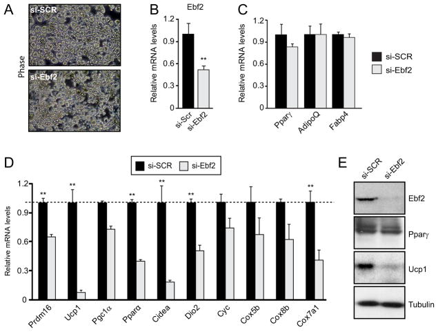
The complete differentiation block caused by shRNA-mediated loss of Ebf2 in preadipocytes confounded a meaningful analysis of brown-specific pathways (that are only expressed in mature adipocytes). Therefore, we performed knock-down experiments in mature brown adipocytes. On day 6 of differentiation, brown adipocytes were electroporated with a control Scr siRNA or an Ebf2 siRNA. 48–72 hr later, Scr- and Ebf2 siRNA- treated cultures were morphologically indistinguishable, each composed of well differentiated adipocytes (Figure 4A). siEbf2 reduced Ebf2 mRNA levels by ~50% (Figure 4B) and caused a much more dramatic loss of Ebf2 protein levels (Figure 4E). Importantly, the depletion of Ebf2 in adipocytes did not affect Pparγ levels (mRNA or protein) or the mRNA levels of general adipocyte genes AdipoQ and Fabp4 (Figure 4C, E). Reduction of Ebf2 did, however, cause a very specific decrease in the expression levels of a wide array of brown-specific and mitochondrial genes, including a ~50% and a ~90% reduction in Prdm16 and Ucp1 mRNA levels, respectively (Figure 4D). Loss of Ebf2 also caused a dramatic reduction in Ucp1 protein levels (Figure 4E).
Figure 4. Ebf2 is required to maintain the expression of brown-specific genes in brown adipocytes.
(A) Phase contrast micrographs of brown adipocyte cultures 2 days after treatment with an Ebf2-specific siRNA or a siSCR control. (B–D) mRNA levels of: Ebf2 (B); general fat markers (C); brown-specific and mitochondrial genes (D). (E) Western blot analysis of Ebf2, Pparγ, Ucp1 and Tubulin (loading control) protein levels. Expression data are mean ± SD; n=3; **p<0.01.
Ebf2 and Pparγ cooperate to activate transcription of Prdm16
The binding of Ebf2 at Pparγ target sites suggested a functional interaction between these two factors. Prdm16 is a transcriptional co-activator that plays a pivotal role in brown fat cell differentiation (Kajimura et al., 2009; Kajimura et al., 2008; Seale et al., 2008; Seale et al., 2007). We noted that ectopic expression of Ebf2 greatly increased Prdm16 levels in fat cells (Figure 3) whereas loss of Ebf2 reduced Prdm16 levels (Figure 4); this raised the question of whether Ebf2 directly regulates Prdm16 transcription. Upon re-examination of the Pparγ ChIP-seq data, we uncovered a strong BAT-specific Pparγ binding site located ~ 25 kb upstream of the Prdm16 promoter (Figure 5A). Using ChIP qPCR, we found that both Pparγ and Ebf2 bind to this region in BAT but not in eWAT (Figure 5B).
The Pparγ/Ebf2 binding region in Prdm16 was cloned upstream of a minimal Thymidine Kinase promoter and Firefly Luciferase reporter gene. We then expressed Ebf2 and/or Pparγ with its partner, Rxrα in combination with the Prdm16-Luciferase reporter gene in NIH-3T3 cells. The reporter gene had very minimal activity on its own and only a slightly increased activity in response to either Ebf2 alone or Pparγ and Rxrα (Figure 5C). However, co-expression of Ebf2 with Pparγ and Rxrα led to a very dramatic rise in transcription (~70 fold over ctl) (Figure 5C). This synergistic activation of transcription was not due to an increase in protein levels of either factor (Figure 5D).
Ebf2 is expressed in brown preadipocytes prior to the differentiation-linked increase in Pparγ and Prdm16 expression (Figure S1). We thus wondered whether Ebf2 was recruited to the Prdm16 locus before Pparγ. To answer this, we performed ChIP-qPCR for Ebf2 and Pparγ during a time-course of brown fat cell differentiation. We found that Ebf2 was already bound to Prdm16 but not to Fabp4 in preadipocytes (day 0); the binding at Prdm16 increased further during differentiation (Figure 5E). By contrast, Pparγ was not detectable at Prdm16 until day 4. These results suggest that Ebf2 binds to brown fat gene targets before (and independently of) Pparγ.
Ebf2 recruits Pparγ to BAT-selective gene targets
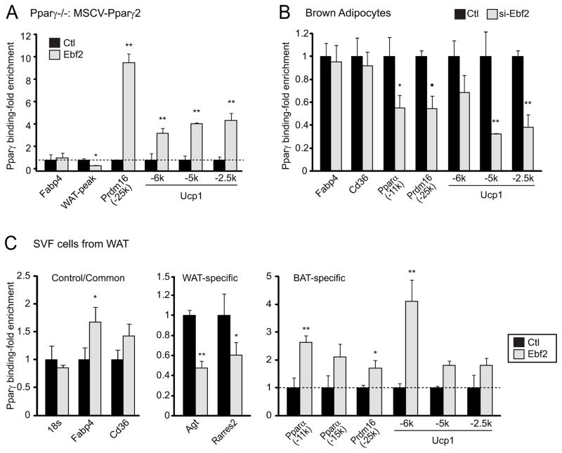
The above experiments raised the question of whether Ebf2 facilitates the binding Pparγ to its BAT-specific target sites. To test this, we used Pparγ-deficient fibroblasts expressing retroviral Pparγ2 in order to hold Pparγ levels constant. These cells were then transduced with control- or Ebf2-expressing retrovirus and induced to undergo adipocyte differentiation for seven days. Gene expression analysis confirmed that Ebf2 robustly stimulated the brown-specific gene program in these cells (not shown). Remarkably, ChIP-qPCR analysis showed that exogenous Pparγ2 interacted strongly with BAT-specific targets, such as Prdm16 and Ucp1 in Ebf2-expressing but not in control-expressing cultures (Figure 6A). Conversely, we found that siRNA-mediated reduction of Ebf2 in brown adipocytes caused a significant decrease in Pparγ binding at several BAT-specific sites, including Ucp1, Pparα and Prdm16, with no change in binding at BAT/WAT common sites or WAT-specific sites (Figure 6B).
Figure 6. Ebf2 promotes the binding of Pparγ to its brown fat-selective gene targets.
(A) ChIP-qPCR analysis of Pparγ binding in adipocytes from Pparγ-deficient cells that were first transduced with Pparγ retrovirus, followed by transduction with either a control (ctl) or Ebf2-expressing retrovirus. (B) ChIP-qPCR analysis of Pparγ binding in Scrambled (Ctl) or Si-Ebf2 treated brown adipocytes. (C) ChIP-qPCR analysis of Pparγ binding in mature adipocytes from control (Ctl) and Ebf2-expressing white preadipocytes. All data are normalized to background binding (at non-specific Insulin and 18s rRNA loci) and presented as mean ± SD, n > 3.
We also examined the effect of Ebf2 expression on Pparγ localization in a primary SVF cells from WAT. Here, we found that Ebf2 expression caused a slight increase in the binding of Pparγ to Fabp4, a target in both BAT and WAT (Figure 6C). However, Ebf2 expression increased Pparγ binding by ~2–4 fold at brown-specific targets, including Prdm16, while significantly reducing Pparγ binding at WAT-specific target sites (Agt, Rarres2) (Figure 6C). Altogether, these results suggest that Ebf2 enables Pparγ to bind to its BAT-specific target sites.
Ebf2 is required for BAT development in mice
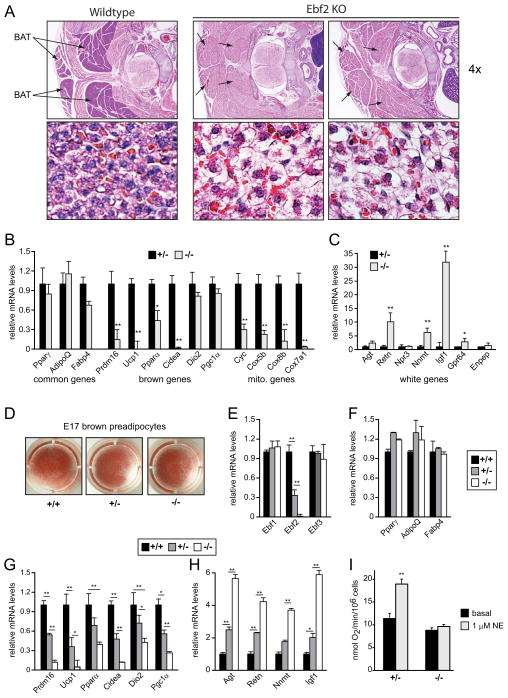
To determine whether Ebf2 is important for brown fat development in vivo, we analyzed BAT in wildtype and Ebf2-deficient mouse embryos. Ebf2-deficient mice are born in normal Mendelian ratios but fail to thrive and typically die soon after birth (Corradi et al., 2003; Ukropec et al., 2006). Due to the systemic illness that occurs postnatally in Ebf2-knockout (KO) animals, we analyzed BAT in late stage mouse embryos. By hematoxylin and eosin (H&E) staining, the presumptive BAT from the Ebf2-deficient embryos at E18 days post-coitum (dpc) was reduced in size and had a very unusual morphology (Figure 7A). Specifically, the KO tissue was very weakly stained with eosin, suggesting a dramatic reduction in mitochondrial density.
Figure 7. Ebf2 is required for BAT development in mice.
(A) Hematoxylin and Eosin (H&E) staining of representative sections from the interscapular region of wildtype and Ebf2-deficient embryos at E18 dpc. (B, C) BAT from Ebf2+/− and Ebf2−/− embryos (E18) was examined by real-time PCR analysis for mRNA levels of: general adipocyte genes, brown-specific genes and mitochondrial genes (B); and WAT-selective genes (C) (n=5–8 embryos/genotype). (D) Brown fat precursors from Ebf2+/+, Ebf2+/− and Ebf2−/− embryos were differentiated into adipocytes in culture and stained with oil-red-o. (E–H) Above cultures were analyzed by real-time qPCR for their expression levels of: Ebfs (E); general adipocyte markers (F); brown-specific genes (G); and white-selective genes (H) (n=3 pools [> 5 mice per pool]). (I) Oxygen consumption in cultured brown adipocytes from Ebf2+/− and Ebf2−/− before and after stimulation with norepinephrine (NE) (mean ± SD; n=3). Expression data are: mean ± SD, * p <0.05; ** p <0.01.
Gene expression and western blot analysis confirmed that Ebf2 mRNA (not shown) and protein (Figure S6A) were ablated in the knockout tissue. Interestingly, Ebf2 KO tissue expressed normal levels of the pan-adipocyte markers Pparγ, AdipoQ and Fabp4 (Figure 7B, left side). However, there was an almost complete loss of brown fat-specific gene expression in Ebf2-KO tissue, including a ~85% reduction in Prdm16 levels and over 95% reduction in Ucp1 and Cidea levels (Figure 7B, center). Mitochondrial gene expression was also severely decreased in Ebf2-deficient BAT (Figure 7B, right). Interestingly, we noted that Ebf2-deficiency also caused a milder decrease in mitochondrial gene levels in embryonic heart, but not in skeletal muscle or brain (Figure S6B). Finally, we found that Ebf2-deficient BAT expressed higher levels of many white-specific gene markers, including Resistin (Retn), Nnmt, Igf1, and Gpr64 (Figure 7C). These results reveal a profound genetic requirement for Ebf2 in determining BAT identity.
We then assessed whether the BAT phenotype in Ebf2-deficient mice was due to fat cell-autonomous defects. To address this, we isolated brown preadipocytes from Ebf2+/+, Ebf2+/− and Ebf2−/− embryos at E17 dpc and subjected them to in vitro differentiation. Cultures of all three genotypes underwent efficient morphological differentiation into lipid-containing adipocytes as shown by oil-red-o staining (Figure 7D). Gene expression analysis showed that Ebf2 mRNA was undetectable in Ebf2−/− cells and reduced by ~60% in Ebf2+/− cultures (Figure 7E). Reduction of Ebf2 did not alter the levels of either Ebf1 or Ebf3 (Figure 7E). Ebf2+/+, Ebf2+/− and Ebf2−/− cultures expressed similar levels of general adipocyte markers (Figure 7F). However, the expression of brown-specific and mitochondrial genes was severely reduced by the decrease or loss of Ebf2, including an 85–90% reduction in the levels of Prdm16, Ucp1 and Cidea in KO relative to wildtype cells (Figure 7G; S6C). Ebf2-deficient cultures also expressed greatly elevated levels (4–6 fold higher than wildtype) of white adipocyte-specific genes (Figure 7H). Interestingly, ectopic over-expression of Ebf1, Ebf2 or Ebf3 into Ebf2−/− brown preadipocytes rescued the differentiation-linked expression of brown genes to levels ~5x higher than seen in uninfected, wildtype cells (Figure S7). These results demonstrate that Ebf2 regulates brown adipose identity in a cell-autonomous manner.
Brown adipocytes produce heat through a catecholamine-stimulated activation of Ucp1 and a resulting increase in uncoupled respiration (Cannon and Nedergaard, 2004). To examine whether Ebf2 is critical for brown adipocyte function, we measured the respiratory activity of cultured Ebf2+/− and Ebf2−/− brown adipocytes using a Clark electrode. At day 7 of differentiation, Ebf2+/− cells had slightly higher levels of basal and maximal (FCCP-stimulated) respiration (Figure 7I, S6D). Importantly however, norepinephrine (NE) stimulation elicited a very dramatic rise (~ doubling) in the O2 consumption of Ebf2+/− cells and had no effect in Ebf2−/− cells (Figure 7I). This NE-stimulated respiration in Ebf2+/− cells was largely insensitive to oligomycin, and was thus uncoupled (Figure S6D). These studies indicate that Ebf2 is essential for the proper respiratory function of brown adipocytes.
Discussion
Pparγ is the master regulator of adipocyte differentiation in brown and white fat cells, even though these cells belong to separate lineages. This raised the intriguing question of whether Pparγ played a major role in lineage-specific programming and if so, how?
We found that Pparγ binds to thousands of loci differentially in BAT and eWAT. Consistent with our results, the Mandrup lab recently reported that in vitro differentiated adipocytes also display depot-selective Pparγ binding profiles (Siersbaek et al., 2012). In both studies, Pparγ binding is highly correlated with depot-selective gene expression, suggesting that Pparγ plays a broad and unanticipated role in executing brown and white fat cell-selective gene programs.
BAT-specific target sites of Pparγ are also specifically bound by Ebf2 in brown adipocytes. Our studies suggest that Ebf2 acts as a pioneer factor in brown fat cells to “mark” genes for the later recruitment of Pparγ and transcriptional activation. This is reminiscent of the role of Ebf1 in B-cells which has been found to promote the de-methylation of some of its target genes to poise genes for their later activation by Pax5 and other transcription factors (Maier et al., 2004; Treiber et al., 2010). In fat cells, we also noted that Ebf2 expression reduced Pparγ binding at white fat-specific loci which could explain the repressive effects of Ebf2 on white-specific gene transcription. Whether this is due to squelching because Pparγ protein is limiting or occurs via the action of a downstream Ebf2-target gene(s) is not yet known.
Interestingly, neither Ebf2 nor Pparγ alone could efficiently activate transcription from a putative enhancer in Prdm16 which contains both Ebf and Pparγ binding motifs. Ebf2-binding may induce structural changes to DNA and/or chromatin that facilitate or stabilize Pparγ binding. It is also possible that Ebf2 has additional effects on the Pparγ transcriptional complex, such as promoting co-activator recruitment and/or displacing co-repressors, to stimulate transcriptional activity. Interestingly, Ebf1 interacts with the Swi/Snf chromatin remodelling complex, which is necessary for Ebf-mediated transcriptional activation from certain B cell-specific enhancers (Gao et al., 2009). Further studies will be needed to unravel the mechanistic details of how Ebf2 and Pparγ synergize to drive transcription in fat cells.
Prdm16 drives brown fat-specific gene expression in all known types of thermogenic adipocytes (Seale et al., 2011; Seale et al., 2007). However, molecular events that control the depot-selective and differentiation-linked expression of Prdm16 had not been identified. We reveal here that Ebf2 and Pparγ act upstream of Prdm16 in the transcriptional program of classic brown fat cells. It seems likely that activation of Prdm16 is critical for the action of Ebf2 in brown fat determination. However, Ebf2 also interacts directly with other major brown fat-specific genes, including Ucp1, suggesting that Ebf2 controls both Prdm16-dependent and -independent pathways. We propose that Prdm16, once induced by Ebf2, provides a feed-forward mechanism to amplify and reinforce the expression of brown fat-specific genes.
The presence of fatty tissue in Ebf2−/− animals, rather than a complete absence of BAT, suggests that other, unknown factors specify preadipose cell fate. One possibility is that Ebf1 and/or Ebf3 preserve preadipocyte commitment in the absence of Ebf2 in vivo. Surprisingly, in cultured brown preadipocytes, shRNA-mediated reduction of Ebf2 completely blocked adipogenesis. This result is consistent with previous studies showing that Ebf2 is required for early events during the differentiation of 3T3-L1 cells (Jimenez et al., 2007) and raises the possibility that other Ebfs can compensate for the chronic but not acute loss of Ebf2 in preadipocytes.
Importantly, the fat tissue in Ebf2-null animals was completely devoid of brown character. This is despite the fact that Ebf1 and especially Ebf3 are expressed in brown adipocytes and have the ability to stimulate the brown fat gene program when ectopically expressed in cultured precursors. The specific requirement for Ebf2 is likely dictated by the timing and levels of its expression during brown adipocyte development/differentiation. Alternatively, Ebf2 may have isoform-specific actions in brown fat cells which can be conferred by other isofroms through high (super-physiological) levels of expression.
The loss of thermogenic gene programming in Ebf2-deficient brown adipocytes was associated with a profound defect in respiratory function, much like that seen in Ucp1-deficient brown adipocytes (Matthias et al., 2000). The loss of brown fat-like respiratory activity predicts that Ebf2-deficient BAT would be incapable of adaptive thermogenesis in response to high fat diet or cold. Unfortunately however, we are not yet able to assess the physiological consequence of Ebf2 action in adult BAT, including effects on energy balance, due to the severe systemic effects caused by whole-body Ebf2 deficiency in our mouse model.
Genetic and developmental studies have shown that there are at least two lineages of thermogenic adipocytes: (1) classic brown adipocytes (in BAT); and (2) beige adipocytes (a.k.a. brite [brown in white] adipocytes) that develop within WAT (Petrovic et al., 2010; Seale et al., 2008; Xue et al., 2007). In inguinal WAT, beige adipocytes develop from committed preadipocytes and only express high levels of thermogenic genes upon stimulation with β-adrenergic agonists (Wu et al., 2012). We found that Ebf2 expression was enriched in brown but not beige adipocytes relative to white adipocytes. Moreover, Ebf2 expression in beige or white adipocyte lines increased thermogenic gene levels in the absence of β-adrenergic stimulation. Ebf2 may thus provide a brown fat-specific mechanism for inducing high levels of thermogenic gene/protein levels during differentiation under basal conditions.
There is great hope that the thermogenic action of brown/beige adipocytes can be used therapeutically to reduce obesity and/or metabolic disease. To effectively increase brown fat mass and/or function, it will be important to identify treatments that stimulate a broad and specific program of differentiation. Our work suggests that the binding of Pparγ to its brown fat-specific gene targets is fundamental for proper brown adipocyte development. Thus, searching for the upstream signals and factors that induce Ebf2 expression and activity in fat precursors will reveal important biological insights and may identify new therapeutic options.
Experimental Procedures
Animals
All animal experiments were performed according to procedures approved by the University of Pennsylvania’s Institutional Animal Care and Use Committee. Ebf2-deficient mice were described before.
ChIP-seq
High throughput sequencing was done by the Functional Genomics Core of the Penn Diabetes Research Center using the Illumina Genome Analyzer IIx or HiSeq 2000, and the reads were aligned to the mm8 genome assembly genome using Bowtie. In each ChIP-seq sample, all the duplicate reads were removed except for one before downstream analysis.
Peak calling and motif analysis
To identify depot-specific Pparγ peaks, we performed peak calling for one depot sample as foreground and the other sample as background, and vice versa. All the peak calling were performed using Homer with options, peak size 200bp and minimum distance 200bp (-size 200 -minDist 200), then 1 rpm cutoff was applied. Common Pparγ peaks were defined as follows. First, Pparγ peaks were called for each depot using a matching input sample, then all the peaks were pooled and overlapping peaks were merged. Among these peaks, any peak that overlaps with the depot-specific Pparγ peaks were eliminated, and the remaining peaks were defined as common Pparγ peaks. It should be noted that our definition of common peak here is naïve in the sense that some peaks might show different peak height between different depots. However, this should not be a problem in this work because our primary target is to identify depot-specific (especially BAT-specific) Pparγ binding sites.
Cell culture
Primary brown and white preadipocytes were fractionated from mice fat depots using collagenase and dispase digestion as described before (Seale et al., 2007). Immortalized brown fat preadipocytes and PPARγ−/− fibroblasts have been described elsewhere. Murine clonal beige and white cells from inguinal WAT were cultured and differentiated as described (Wu et al., 2012). For adipocyte differentiation assays, immortalized brown fat or primary preadipocytes were grown to confluence and cells were treated with induction medium containing 10% FBS, 0.5 mM isobutylmethylxanthine, 125 nM indomethacin, 1 μM dexamethosone, 20 nM insulin, and 1 nM T3. After 48 h, cells were switched to medium containing 10% FBS, 1 nM T3 and 20 nM insulin until harvest. To stimulate thermogenesis, cells were incubated with 10 μM isoproterenol for 3 hours.
Oxygen Consumption Assays
Primary brown preadipocytes were induced to undergo adipogenesis. At day 7 of differentiation, adipocytes were trypisinized, counted and oxygen consumption was measured using a Strathkelvin Clark-type electrode. 1 μM of norepinephrine (Sigma), 1 μM oligomycin (Sigma) or 2 μg of FCCP (Sigma) was added acutely to the chamber to activate Ucp1, block state III respiration and induce uncoupling respectively.
Viral vectors and production
Full-length coding sequences for mouse Ebf1 (GeneID 13591), Ebf2 (GeneID 13592) and Ebf3 (GeneID 13593) were amplified from BAT mRNA, and cloned into pMSCV-puro retroviral vector (Stratagene) or pcDNA3.1 (Invitrogen). The siRNA and double-stranded DNA sequence encoding the shRNA targeting Ebf2-knockdown were purchased from (Dharmacon). For retrovirus production, 293T cells (Kinsella and Nolan, 1996) were transfected with 15 μg retroviral vectors along with packaging plasmids at 70% confluence by calcium phosphate co-precipitation. The viral supernatant was harvested every 24 hours for 3 days and concentrated using SW28 ultracentrifuge rotor at 26k rpm. For retroviral transduction, cells were incubated overnight with viral supernatant supplemented with 8 μg/ml polybrene.
Real-time PCR
Total RNA from cultured cells or tissues was isolated using the TRIzol method (Invitrogen) combined with Qiagen RNeasy mini columns. For real-time PCR analysis, RNA was reverse transcribed using the ABI high capacity cDNA synthesis kit and used in quantitative PCR reactions containing SYBR-green fluorescent dye (ABI) using an ABI9300 PCR machine. Tata-binding protein (Tbp) served as an internal control to normalize samples. Primers are listed in Supplemental Table 1.
Western blot analysis
Cells or tissues were lysed in radioimmunoprecipitation assay (RIPA) lysis buffer containing 0.5% NP-40, 0.1% sodium deoxycholate, 150 mM NaCl, 50 mM Tris-Cl, pH 7.5, protease inhibitor cocktail (Complete; Roche) and 1 mM phenylmethylsulfonyl fluoride (PMSF). The protein content was measured using a detergent-compatible (DC) protein assay kit (Bio-Rad). Lysates or nuclear fractions were resolved onto bis-Tris NuPAGE gels (Invitrogen), transferred to PVDF membrane (Millipore), and probed with anti-Ebf1(Santa Cruz Biotechnology), anti-Ebf2 (R&D systems), anti-Ebf3 (Santa Cruz Biotechnology), anti-EBF (D-8) (Santa Cruz Biotechnology), anti-Pparγ (E-8) (Santa Cruz Biotechnology), anti-Ucp1 (R&D systems), anti-FLAG (Mouse polyclonal), anti-TBP (Abcam), and anti–tubulin (Santa Cruz Biotechnology) antibodies.
Oil-red-O staining
After 6–8 days of differentiation, cells were washed once in PBS and fixed with freshly prepared 4% formaldehyde for 15 min and stained using oil-red-o solution for 30 min.
Chromatin Immunoprecipitation (ChIP)
Fat depots from 129 × 1/SvJ male mice (8 weeks old) were harvested and washed with PBS. Fat tissues were minced in DMEM and cross-linked with 1% formaldehyde for 15 min at room temperature. Cross-linking was quenched using 125 mM glycine for 5 min. Tissues were then washed with PBS and ChIP dilution buffer containing 0.1% SDS, 50 mM Tris, 140 mM NaCl, 1% triton X-100, 0.1% Na-Deoxycholate, 1mM PMSF complete protease inhibitor cocktail (Roche), and 1 mM EDTA. Chromatin was sheared by sonication and immunoprecipitation was carried out as described (Ishibashi et al., 2012). The protein-DNA complexes were immunoprecipitated using 2.5 μg of antibody (sc-2027; Santa Cruz) and isolated with protein A-Sepharose beads, washed and eluted with 1% SDS–0.1 M NaHCO3. Reverse cross-linking was performed for overnight incubation at 65°C, followed by proteinase K digestion and purification of DNA fragments using PCR purification columns (Qiagen). Eluted DNA was analyzed by real-time PCR on a 7900 machine (ABI) using SYBR green chemistry (ABI, Affymetrix). Target enrichment was calculated as percent input and normalized to 18S for nonspecific background signal. Primer sequences used for this study are found in Table S2 in the supplemental material.
Transcription assays
Luciferase reporter plasmids were constructed by PCR cloning C57Bl/6 genomic fragments corresponding to Pparγ-bound regions into the pGL4.24 vector (Promega). pGL4-based reporters were co-transfected into NIH3T3 cells (ATCC) in multi-well plates along with Renilla luciferase (internal control), Ebf2, and/or Pparγ2 expression plasmids using Lipofectamine 2000 (Invitrogen). Cells were harvested into passive lysis buffer (Promega) at 48 hours post-transfection and dual luciferase activity was assayed with a Synergy HT plate reader (BioTek). Reporter luciferase activity was normalized to the internal Renilla control activity.
Statistical Analysis
All data reported as mean ± SD unless otherwise noted in figure legend. Student’s t-test was used to calculate significance (*p < 0.05; **p<0.01) using Excel or Prism software packages.
Supplementary Material
Ebf2 is selectively expressed in brown relative to white adipocytes and binds to chromatin at brown fat-specific target sites of Pparγ.
Ebf2 expression in white fat or muscle precursor cells recruits Pparγ to its brown fat-specific gene targets and drives a complete program of brown adipocyte differentiation.
Brown adipose cells and tissue from Ebf2-deficient mice display a complete loss of thermogenic characteristics while gaining molecular attributes of white adipose.
Acknowledgments
We thank Mitch Lazar and Dave Steger for expert guidance with ChIP studies; Dr. Mary Selak for help with oxygen consumption assays; Li Huang for expert technical assistance and members of the Seale lab for helpful discussions. We are grateful to the Functional Genomics Core of the Penn Diabetes and Endocrinology Research Center (DK19525) for high throughput sequencing and data analyses. J.W. is supported by an NIH grant (1K01DK094824). This work was supported by NIDDK/NIH award R00 DK081605, NIGMS/NIH award DP2OD007288 and a Searle Scholars Award to P.S.
Footnotes
Publisher's Disclaimer: This is a PDF file of an unedited manuscript that has been accepted for publication. As a service to our customers we are providing this early version of the manuscript. The manuscript will undergo copyediting, typesetting, and review of the resulting proof before it is published in its final citable form. Please note that during the production process errors may be discovered which could affect the content, and all legal disclaimers that apply to the journal pertain.
References
- Akerblad P, Lind U, Liberg D, Bamberg K, Sigvardsson M. Early B-cell factor (O/E-1) is a promoter of adipogenesis and involved in control of genes important for terminal adipocyte differentiation. Mol Cell Biol. 2002;22:8015–8025. doi: 10.1128/MCB.22.22.8015-8025.2002. [DOI] [PMC free article] [PubMed] [Google Scholar]
- Barak Y, Nelson MC, Ong ES, Jones YZ, Ruiz-Lozano P, Chien KR, Koder A, Evans RM. PPAR gamma is required for placental, cardiac, and adipose tissue development. Mol Cell. 1999;4:585–595. doi: 10.1016/s1097-2765(00)80209-9. [DOI] [PubMed] [Google Scholar]
- Bostrom P, Wu J, Jedrychowski MP, Korde A, Ye L, Lo JC, Rasbach KA, Bostrom EA, Choi JH, Long JZ, et al. A PGC1-alpha-dependent myokine that drives brown-fat-like development of white fat and thermogenesis. Nature. 2012;481:463–468. doi: 10.1038/nature10777. [DOI] [PMC free article] [PubMed] [Google Scholar]
- Cannon B, Nedergaard J. Brown adipose tissue: function and physiological significance. Physiol Rev. 2004;84:277–359. doi: 10.1152/physrev.00015.2003. [DOI] [PubMed] [Google Scholar]
- Cederberg A, Gronning LM, Ahren B, Tasken K, Carlsson P, Enerback S. FOXC2 is a winged helix gene that counteracts obesity, hypertriglyceridemia, and diet-induced insulin resistance. Cell. 2001;106:563–573. doi: 10.1016/s0092-8674(01)00474-3. [DOI] [PubMed] [Google Scholar]
- Chiang SH, Bazuine M, Lumeng CN, Geletka LM, Mowers J, White NM, Ma JT, Zhou J, Qi N, Westcott D, et al. The protein kinase IKKepsilon regulates energy balance in obese mice. Cell. 2009;138:961–975. doi: 10.1016/j.cell.2009.06.046. [DOI] [PMC free article] [PubMed] [Google Scholar]
- Corradi A, Croci L, Broccoli V, Zecchini S, Previtali S, Wurst W, Amadio S, Maggi R, Quattrini A, Consalez GG. Hypogonadotropic hypogonadism and peripheral neuropathy in Ebf2-null mice. Development. 2003;130:401–410. doi: 10.1242/dev.00215. [DOI] [PubMed] [Google Scholar]
- Cypess AM, Lehman S, Williams G, Tal I, Rodman D, Goldfine AB, Kuo FC, Palmer EL, Tseng YH, Doria A, et al. Identification and importance of brown adipose tissue in adult humans. N Engl J Med. 2009;360:1509–1517. doi: 10.1056/NEJMoa0810780. [DOI] [PMC free article] [PubMed] [Google Scholar]
- Feldmann HM, Golozoubova V, Cannon B, Nedergaard J. UCP1 ablation induces obesity and abolishes diet-induced thermogenesis in mice exempt from thermal stress by living at thermoneutrality. Cell Metab. 2009;9:203–209. doi: 10.1016/j.cmet.2008.12.014. [DOI] [PubMed] [Google Scholar]
- Gao H, Lukin K, Ramirez J, Fields S, Lopez D, Hagman J. Opposing effects of SWI/SNF and Mi-2/NuRD chromatin remodeling complexes on epigenetic reprogramming by EBF and Pax5. Proc Natl Acad Sci U S A. 2009;106:11258–11263. doi: 10.1073/pnas.0809485106. [DOI] [PMC free article] [PubMed] [Google Scholar]
- Guerra C, Koza RA, Yamashita H, Walsh K, Kozak LP. Emergence of brown adipocytes in white fat in mice is under genetic control. Effects on body weight and adiposity. The Journal of clinical investigation. 1998;102:412–420. doi: 10.1172/JCI3155. [DOI] [PMC free article] [PubMed] [Google Scholar]
- Hondares E, Rosell M, Diaz-Delfin J, Olmos Y, Monsalve M, Iglesias R, Villarroya F, Giralt M. Peroxisome proliferator-activated receptor alpha (PPARalpha) induces PPARgamma coactivator 1alpha (PGC-1alpha) gene expression and contributes to thermogenic activation of brown fat: involvement of PRDM16. J Biol Chem. 2011;286:43112–43122. doi: 10.1074/jbc.M111.252775. [DOI] [PMC free article] [PubMed] [Google Scholar]
- Ishibashi J, Firtina Z, Rajakumari S, Wood KH, Conroe HM, Steger DJ, Seale P. An Evi1-C/EBPbeta complex controls peroxisome proliferator-activated receptor gamma2 gene expression to initiate white fat cell differentiation. Mol Cell Biol. 2012;32:2289–2299. doi: 10.1128/MCB.06529-11. [DOI] [PMC free article] [PubMed] [Google Scholar]
- Jimenez MA, Akerblad P, Sigvardsson M, Rosen ED. Critical role for Ebf1 and Ebf2 in the adipogenic transcriptional cascade. Mol Cell Biol. 2007;27:743–757. doi: 10.1128/MCB.01557-06. [DOI] [PMC free article] [PubMed] [Google Scholar]
- Kajimura S, Seale P, Kubota K, Lunsford E, Frangioni JV, Gygi SP, Spiegelman BM. Initiation of myoblast to brown fat switch by a PRDM16-C/EBP-beta transcriptional complex. Nature. 2009;460:1154–1158. doi: 10.1038/nature08262. [DOI] [PMC free article] [PubMed] [Google Scholar]
- Kajimura S, Seale P, Tomaru T, Erdjument-Bromage H, Cooper MP, Ruas JL, Chin S, Tempst P, Lazar MA, Spiegelman BM. Regulation of the brown and white fat gene programs through a PRDM16/CtBP transcriptional complex. Genes Dev. 2008;22:1397–1409. doi: 10.1101/gad.1666108. [DOI] [PMC free article] [PubMed] [Google Scholar]
- Kinsella TM, Nolan GP. Episomal vectors rapidly and stably produce high-titer recombinant retrovirus. Hum Gene Ther. 1996;7:1405–1413. doi: 10.1089/hum.1996.7.12-1405. [DOI] [PubMed] [Google Scholar]
- Kleiner S, Mepani RJ, Laznik D, Ye L, Jurczak MJ, Jornayvaz FR, Estall JL, Chatterjee Bhowmick D, Shulman GI, Spiegelman BM. Development of insulin resistance in mice lacking PGC-1alpha in adipose tissues. Proc Natl Acad Sci U S A. 2012;109:9635–9640. doi: 10.1073/pnas.1207287109. [DOI] [PMC free article] [PubMed] [Google Scholar]
- Kopecky J, Hodny Z, Rossmeisl M, Syrovy I, Kozak LP. Reduction of dietary obesity in aP2-Ucp transgenic mice: physiology and adipose tissue distribution. Am J Physiol. 1996;270:E768–775. doi: 10.1152/ajpendo.1996.270.5.E768. [DOI] [PubMed] [Google Scholar]
- Lefterova MI, Zhang Y, Steger DJ, Schupp M, Schug J, Cristancho A, Feng D, Zhuo D, Stoeckert CJ, Jr, Liu XS, Lazar MA. PPAR{gamma} and C/EBP factors orchestrate adipocyte biology via adjacent binding on a genome-wide scale. Genes Dev. 2008;22:2941–2952. doi: 10.1101/gad.1709008. [DOI] [PMC free article] [PubMed] [Google Scholar]
- Lepper C, Fan CM. Inducible lineage tracing of Pax7-descendant cells reveals embryonic origin of adult satellite cells. Genesis. 2010;48:424–436. doi: 10.1002/dvg.20630. [DOI] [PMC free article] [PubMed] [Google Scholar]
- Lin J, Wu PH, Tarr PT, Lindenberg KS, St-Pierre J, Zhang CY, Mootha VK, Jager S, Vianna CR, Reznick RM, et al. Defects in adaptive energy metabolism with CNS-linked hyperactivity in PGC-1alpha null mice. Cell. 2004;119:121–135. doi: 10.1016/j.cell.2004.09.013. [DOI] [PubMed] [Google Scholar]
- Lowell BB, VSS, Hamann A, Lawitts JA, Himms-Hagen J, Boyer BB, Kozak LP, Flier JS. Development of obesity in transgenic mice after genetic ablation of brown adipose tissue. Nature. 1993;366:740–742. doi: 10.1038/366740a0. [DOI] [PubMed] [Google Scholar]
- Maier H, Ostraat R, Gao H, Fields S, Shinton SA, Medina KL, Ikawa T, Murre C, Singh H, Hardy RR, Hagman J. Early B cell factor cooperates with Runx1 and mediates epigenetic changes associated with mb-1 transcription. Nat Immunol. 2004;5:1069–1077. doi: 10.1038/ni1119. [DOI] [PubMed] [Google Scholar]
- Matthias A, Ohlson KB, Fredriksson JM, Jacobsson A, Nedergaard J, Cannon B. Thermogenic responses in brown fat cells are fully UCP1-dependent. UCP2 or UCP3 do not substitute for UCP1 in adrenergically or fatty scid-induced thermogenesis. J Biol Chem. 2000;275:25073–25081. doi: 10.1074/jbc.M000547200. [DOI] [PubMed] [Google Scholar]
- Mikkelsen TS, Xu Z, Zhang X, Wang L, Gimble JM, Lander ES, Rosen ED. Comparative epigenomic analysis of murine and human adipogenesis. Cell. 2010;143:156–169. doi: 10.1016/j.cell.2010.09.006. [DOI] [PMC free article] [PubMed] [Google Scholar]
- Nedergaard J, Bengtsson T, Cannon B. Unexpected evidence for active brown adipose tissue in adult humans. Am J Physiol Endocrinol Metab. 2007;293:E444–452. doi: 10.1152/ajpendo.00691.2006. [DOI] [PubMed] [Google Scholar]
- Nedergaard J, Petrovic N, Lindgren EM, Jacobsson A, Cannon B. PPARgamma in the control of brown adipocyte differentiation. Biochim Biophys Acta. 2005;1740:293–304. doi: 10.1016/j.bbadis.2005.02.003. [DOI] [PubMed] [Google Scholar]
- Nielsen R, Pedersen TA, Hagenbeek D, Moulos P, Siersbaek R, Megens E, Denissov S, Borgesen M, Francoijs KJ, Mandrup S, Stunnenberg HG. Genome-wide profiling of PPAR{gamma}:RXR and RNA polymerase II occupancy reveals temporal activation of distinct metabolic pathways and changes in RXR dimer composition during adipogenesis. Genes Dev. 2008;22:2953–2967. doi: 10.1101/gad.501108. [DOI] [PMC free article] [PubMed] [Google Scholar]
- Ouellet V, Labbe SM, Blondin DP, Phoenix S, Guerin B, Haman F, Turcotte EE, Richard D, Carpentier AC. Brown adipose tissue oxidative metabolism contributes to energy expenditure during acute cold exposure in humans. J Clin Invest. 2012;122:545–552. doi: 10.1172/JCI60433. [DOI] [PMC free article] [PubMed] [Google Scholar]
- Petrovic N, Walden TB, Shabalina IG, Timmons JA, Cannon B, Nedergaard J. Chronic peroxisome proliferator-activated receptor gamma (PPARgamma) activation of epididymally derived white adipocyte cultures reveals a population of thermogenically competent, UCP1-containing adipocytes molecularly distinct from classic brown adipocytes. J Biol Chem. 2010;285:7153–7164. doi: 10.1074/jbc.M109.053942. [DOI] [PMC free article] [PubMed] [Google Scholar]
- Puigserver P, Wu Z, Park CW, Graves R, Wright M, Spiegelman BM. A cold-inducible coactivator of nuclear receptors linked to adaptive thermogenesis. Cell. 1998;92:829–839. doi: 10.1016/s0092-8674(00)81410-5. [DOI] [PubMed] [Google Scholar]
- Rosen ED, Hsu CH, Wang X, Sakai S, Freeman MW, Gonzalez FJ, Spiegelman BM. C/EBPalpha induces adipogenesis through PPARgamma: a unified pathway. Genes Dev. 2002;16:22–26. doi: 10.1101/gad.948702. [DOI] [PMC free article] [PubMed] [Google Scholar]
- Rosen ED, Sarraf P, Troy AE, Bradwin G, Moore K, Milstone DS, Spiegelman BM, Mortensen RM. PPAR gamma is required for the differentiation of adipose tissue in vivo and in vitro. Mol Cell. 1999;4:611–617. doi: 10.1016/s1097-2765(00)80211-7. [DOI] [PubMed] [Google Scholar]
- Rothwell NJ, Stock MJ. A role for brown adipose tissue in diet-induced thermogenesis. Nature. 1979;281:31–35. doi: 10.1038/281031a0. [DOI] [PubMed] [Google Scholar]
- Saito M, Okamatsu-Ogura Y, Matsushita M, Watanabe K, Yoneshiro T, Nio-Kobayashi J, Iwanaga T, Miyagawa M, Kameya T, Nakada K, et al. High incidence of metabolically active brown adipose tissue in healthy adult humans: effects of cold exposure and adiposity. Diabetes. 2009;58:1526–1531. doi: 10.2337/db09-0530. [DOI] [PMC free article] [PubMed] [Google Scholar]
- Scime A, Grenier G, Huh MS, Gillespie MA, Bevilacqua L, Harper ME, Rudnicki MA. Rb and p107 regulate preadipocyte differentiation into white versus brown fat through repression of PGC-1alpha. Cell Metab. 2005;2:283–295. doi: 10.1016/j.cmet.2005.10.002. [DOI] [PubMed] [Google Scholar]
- Seale P, Bjork B, Yang W, Kajimura S, Chin S, Kuang S, Scime A, Devarakonda S, Conroe HM, Erdjument-Bromage H, et al. PRDM16 controls a brown fat/skeletal muscle switch. Nature. 2008;454:961–967. doi: 10.1038/nature07182. [DOI] [PMC free article] [PubMed] [Google Scholar]
- Seale P, Conroe HM, Estall J, Kajimura S, Frontini A, Ishibashi J, Cohen P, Cinti S, Spiegelman BM. Prdm16 determines the thermogenic program of subcutaneous white adipose tissue in mice. J Clin Invest. 2011;121:96–105. doi: 10.1172/JCI44271. [DOI] [PMC free article] [PubMed] [Google Scholar]
- Seale P, Kajimura S, Yang W, Chin S, Rohas LM, Uldry M, Tavernier G, Langin D, Spiegelman BM. Transcriptional control of brown fat determination by PRDM16. Cell Metab. 2007;6:38–54. doi: 10.1016/j.cmet.2007.06.001. [DOI] [PMC free article] [PubMed] [Google Scholar]
- Siersbaek MS, Loft A, Aagaard MM, Nielsen R, Schmidt SF, Petrovic N, Nedergaard J, Mandrup S. Genome-wide profiling of peroxisome proliferator-activated receptor gamma in primary epididymal, inguinal, and brown adipocytes reveals depot-selective binding correlated with gene expression. Mol Cell Biol. 2012;32:3452–3463. doi: 10.1128/MCB.00526-12. [DOI] [PMC free article] [PubMed] [Google Scholar]
- Tontonoz P, Hu E, Spiegelman BM. Stimulation of adipogenesis in fibroblasts by PPAR gamma 2, a lipid- activated transcription factor. Cell. 1994;79:1147–1156. doi: 10.1016/0092-8674(94)90006-x. [DOI] [PubMed] [Google Scholar]
- Treiber T, Mandel EM, Pott S, Gyory I, Firner S, Liu ET, Grosschedl R. Early B cell factor 1 regulates B cell gene networks by activation, repression, and transcription- independent poising of chromatin. Immunity. 2010;32:714–725. doi: 10.1016/j.immuni.2010.04.013. [DOI] [PubMed] [Google Scholar]
- Ukropec J, Anunciado RV, Ravussin Y, Kozak LP. Leptin is required for uncoupling protein-1-independent thermogenesis during cold stress. Endocrinology. 2006;147:2468–2480. doi: 10.1210/en.2005-1216. [DOI] [PubMed] [Google Scholar]
- Uldry M, Yang W, St-Pierre J, Lin J, Seale P, Spiegelman BM. Complementary action of the PGC-1 coactivators in mitochondrial biogenesis and brown fat differentiation. Cell Metab. 2006;3:333–341. doi: 10.1016/j.cmet.2006.04.002. [DOI] [PubMed] [Google Scholar]
- van Marken Lichtenbelt WD, Vanhommerig JW, Smulders NM, Drossaerts JM, Kemerink GJ, Bouvy ND, Schrauwen P, Teule GJ. Cold-activated brown adipose tissue in healthy men. N Engl J Med. 2009;360:1500–1508. doi: 10.1056/NEJMoa0808718. [DOI] [PubMed] [Google Scholar]
- Virtanen KA, Lidell ME, Orava J, Heglind M, Westergren R, Niemi T, Taittonen M, Laine J, Savisto NJ, Enerback S, Nuutila P. Functional brown adipose tissue in healthy adults. N Engl J Med. 2009;360:1518–1525. doi: 10.1056/NEJMoa0808949. [DOI] [PubMed] [Google Scholar]
- Wang SS, Lewcock JW, Feinstein P, Mombaerts P, Reed RR. Genetic disruptions of O/E2 and O/E3 genes reveal involvement in olfactory receptor neuron projection. Development (Cambridge, England) 2004;131:1377–1388. doi: 10.1242/dev.01009. [DOI] [PubMed] [Google Scholar]
- Wu J, Bostrom P, Sparks LM, Ye L, Choi JH, Giang AH, Khandekar M, Virtanen KA, Nuutila P, Schaart G, et al. Beige adipocytes are a distinct type of thermogenic fat cell in mouse and human. Cell. 2012;150:366–376. doi: 10.1016/j.cell.2012.05.016. [DOI] [PMC free article] [PubMed] [Google Scholar]
- Xue B, Rim JS, Hogan JC, Coulter AA, Koza RA, Kozak LP. Genetic variability affects the development of brown adipocytes in white fat but not in interscapular brown fat. J Lipid Res. 2007;48:41–51. doi: 10.1194/jlr.M600287-JLR200. [DOI] [PubMed] [Google Scholar]
Associated Data
This section collects any data citations, data availability statements, or supplementary materials included in this article.


