Abstract
To date, a laboratory animal model for the study of Sin Nombre virus (SNV) infection or associated disease has not been described. Unlike infection with Andes virus, which causes lethal hantavirus pulmonary syndrome (HPS)-like disease in hamsters, SNV infection is short-lived, with no viremia and little dissemination. Here we investigated the effect of passaging SNV in hamsters. We found that a host-adapted SNV achieves prolonged and disseminated infection in hamsters, including efficient replication in pulmonary endothelial cells, albeit without signs of disease.
TEXT
Sin Nombre virus (SNV) is the predominant etiological agent of hantavirus pulmonary syndrome (HPS) in North America. Until now, no disease model has been documented for the study of HPS caused by SNV, and with the exception of experimental infection of deer mice (the natural rodent host of SNV), no animal model has been described which would allow in vivo testing of vaccines or antiviral agents against SNV infection (1–3). The Andes virus (ANDV) Syrian hamster model remains the only disease model for studying the pathogenesis of HPS or testing potential medical countermeasures to treat or prevent this rare but frequently fatal disease of humans (4). To date, most hantaviruses studied in Syrian hamsters, including Hantaan, Puumala, Seoul, Dobrava, and Choclo viruses, have been shown to infect these animals, resulting in disseminated and prolonged, although subclinical, infections (3–6). The exceptions to this are the aforementioned ANDV disease model as well as Maporal virus (MAPV), which causes HPS-like disease in approximately 30% of infected hamsters (7), and SNV. SNV is highly infectious for hamsters (50% infectious dose of 2 PFU), but the resulting infection is self-limiting, with no detectable viremia and little dissemination, and antigen-positive cells are only occasionally detected in organs analyzed (8).
A hamster model for SNV infection would allow comparisons of infection kinetics and/or pathogenicity of HPS associated with North and South American hantaviruses as well as providing a convenient in vivo option for assessing potential vaccines or antivirals. Therefore, to further characterize SNV infection in Syrian hamsters, we performed 20 serial passages of the virus through hamsters. The initial infection (passage 0 [p0]) was conducted with a 10% (wt/vol) lung homogenate containing SNV that had been passaged only in deer mice, as previously described for experimental infection of deer mice (1). Five hamsters were simultaneously infected by two routes (intranasal [25 μl per naris] and intraperitoneal [400 μl]) in order to increase the efficiency of infection, especially in the lungs. On days 5, 7, 9, 11, and 13 postinfection (p.i.), a single hamster was euthanized, and samples were collected for histology and virological analysis using a previously described SNV specific real-time reverse transcriptase PCR (RT-PCR) assay with in vitro-transcribed RNA standards (1). Subsequent passages utilized RT-PCR-positive homogenates of lung and liver samples (10%, wt/vol) collected from a single hamster during the previous passage with hamsters infected via two routes as outlined above. The first three passages (p0, p1, and p2) were done using groups of five hamsters aged 3 to 4 weeks. The remaining passages (p3 through p20) were conducted with groups of four animals approximately 6 to 8 weeks of age, with a single animal being euthanized and samples being collected on days 7 and 9 p.i. and the remaining two animals being monitored for up to 60 days for disease progression and survival.
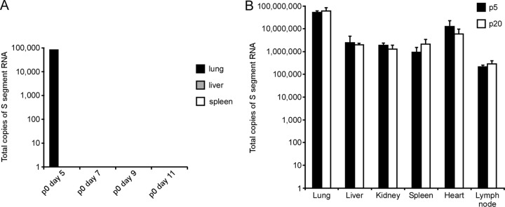
In the initial infection (p0), SNV was rapidly cleared by hamsters, with low levels of virus detected only in lung samples collected at day 5 p.i. (Fig. 1). Although the low-level detection of SNV at p0 is similar to that described by Wahl-Jensen and colleagues, the virus that had been passaged only in deer mice was detectable 7 days early than the Vero-adapted isolated used in previous studies (8). These findings suggest that a non-Vero-adapted hantavirus might be inherently better able to infect hamsters, although further studies would be required to address this possibility. Over the following five passages, SNV replicated with increasing efficiency in hamsters with prolonged detection of viral RNA in tissue samples and, after p2, whole blood samples. After p5, SNV replication appeared to be uniform across the remaining passages (Fig. 1).
Fig 1.

Serial passaging of Sin Nombre virus in hamsters results in prolonged and systemic detection of viral RNA. Quantitative RT-PCR analysis was utilized to determine the total number of copies of S-segment RNA in organs from infected hamsters. In the initial infection, Sin Nombre virus was rapidly cleared by hamsters, with viral RNA being detected only in lung samples collected on day 5 postinfection (A). After five serial passages in hamsters, Sin Nombre virus established a systemic and prolonged infection, with viral RNA being readily detectable in several organs (B). Results are values obtained from a single animal per time point in the initial infection (A) or from tissues collected 9 days postinfection from groups of three animals infected with p5 and p20 SNV (B). Error bars represent standard deviations.
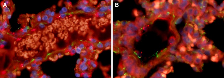
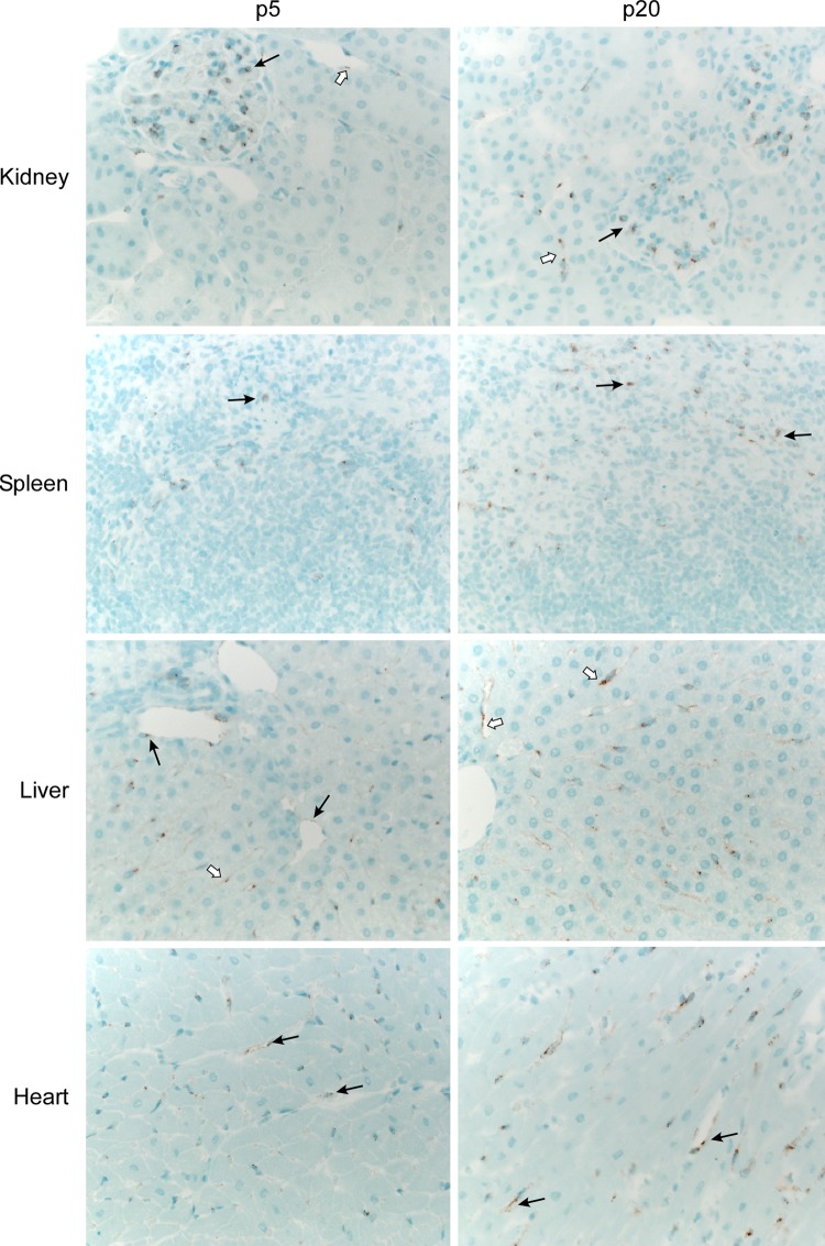
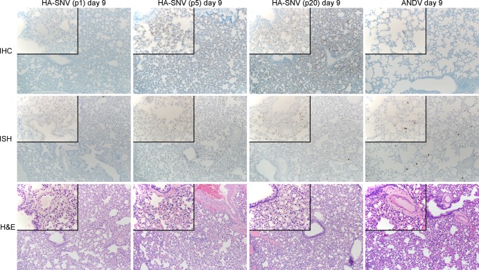
Immunohistochemistry (IHC) was performed on tissue samples from each passage using a polyclonal rabbit antiserum generated against the SNV nucleoprotein (2) essentially as previously described (9). Supporting the RT-PCR results, extensive viral antigen was detected in all organs analyzed (lung, spleen, liver, kidney, and heart) but most notably in lung specimens collected after p5, which were nearly indistinguishable from matched samples collected from ANDV infected hamsters (Fig. 2 and 3). In situ hybridization (ISH) was also performed on lung samples from hamster-adapted (HA)-SNV- and ANDV-infected hamsters with virus-specific probes targeting the respective nucleoproteins (SNV nucleotides 151 to 1194 and ANDV nucleotides 30 to 981) as described previously (10) and confirmed that both viruses replicated similarly in the pulmonary endothelium (Fig. 3). To confirm the presence of virus in endothelial cells, lung specimens were costained with monoclonal antibodies targeting the nucleoprotein (anti-SNV NP clone 5F1/F7 and anti-ANDV NP clone 1A8/F6, 1:1,000 dilution each; Austral Biologicals) and polyclonal rabbit anti-CD31 antibodies (ab28364, 1:50 dilution; Abcam) with Alexa Fluor 594 (anti-mouse) and 488 (anti-rabbit) secondary antibodies (A11005 and A11034, respectively, each at a 1:200 dilution; Invitrogen). Slides were treated with ProLong Gold antifade reagent with DAPI (4′,6-diamidino-2-phenylindole) stain and coverslipped. Hantaviral antigen was predominantly observed in CD31-positive cells in lung samples collected from hamsters infected with either HA-SNV or ANDV, which confirmed that both viruses replicate in pulmonary endothelial cells (Fig. 4).
Fig 2.
A serially passaged Sin Nombre virus establishes disseminated infection in hamsters. Immunohistochemistry revealed the presence of hantaviral antigen in all organs collected from hamsters infected with the passaged (hamster-adapted) Sin Nombre virus. Shown are kidney, spleen, liver, and heart samples from a representative animal infected with the p5 (left) and p20 (right) variants. Viral antigen was detected in glomerular endothelium (arrow) and endothelial cells lining capillaries in the renal interstitial space between proximal tubules (open arrow) of the kidneys, numerous mononuclear cells within the splenic marginal zones (arrow), in endothelial cells of portal veins (arrow) and endothelial cells lining hepatic sinusoids (open arrows) of the liver, and in endothelial cells lining capillaries between cardiomyofibers (arrows) of the heart.
Fig 3.
Serially passaging Sin Nombre virus results in increased viral replication in hamster lungs without histological changes associated with HPS. Histological analysis was carried out on lung samples from hamsters infected with passaged Sin Nombre virus and compared to time-matched samples from Andes virus-infected animals. In the initial passage (p1) of Sin Nombre virus, nucleoprotein antigen and RNA were undetectable by immunohistochemistry and in situ hybridization. However, by p5 and continuing through p20, viral antigen and RNA were readily detectable in pulmonary endothelial cells from hamsters infected with the passaged virus, with a distribution and intensity similar to that observed in Andes-infected hamsters. Despite increased replication, no histological lesions were noted in the lungs of hamsters infected with the passaged virus. By comparison, time-matched samples collected from Andes-infected hamsters demonstrated perivascular edema, which ultimately leads to diffuse pulmonary edema and death.
Fig 4.

Immunofluorescence staining for endothelial cell markers and hantaviral antigen in hamster lungs. Lung samples collected 9 days postinfection were costained for endothelial cell markers (CD31, green) and hantavirus nucleoprotein antigen (red). Lung specimens from hamsters infected with a hamster-adapted (passage 5) Sin Nombre virus (A) and Andes virus (B) exhibited the presence of hantaviral antigen in cells positive for CD31, supporting the notion that both viruses replicate in pulmonary endothelial cells.
Despite the similarities to ANDV infection, lung specimens from HA-SNV-infected hamsters revealed no histological changes (Fig. 3), and throughout the course of the passaging studies, HA-SNV-infected hamsters demonstrated only mild signs of infection, including lethargy, with no apparent breathing deficiencies or radiographic changes. Interestingly, similar to recent findings with Puumala virus (6), hamsters appeared to be unable to clear HA-SNV infection, with viral RNA and antigen being readily detectable in lung specimens collected at 60 days p.i. (data not shown).
Genetically, SNV was remarkably stable throughout the course of serial passaging. Sanger sequence analysis revealed no amino acid changes in the nucleoprotein- or polymerase-coding regions of HA-SNV p5 or p20 compared to the original SNV inoculum obtained from deer mice. Analysis of the glycoprotein sequences revealed two amino acid changes in HA-SNV p20, one in glycoprotein N (Gn) at position 239 (S239E) and a second in glycoprotein C (Gc) at position 1058 (G1058S). Interestingly, only the G1058S mutation was present in HA-SNV p5. Although neither mutation occurs in a known functional domain or immunological epitope within the glycoproteins, the appearance of the G1058S mutation on or before p5 suggests it might increase the virus's fitness in hamsters, allowing increased and consistent replication thereafter.
The pathophysiology of HPS in humans and hamsters is complex and not completely understood (3, 11, 12). An enduring question regarding the Syrian hamster model of HPS is why ANDV and to a lesser extent MAPV infection results in severe respiratory disease in hamsters, while other prominent agents of HPS in humans do not cause any signs of illness in these animals (4, 7). Unlike other hantaviruses studied to date, SNV results in a short infection which is quickly cleared by hamsters and results in a robust neutralizing and nonneutralizing antibody response (8, 13). Our results suggest that the apparent block for SNV infection in hamsters is the glycoproteins, although further characterization of the effect of the two mutations identified in these studies would require a reverse genetics system, which to date has not been described for hantaviruses (14). It should also be noted that the results of the present study cannot account for potential genetic changes in noncoding regions of HA-SNV. Regardless, by serially passaging SNV in hamsters, we have demonstrated that pulmonary edema and HPS-like disease manifestations in hamsters are not due solely to viremia and viral dissemination or replication in pulmonary endothelium.
To date, the only in vivo model described for SNV infection is the deer mouse model, which relies on maintaining an in-house breeding colony of mice (1). Although serially passaging SNV did not result in a disease model, the prolonged and systemic infection associated with HA-SNV in hamsters provides an important and convenient in vivo model which is well suited to the evaluation of vaccines and antiviral agents against this prominent agent of HPS.
ACKNOWLEDGMENTS
Animal experiments were approved by the Institutional Animal Care and Use Committee of the Rocky Mountain Laboratories (RML) and performed following the guidelines of the Association for Assessment and Accreditation of Laboratory Animal Care, International (AAALAC), by certified staff in an AAALAC-approved facility. Experiments were conducted in the Biosafety Level 4 facility at the RML.
We thank Rebecca Rosenke and Dan Long (Division of Intramural Research, National Institute of Allergy and Infectious Disease, National Institutes of Health, [DIR, NIAID, NIH]) for processing the tissue samples for histological analysis and Anita Mora (DIR, NIAID, NIH) for help with the artwork.
This work was supported by the Division of Intramural Research, National Institute of Allergy and Infectious Diseases, National Institutes of Health.
Footnotes
Published ahead of print 6 February 2013
REFERENCES
- 1. Botten J, Mirowsky K, Kusewitt D, Bharadwaj M, Yee J, Ricci R, Feddersen RM, Hjelle B. 2000. Experimental infection model for Sin Nombre hantavirus in the deer mouse (Peromyscus maniculatus). Proc. Natl. Acad. Sci. U. S. A. 97:10578–10583 [DOI] [PMC free article] [PubMed] [Google Scholar]
- 2. Medina RA, Mirowsky-Garcia K, Hutt J, Hjelle B. 2007. Ribavirin, human convalescent plasma and anti-beta3 integrin antibody inhibit infection by Sin Nombre virus in the deer mouse model. J. Gen. Virol. 88:493–505 [DOI] [PubMed] [Google Scholar]
- 3. Safronetz D, Ebihara H, Feldmann H, Hooper JW. 2012. The Syrian hamster model of hantavirus pulmonary syndrome. Antivir. Res. 95:282–292 [DOI] [PMC free article] [PubMed] [Google Scholar]
- 4. Hooper JW, Larsen T, Custer DM, Schmaljohn CS. 2001. A lethal disease model for hantavirus pulmonary syndrome. Virology 289:6–14 [DOI] [PubMed] [Google Scholar]
- 5. Eyzaguirre EJ, Milazzo ML, Koster FT, Fulhorst CF. 2008. Choclo virus infection in the Syrian golden hamster. Am. J. Trop. Med. Hyg. 78:669–674 [PMC free article] [PubMed] [Google Scholar]
- 6. Sanada T, Kariwa H, Nagata N, Tanikawa Y, Seto T, Yoshimatsu K, Arikawa J, Yoshii K, Takashima I. 2011. Puumala virus infection in Syrian hamsters (Mesocricetus auratus) resembling hantavirus infection in natural rodent hosts. Virus Res. 160:108–119 [DOI] [PubMed] [Google Scholar]
- 7. Milazzo ML, Eyzaguirre EJ, Molina CP, Fulhorst CF. 2002. Maporal viral infection in the Syrian golden hamster: a model of hantavirus pulmonary syndrome. J. Infect. Dis. 186:1390–1395 [DOI] [PubMed] [Google Scholar]
- 8. Wahl-Jensen V, Chapman J, Asher L, Fisher R, Zimmerman M, Larsen T, Hooper JW. 2007. Temporal analysis of Andes virus and Sin Nombre virus infections of Syrian hamsters. J. Virol. 81:7449–7462 [DOI] [PMC free article] [PubMed] [Google Scholar]
- 9. Safronetz D, Zivcec M, Lacasse R, Feldmann F, Rosenke R, Long D, Haddock E, Brining D, Gardner D, Feldmann H, Ebihara H. 2011. Pathogenesis and host response in Syrian hamsters following intranasal infection with Andes virus. PLoS Pathog. 7:e1002426 doi:10.1371/journal.ppat.1002426 [DOI] [PMC free article] [PubMed] [Google Scholar]
- 10. Wang F, Flanagan J, Su N, Wang LC, Bui S, Nielson A, Wu X, Vo HT, Ma XJ, Luo Y. 2012. RNAscope: a novel in situ RNA analysis platform for formalin-fixed, paraffin-embedded tissues. J. Mol. Diagn. 14:22–29 [DOI] [PMC free article] [PubMed] [Google Scholar]
- 11. Jonsson CB, Hooper J, Mertz G. 2008. Treatment of hantavirus pulmonary syndrome. Antivir. Res. 78:162–169 [DOI] [PMC free article] [PubMed] [Google Scholar]
- 12. Macneil A, Nichol ST, Spiropoulou CF. 2011. Hantavirus pulmonary syndrome. Virus Res. 162:138–147 [DOI] [PubMed] [Google Scholar]
- 13. McElroy AK, Smith JM, Hooper JW, Schmaljohn CS. 2004. Andes virus M genome segment is not sufficient to confer the virulence associated with Andes virus in Syrian hamsters. Virology 326:130–139 [DOI] [PubMed] [Google Scholar]
- 14. Brown KS, Ebihara H, Feldmann H. 2012. Development of a minigenome system for Andes virus, a New World hantavirus. Arch. Virol. 157:2227–2233 [DOI] [PMC free article] [PubMed] [Google Scholar]


