Abstract
Small ruminants infected with peste des petits ruminants virus exhibit lesions typical of epithelial infection and necrosis. However, the only established host receptor for this virus is the immune cell marker signaling lymphocyte activation molecule (SLAM). We have confirmed that the ovine Nectin-4 protein, when overexpressed in epithelial cells, permits efficient replication of PPRV. Furthermore, this gene was predominantly expressed in epithelial tissues and encoded by multiple haplotypes in sheep breeds from around the world.
TEXT
Peste des petits ruminants virus (PPRV), a Morbillivirus of sheep and goats, causes a debilitating disease in the host typified by respiratory and gastrointestinal pathology (1). Like all morbilliviruses, PPRV has an established lymphatic and epithelial tropism (2, 3). The signaling lymphocyte activation molecule (SLAM) is well recognized as the universal receptor for morbillivirus infection of immune cells, and this receptor tropism results in the leukopenia observed during infection (4). Recently, the Nectin-4 protein was identified as an epithelial receptor for both measles virus (MeV) and canine distemper virus (CDV), and this has provided important new insights into how these viruses spread through the host (5–7). In contrast, no epithelial receptor for PPRV has been identified to date.
The emerging model for MeV replication is that SLAM-positive macrophages and dendritic cells of the human respiratory airway are the first cells to become infected after viral inhalation (5, 8, 9). These cells then migrate across the epithelium, leading to infection of local lymph tissues (e.g., bronchial lymph node) and further spread of the virus. MeV release into the airway epithelium and subsequent infection of new hosts occur subsequently in a process mediated by the human Nectin-4 receptor, a major component of the adherens junctions found in the epithelium. Adherens junctions are located below the tight junctions in the epithelium, and basolateral infection via these junctions is thought to facilitate MeV transmission (5, 10). This model has led to Nectin-4 being referred to as an exit receptor (5).
The aim of this study was to determine whether ovine Nectin-4 can act as an efficient receptor for PPRV and to characterize its host variation and tissue distribution.
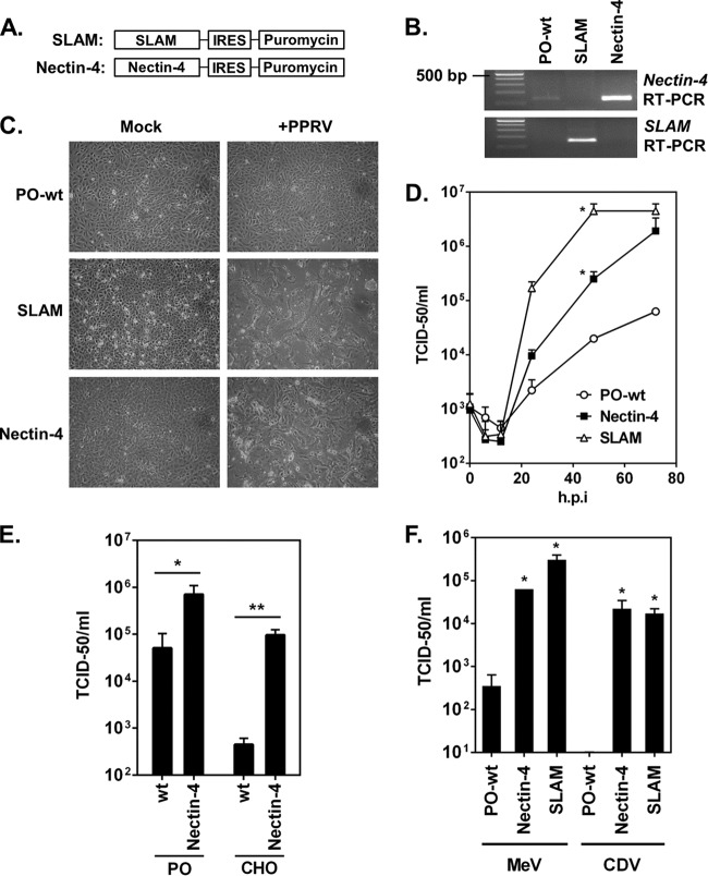
The Nectin-4 and SLAM mRNA open reading frames (ORFs) were amplified, by reverse transcriptase PCR (RT-PCR), from total cell RNA extracted from ovine mammary tissue and peripheral blood monocytes, respectively. Primers (sequences available on request) to amplify SLAM were based on the GenBank sequence DQ228866 (Ovis aries SLAM), while Nectin primers were based on BLAST search results of the International Sheep Genomics Consortium database using the bovine (Bos taurus) Nectin-4 sequence (NM_001024494). The ORF of the previously uncharacterized ovine Nectin-4 gene (JX944709) was sequenced, and the predicted amino acid sequence was shown to be closely related to both bovine (Bos taurus) and human Nectin-4 proteins (98% and 92% identity, respectively). Lentiviruses were constructed encoding ovine SLAM or Nectin-4 ORFs (Fig. 1A), and these were used to transduce sheep kidney epithelial cells (PO cell line), followed by puromycin selection. Successful integration of the lentivirus cassette and expression of SLAM or Nectin-4 mRNA were confirmed by RT-PCR (Fig. 1B). A low level of Nectin-4 mRNA expression was also detected in nontransduced PO cells. Experimental infection of the transduced cell lines with PPRV (Ivory Coast 1989 strain [11]) resulted in large syncytia that formed 24 to 48 h postinfection (Fig. 1C). In contrast, no syncytia or visible cytopathic effects were seen in the wild-type cells, demonstrating that lentivirus-encoded expression of ovine SLAM or Nectin-4 proteins permits efficient replication of PPRV (Fig. 1C). Comparison of the replication kinetics of PPRV in a single-step growth curve (multiplicity of infection [MOI] of 4) demonstrated significantly faster replication in cell lines expressing SLAM or Nectin-4 compared to nontransduced cells (P < 0.05; t test comparison of mean viral growth rates) (Fig. 1D). Of note, a low level of viral replication was detected in nontransduced cells, presumably because of the endogenous Nectin-4 (Fig. 1B and D). To confirm the role of ovine Nectin-4 in PPRV entry, a Chinese hamster ovary (CHO) cell line was also transduced with the Nectin-4-based lentivirus and selected with puromycin. These non-host-derived cell lines were previously used in MeV studies because they are refractory to morbillivirus infection and do not express detectable levels of Nectin-4 (5, 6). Similarly to the PO cell lines, CHO cells expressing Nectin-4 exhibited extensive syncytia after infection while wild-type cells did not (data not shown). Infection of these cells at an MOI of 4 resulted in significantly less background replication in the nontransduced CHO cells (P < 0.005) than the equivalent PO cells (P < 0.05) (Fig. 1E), providing additional evidence for the role of ovine Nectin-4 in PPRV entry.
Fig 1.
Ovine Nectin-4 can function as a receptor for PPRV. (A) Schematic representation of the lentivirus constructs encoding ovine SLAM or Nectin-4 ORFs. The bicistronic constructs encoded the encephalomyocarditis virus internal ribosome entry site (IRES) and a puromycin resistance gene. (B) Oligo(dT)-primed RT-PCR analysis of total cell RNA extracted from transduced or wild-type (wt) PO cell lines confirming SLAM or Nectin-4 mRNA expression. (C) PPRV infection in PO cell lines. Wild-type or lentivirus-transduced cells were infected at high MOI (4) with PPRV (Ivory Coast 1989 strain). Cytopathic effect was observed by phase-contrast microscopy at 48 h p.i. All images were equally manipulated for brightness and contrast using MS Powerpoint. (D) High-MOI single-step growth curve of PPRV in PO cell lines. Wild-type or transduced PO cells were infected at an MOI of 4. Virus was harvested at 0, 6, 12, 24, 48, and 72 h p.i. by freeze-thawing. PPRV replication rates were compared using an unpaired two-tailed t test comparison of the mean rates of viral exponential growth (12 to 48 h p.i.). Individual slopes were calculated by fitting a first order polynomial straight line to each data set; *, P < 0.05. (E) PPRV infection in PO and CHO cells expressing ovine Nectin-4. Wild-type or transduced PO or CHO cell lines were infected at high MOI (4) with PPRV (Ivory Coast 1989 strain). Virus was harvested at 48 h p.i. by freeze-thawing. (F) MeV and CDV replicate efficiently in PO cell lines expressing ovine SLAM or Nectin-4. Wild-type or transduced PO cell lines were infected at low MOI (0.1) with MeV (Dublin strain) or CDV (5804P strain). Virus was harvested at 72 h p.i. by freeze-thawing. (E and F) An unpaired two-tailed t test was used to compare the viral titers; *, P < 0.05; **, P < 0.005. (D, E, and F) Virus titration was performed on Vero cells expressing canine SLAM with a detection limit of 10 TCID50. All infections were performed in triplicate; the error bars denote standard deviations from the means.
SLAM proteins are well established as universal receptors for morbilliviruses, and many research laboratories use cell lines overexpressing canine SLAM to propagate these viruses (12, 13). At 72 h after low-MOI (0.1 50% tissue culture infective dose [TCID50]/cell) infection, significantly more (P < 0.05; t test) MeV and CDV replication was recorded in PO cells expressing ovine SLAM or Nectin-4 compared to nontransduced cells (Fig. 1F). The efficient replication of MeV and CDV in Nectin-4-expressing cells indicates that this protein, like SLAM, is also a universal morbillivirus receptor. This is the first recorded use of lentiviruses to facilitate the production of a PPRV-permissive cell line and the first development of a highly permissive host-derived cell line for PPRV.
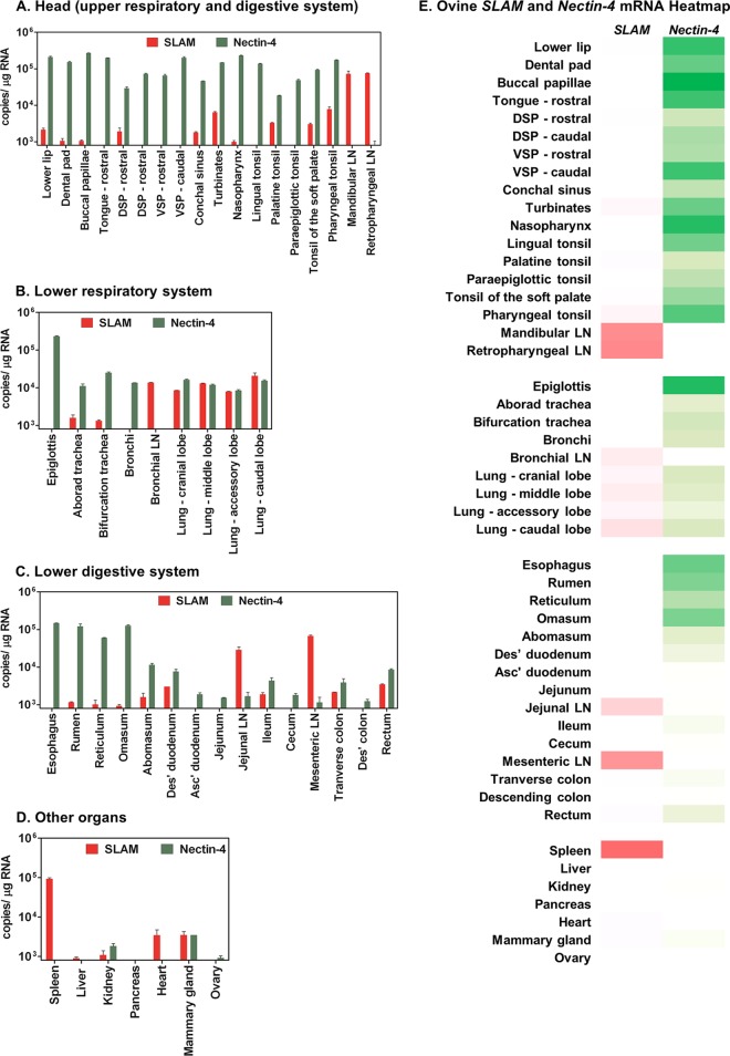
Previously, the in vivo expression profile of caprine (goat) SLAM was demonstrated through analysis of a range of tissue samples taken from healthy animals (14). SLAM mRNA expression was highest in lymphatic tissues such as the lymph node and spleen; however, expression in tissues where PPRV replication is common, such as the lungs and lower intestinal tract, was very low (14). Our identification of ovine Nectin-4 as a receptor for PPRV led us to investigate the expression profile of this gene, and ovine SLAM, in vivo. Samples (49 separate tissues) were taken from a healthy sheep (2-year-old ewe; Texel/North Country Mule cross), focusing on the respiratory and gastrointestinal tracts but including representative tissues from other major organs. This research conformed to the ethical guidelines in place, both nationally and institutionally. RNA was extracted from each of these tissues using the MagNA Pure LC platform (Roche). RNA purity and quality were determined by spectrophotometry (A260/A280 ratio of 1.9 to 2.1) and β-actin quantitative PCR (qPCR) (data not shown). The mRNA expression levels of SLAM and Nectin-4 were quantified using TaqMan assays developed and optimized for the purpose of this study (SLAM: forward primer, CCCAAGTCCAGAAATCAGGT, reverse primer, GCGTCACACTGGCATAGACT, and probe, FAM-CGCTGCCACAGAGCCTGTCC-TAMRA; Nectin-4: forward primer, TACCTGGGACACAGAGGTCA, reverse primer, GGGATACCACGCAGGTAAGT, and probe, FAM-CACACTCCCGCTCAGCTGCC-TAMRA [where FAM is 6-carboxyfluorescein and TAMRA is 6-carboxytetramethylrhodamine]). RT-qPCR analysis of oligo(dT)-primed cDNA libraries demonstrated high levels of expression of Nectin-4 in epithelial tissues taken from the mouth and upper respiratory tract as well as the stomach (rumen, reticulum, and omasum) (Fig. 2). Similarly to the caprine study (14), ovine SLAM expression predominated in the lymphatic tissue (the spleen and the mandibular, retropharyngeal, jejunal, and mesenteric lymph nodes). Interestingly, expression of SLAM and Nectin-4 mRNA together was detected most commonly in the lungs, albeit at lower levels (Fig. 2).
Fig 2.
Endogenous mRNA expression profile of ovine SLAM and Nectin-4 in various Ovis aries tissues. A total of 49 separate tissues were isolated from a 2-year-old ewe (Texel/North Country Mule cross). Total cell RNA was extracted from tissue, and 100 ng of this RNA was used to generate an oligo(dT)-primed cDNA library. mRNA copies of SLAM (red) and Nectin-4 (green) were calculated by duplicate TaqMan qPCR analysis of these cDNA libraries (using standard curve quantification). The detection limit of this qPCR was set at 10 copies. The data were normalized to copies/μg RNA and grouped anatomically, as follows: the head (upper respiratory and digestive system) (A), the lower respiratory system (B), the lower digestive system (C), and the other major organs (D). (E) Heat map of the mRNA data in panels A to D representing the relative levels of SLAM (red) and Nectin-4 (green) expression in the 49 tissues (darker shading indicates higher expression). Abbreviations: LN, lymph nodes; DSP, dorsal surface of the soft palate; VSP, ventral surface of the soft palate; Asc′, ascending; Des′, descending. This data set is representative of 3 individually performed RT-qPCR analyses.
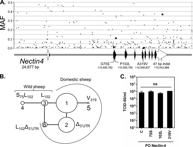
The role of host-encoded genetic variation in susceptibility to viral infection and disease has become increasingly important in both medical and veterinary communities (15, 16). However, little is known of the genetic variation in mammalian morbillivirus receptors since, to date, limited genetic information has been accessible. The availability of whole-genome sequence data from the International Sheep Genomics Consortium (ISGC) provided the opportunity to identify polymorphisms that may affect the polypeptide sequence and mRNA splicing of Nectin-4 in 75 diverse sheep sampled from 39 breeds and 2 wild species from around the world (17). Single nucleotide polymorphism (SNP) analysis of a 24,877-bp region containing Nectin-4 identified 317 SNPs and approximately 50 insertion/deletion polymorphisms (indels). The SNPs were predicted to alter the Nectin-4 coding sequence at G75S, P102L, and A319V and had minor allele frequencies (MAF) of 0.02, 0.11, and 0.01, respectively (Fig. 3A). There were no SNPs or deletions identified in any of the 5′ or 3′ exon splice sites where conserved GT or AG sequences are found. There was, however, a common 47-bp indel in the 3′ untranslated region (UTR) where the deleted allele had an MAF of 0.37. The 47-bp ovine sequence was present in wild sheep and cattle, suggesting that the insertion form of this indel may be the ancestral state. An unrooted median-joining network of haplotypes comprising G75S, P102L, and A319V and 47-bp 3′ UTR indel variants was generated as previously described (16) to provide a framework for investigating the potential effects of these polymorphisms on morbillivirus infection in sheep. Of particular interest is the relatively high frequency and distribution of the 47-bp deletion in domesticated sheep (haplotypes 2 and 6) (Fig. 3B; see Table S1 in the supplemental material). To test the effect of haplotypes encoding different Nectin-4 polypeptides, additional lentivirus-transduced PO cell lines were produced to overexpress isoforms of the Nectin-4 protein varying at residues 75, 102, and 319. Replication of PPRV in these cells was then compared to that in the cell line described above (Fig. 1, Nectin-4), which overexpressed haplotype 1 Nectin-4 protein (G75, P102, A319). Experimental infection at high MOI (4) resulted in similar cytopathic effects (data not shown) and virus yields (Fig. 3C). Equivalent results were also observed after a low-MOI (0.1) infection (72 h postinfection [p.i.]; data not shown), indicating that these three naturally occurring, nonsynonymous SNPs in Nectin-4 do not have a major effect on PPRV infection in this system. The effects of ovine Nectin-4 haplotypes encoding the 47-bp deletion in the 3′ UTR were not tested and thus are presently unknown.
Fig 3.
Genomic structure, polymorphisms, unrooted median-joining network, and effects of allelic variants of ovine Nectin-4. (A) Genomic map of Nectin-4: white arrows, 5′ and 3′ UTR of exons; black arrows, exon coding regions; gray rectangles, introns or intergenic regions; small diamonds, positions and minor allele frequencies (MAF) of SNPs in an international panel of 75 sheep; large squares, position of Nectin-4 missense polymorphisms and 47-bp indel in the 3′ UTR. The codon polymorphism sequences are as follows: G75S, GGC→AGC; P102L, CCC→CTC; A319V, GCA→GTA. The major and minor alleles for the 47-bp indel in the 3′ UTR are 5′-TAACA[AGAGGACAAGTAAGGTCCTTGGCCTTGCGGTTCCTCGGCTCTTCCCC]TAGGC-3′ and 5′-TAACA[]TAGGC-3′, respectively. (B) Unrooted median-joining network of haplotypes comprising G75S, P102L, A319V, and 47-bp 3′ UTR indel variants. The areas of circles are proportional to the frequencies in the 75 ISGC sheep (see Table S1 in the supplemental material). The predicted sequence of variable sites for the most frequent haplotype (1) was G75, P102, and A319 and contained the 47-bp insertion in the 3′ UTR. The difference from and relationship to haplotype 1 are indicated for each additional haplotype. The delta symbol indicates the deleted form of the 47-bp indel polymorphism. (C) PPRV infection of transduced PO cell lines overexpressing Nectin-4 haplotype isoforms. Abbreviations for cell lines: C, control line overexpressing the most common Nectin-4 haplotype in domestic sheep (haplotype 1: G75, P102, and A319); S75, line with S75, P102, and A319; L102, line with haplotype 3 (G75, L102, and A319); 319V, line with haplotype 5 (G75, P102, and V319). All four cell lines were infected at an MOI of 4 with PPRV (Ivory Coast 1989 strain). Virus was harvested at 48 h p.i. by freeze-thawing. Virus titration was performed on Vero cells expressing canine SLAM with a detection limit of 10 TCID50. All infections were performed in triplicate; the error bars denote standard deviations from the means. PPRV replication rates were compared using an unpaired two-tailed t test (ns, nonsignificant).
The identification of ovine Nectin-4 protein as a permissive receptor for PPRV and the high level of expression of its mRNA in epithelial tissues of the respiratory tract are supportive of a role for this protein in PPRV epithelial pathogenesis. PPRV infection is often associated with the formation of lesions inside the mouth, nose, and upper respiratory tract, despite the absence of high levels of SLAM expression in these tissues (references 2 and 3 and Fig. 2). These lesions typically form after the onset of viremia, and Nectin-4-mediated infection of these tissues may therefore contribute directly to the onward transmission of PPRV, in a manner similar to the model proposed for MeV (5). The identification of Nectin-4 as the likely PPRV epithelial receptor, in addition to the well-established lymphotropic receptor SLAM, does not explain all aspects of PPRV pathogenesis. Disease in sheep and goats is often associated with gross pathology in the intestine (duodenum, ileum, colon, and rectum) (2, 3); however, the level of Nectin-4 and SLAM expression in these tissues is relatively low (Fig. 2). In contrast, pathology in the stomach (rumen, reticulum, and omasum) is rare, despite higher levels of Nectin-4 expression in these tissues. The intestinal pathology may therefore be the result of host-mediated effects or infection via alternative routes, and this represents an obvious and interesting area for continued investigation.
Differential SLAM and/or Nectin-4 expression in varied hosts may also play a role in determining the individual characteristics of morbillivirus disease, such as the primary viral bronchopneumonia caused by PPRV. Previously, a high level of Nectin-4 expression was recorded in the trachea of humans (5, 6), but in our study expression levels in this tissue were markedly low in sheep. However, our study did show a broad expression profile for SLAM and Nectin-4 mRNA throughout the lung, which may be a factor in PPRV-induced pneumonia. The expression of SLAM mRNA in these predominantly nonimmune epithelial tissues is likely due to the presence of SLAM-positive immune cells such as macrophages and dendritic cells, rather than epithelial cells, expressing this gene. Importantly, it is worth noting that the mRNA expression profile of ovine Nectin-4 recorded here correlates well with the expression profile of human Nectin-4 protein reported elsewhere using immunohistochemistry (Human Protein Atlas [18]). This provides supportive evidence that the expression profile of this gene/protein is probably highly conserved in mammals.
The identification of ovine Nectin-4 as a PPRV receptor, in addition to earlier identifications for MeV and CDV (5–7), highlights the important role this protein must play in the morbillivirus life cycle and contributes to our understanding of PPRV as a pathogen of small ruminants. Furthermore, our findings shed new light on potential pathogenesis factors such as the tissue distribution and genetic variation of morbillivirus receptors. In a broader sense, the SLAM and Nectin-4 lentiviruses generated during this study will simplify the generation of sensitive cell lines for PPRV detection and virus isolation in the field. Cell lines of this nature, especially those expressing the most appropriate receptor diplotypes, are integral tools in eradication campaigns and would complement the molecular epidemiology techniques already in place for PPRV.
Supplementary Material
ACKNOWLEDGMENTS
We thank Michael Baron, Louisa Chard, Satya Parida, and Ashley Banyard for useful discussions and reagents. In addition, we thank Tony Smith and the operations staff at The Pirbright Institute for their help during this study.
T.K. is also the CEO of a bioinformatics company, Intrepid Bioinformatics. We do not believe that there is a conflict of interest between the conclusions of this article and the interests of this company.
N.J. is a Wellcome Trust Intermediate Clinical Fellow. D.B. is supported by an independent research fellowship awarded by The Pirbright Institute.
Footnotes
Published ahead of print 6 February 2013
Supplemental material for this article may be found at http://dx.doi.org/10.1128/JVI.02792-12.
REFERENCES
- 1. Banyard AC, Parida S, Batten C, Oura C, Kwiatek O, Libeau G. 2010. Global distribution of peste des petits ruminants virus and prospects for improved diagnosis and control. J. Gen. Virol. 91:2885–2897 [DOI] [PubMed] [Google Scholar]
- 2. Couacy-Hymann E, Bodjo C, Danho T, Libeau G, Diallo A. 2007. Evaluation of the virulence of some strains of peste-des-petits-ruminants virus (PPRV) in experimentally infected West African dwarf goats. Vet. J. 173:178–183 [DOI] [PubMed] [Google Scholar]
- 3. Hammouchi M, Loutfi C, Sebbar G, Touil N, Chaffai N, Batten C, Harif B, Oura C, El Harrak M. 2012. Experimental infection of alpine goats with a Moroccan strain of peste des petits ruminants virus (PPRV). Vet. Microbiol. 160:240–244 [DOI] [PubMed] [Google Scholar]
- 4. Sato H, Yoneda M, Honda T, Kai C. 2012. Morbillivirus receptors and tropism: multiple pathways for infection. Front. Microbiol. 3:75. [DOI] [PMC free article] [PubMed] [Google Scholar]
- 5. Muhlebach MD, Mateo M, Sinn PL, Prufer S, Uhlig KM, Leonard VH, Navaratnarajah CK, Frenzke M, Wong XX, Sawatsky B, Ramachandran S, McCray PB, Jr, Cichutek K, von Messling V, Lopez M, Cattaneo R. 2011. Adherens junction protein nectin-4 is the epithelial receptor for measles virus. Nature 480:530–533 [DOI] [PMC free article] [PubMed] [Google Scholar]
- 6. Noyce RS, Bondre DG, Ha MN, Lin LT, Sisson G, Tsao MS, Richardson CD. 2011. Tumor cell marker PVRL4 (nectin 4) is an epithelial cell receptor for measles virus. PLoS Pathog. 7:e1002240 doi:10.1371/journal.ppat.1002240 [DOI] [PMC free article] [PubMed] [Google Scholar]
- 7. Pratakpiriya W, Seki F, Otsuki N, Sakai K, Fukuhara H, Katamoto H, Hirai T, Maenaka K, Techangamsuwan S, Lan NT, Takeda M, Yamaguchi R. 2012. Nectin4 is an epithelial cell receptor for canine distemper virus and involved in neurovirulence. J. Virol. 86:10207–10210 [DOI] [PMC free article] [PubMed] [Google Scholar]
- 8. Ferreira CS, Frenzke M, Leonard VH, Welstead GG, Richardson CD, Cattaneo R. 2010. Measles virus infection of alveolar macrophages and dendritic cells precedes spread to lymphatic organs in transgenic mice expressing human signaling lymphocytic activation molecule (SLAM, CD150). J. Virol. 84:3033–3042 [DOI] [PMC free article] [PubMed] [Google Scholar]
- 9. Lemon K, de Vries RD, Mesman AW, McQuaid S, van Amerongen G, Yuksel S, Ludlow M, Rennick LJ, Kuiken T, Rima BK, Geijtenbeek TB, Osterhaus AD, Duprex WP, de Swart RL. 2011. Early target cells of measles virus after aerosol infection of non-human primates. PLoS Pathog. 7:e1001263 doi:10.1371/journal.ppat.1001263 [DOI] [PMC free article] [PubMed] [Google Scholar]
- 10. Niessen CM. 2007. Tight junctions/adherens junctions: basic structure and function. J. Invest. Dermatol. 127:2525–2532 [DOI] [PubMed] [Google Scholar]
- 11. Chard LS, Bailey DS, Dash P, Banyard AC, Barrett T. 2008. Full genome sequences of two virulent strains of peste-des-petits ruminants virus, the Cote d'Ivoire 1989 and Nigeria 1976 strains. Virus Res. 136:192–197 [DOI] [PubMed] [Google Scholar]
- 12. Buczkowski H, Parida S, Bailey D, Barrett T, Banyard AC. 2012. A novel approach to generating morbillivirus vaccines: negatively marking the rinderpest vaccine. Vaccine 30:1927–1935 [DOI] [PubMed] [Google Scholar]
- 13. Tatsuo H, Yanagi Y. 2002. The morbillivirus receptor SLAM (CD150). Microbiol. Immunol. 46:135–142 [DOI] [PubMed] [Google Scholar]
- 14. Meng X, Dou Y, Zhai J, Zhang H, Yan F, Shi X, Luo X, Li H, Cai X. 2011. Tissue distribution and expression of signaling lymphocyte activation molecule receptor to peste des petits ruminant virus in goats detected by real-time PCR. J. Mol. Histol. 42:467–472 [DOI] [PubMed] [Google Scholar]
- 15. Chapman SJ, Hill AV. 2012. Human genetic susceptibility to infectious disease. Nat. Rev. Genet. 13:175–188 [DOI] [PubMed] [Google Scholar]
- 16. Heaton MP, Clawson ML, Chitko-Mckown CG, Leymaster KA, Smith TP, Harhay GP, White SN, Herrmann-Hoesing LM, Mousel MR, Lewis GS, Kalbfleisch TS, Keen JE, Laegreid WW. 2012. Reduced lentivirus susceptibility in sheep with TMEM154 mutations. PLoS Genet. 8:e1002467 doi:10.1371/journal.pgen.1002467 [DOI] [PMC free article] [PubMed] [Google Scholar]
- 17. Kijas JW, Lenstra JA, Hayes B, Boitard S, Porto Neto LR, San Cristobal M, Servin B, McCulloch R, Whan V, Gietzen K, Paiva S, Barendse W, Ciani E, Raadsma H, McEwan J, Dalrymple B, International Sheep Genomics Consortium Members 2012. Genome-wide analysis of the world's sheep breeds reveals high levels of historic mixture and strong recent selection. PLoS Biol. 10:e1001258 doi:10.1371/journal.pbio.1001258 [DOI] [PMC free article] [PubMed] [Google Scholar]
- 18. Uhlen M, Oksvold P, Fagerberg L, Lundberg E, Jonasson K, Forsberg M, Zwahlen M, Kampf C, Wester K, Hober S, Wernerus H, Bjorling L, Ponten F. 2010. Towards a knowledge-based Human Protein Atlas. Nat. Biotechnol. 28:1248–1250 [DOI] [PubMed] [Google Scholar]
Associated Data
This section collects any data citations, data availability statements, or supplementary materials included in this article.


