Abstract
Transmission of Yersinia pestis is greatly enhanced after it forms a bacterial biofilm in the foregut of the flea vector that interferes with normal blood feeding. Here we report that the ability to produce a normal foregut-blocking infection depends on induction of the Y. pestis PhoP-PhoQ two-component regulatory system in the flea. Y. pestis phoP-negative mutants achieved normal infection rates and bacterial loads in the flea midgut but produced a less cohesive biofilm both in vitro and in the flea and had a greatly reduced ability to localize to and block the flea foregut. Thus, not only is the PhoP-PhoQ system induced in the flea gut environment, but also this induction is required to produce a normal transmissible infection. The altered biofilm phenotype in the flea was not due to lack of PhoPQ-dependent or PmrAB-dependent addition of aminoarabinose to the Y. pestis lipid A, because an aminoarabinose-deficient mutant that is highly sensitive to cationic antimicrobial peptides had a normal phenotype in the flea digestive tract. In addition to enhancing transmissibility, induction of the PhoP-PhoQ system in the arthropod vector prior to transmission may preadapt Y. pestis to resist the initial encounter with the mammalian innate immune response.
INTRODUCTION
Yersinia pestis, the causative agent of bubonic and pneumonic plague, is unique among the enteric group of Gram-negative bacteria in having adopted an arthropod-borne route of transmission. During its life cycle, Y. pestis alternates between two eukaryotic hosts: a mammal (usually a rodent) and an insect (a flea). Y. pestis faces quite different physiological challenges in these disparate host environments. Prokaryotes have evolved sophisticated systems to detect changes in their environment and to respond appropriately by selective synthesis of adaptive gene products. One archetypal environmental sensing and response mechanism in bacteria is the two-component regulatory system, in which an inner membrane sensor kinase protein detects the presence or absence of an environmental stimulus and transduces a signal by phosphotransfer to a cytoplasmic transcriptional regulator (1). The activated transcription factor then coordinately regulates the expression of genes under its control to adapt to the new environmental condition.
One such two-component signal transduction system, PhoP-PhoQ, has a proven role in adaptation of Gram-negative bacteria to vertebrate, invertebrate, and plant host environments (2–8). PhoP and PhoQ homologs are widely distributed among both pathogenic and nonpathogenic Gram-negative bacteria, and the system is considered to constitute a general stress response (4). The prototypical function of the PhoP-PhoQ system appears to be adaptation to low-Mg2+ environments, which stimulate the system to upregulate genes involved in Mg2+ transport and homeostasis and in the modification of outer membrane components such as lipopolysaccharide (LPS) (4, 9–12). In addition to low Mg2+, other environmental stresses such as low pH or cationic antimicrobial peptide (CAMP) binding to the bacterial surface can induce the PhoP-PhoQ system (13, 14). Loss of functional phoP in Salmonella and other pathogens results in attenuated virulence, because PhoP-activated genes include virulence factors that confer resistance to components of the innate immune response such as antimicrobial peptides and macrophages (2, 15–18). PhoP is considered to be a central element in a complex regulatory hierarchy because it regulates other transcription factors and two-component systems; the expression of approximately 3% of Salmonella genes is directly or indirectly affected by PhoP (19, 20).
The Y. pestis PhoP and PhoQ proteins have 90% and 77% amino acid similarity, respectively, to their S. enterica homologs (21). A Y. pestis phoP mutant was more sensitive to low pH, high osmolarity, and oxidative stress than the wild-type parent strain and was also more susceptible to killing by J774 macrophages in vitro (8, 22, 23). Microarray analyses indicate that the expression of as many as 400 Y. pestis genes is influenced by PhoP under low-Mg2+ conditions in vitro (12, 23–25). These genes include Y. pestis homologs of LPS-modifying and other PhoP-regulated genes of S. enterica that are known to be important for survival within macrophages. In general, however, only limited overlap of PhoP-regulated genes has been observed among the Enterobacteriaceae (11, 12, 26, 27). Closely related species have evolved distinct regulatory pathways in some cases. An example pertinent to this study is the covalent attachment of 4-amino-4-deoxy-l-arabinose (4-aminoarabinose) to the phosphate residues of the lipid A component of LPS, which is required for bacterial resistance to CAMPs that are produced by both insects and mammals (16, 28, 29). Aminoarabinose addition in Salmonella requires upregulation of the PmrA-PmrB two-component system via PhoP-PhoQ, whereas in Y. pestis it can be independently regulated by either PhoP-PhoQ or PmrA-PmrB alone (30). In addition, the type and magnitude of the inducing stress can result in differential regulation of subsets of PhoP-regulated genes (12, 31).
After being taken up in a blood meal, Y. pestis multiplies extracellularly in the lumen of the flea midgut and grows in the form of a biofilm, a dense bacterial aggregate that is enclosed in an extracellular matrix (32). In some fleas, the biofilm adheres to the cuticle-covered spines that line the interior of the proventriculus, a valve in the foregut that connects the midgut to the esophagus. The bacterial growth can interfere with the normal valvular action of the proventriculus during feeding attempts and eventually block the flow of blood into the midgut (33). Partial or complete blockage of the proventriculus by the Y. pestis biofilm greatly enhances transmissibility to a new host (33–35).
In a previous study, we reported that the Y. pestis PhoP-PhoQ system was upregulated during infection of the flea at the stage when blockage-dependent transmission occurs (36). In this study, we investigated the role of the PhoP-PhoQ system in the ability of Y. pestis to produce a transmissible infection in the flea. Because certain PhoP-regulated genes are cooperatively or independently regulated by PmrB in Y. pestis (30), we also examined the role of the PmrA-PmrB two-component system in fleas.
MATERIALS AND METHODS
Bacterial strains and mutagenesis.
Bacterial strains used in this study are listed in Table 1. The wild-type Y. pestis GB strain and the isogenic mutant Y. pestis GB SAI2.2, which has a 31-bp internal deletion in the phoP gene, have been described (22). A 153-bp in-frame deletion of the phoP gene was made in Y. pestis KIM6+ (which lacks the 70-kb Yersinia virulence plasmid) by using a megaprimer mutagenesis strategy and allelic exchange (29, 37, 38). The 353-bp megaprimer was generated by PCR of Y. pestis genomic DNA using an upstream primer and a 43-nt mutagenic primer that consisted of the 24 nt upstream and 19 nt downstream of the desired 153-bp deletion and thus incorporated the exact deletion junction. The megaprimer was used in a second PCR with a downstream primer to generate the mutated phoP allele. This second PCR product was ligated into the pCR4Blunt-TOPO vector (Invitrogen, Carlsbad, CA). Making use of the SacI and XbaI sites in the primers, the mutated phoP allele was removed from the pCR4 vector and ligated into SacI- and XbaI-digested pCVD442. The ligation mixture was used to transform Escherichia coli S17-1 by electroporation. The pCVD442 suicide vector containing the mutated phoP gene was introduced into Y. pestis KIM6+ by conjugation with the transformed E. coli S17-1 clone. An allelic exchange mutant, designated Y. pestis KIM6+ ΔphoP, in which the mutant phoP containing the 153-bp deletion had replaced the wild-type phoP gene, was selected using Congo red agar plates containing 7% sucrose (38). Y. pestis KIM6+ ΔphoP and KIM6+ transformants containing pGFP (Clontech, Palo Alto, CA) were obtained by electroporation.
Table 1.
Bacterial strains and plasmids
| Strain or plasmid | Descriptiona | Reference or source |
|---|---|---|
| Y. pestis strains | ||
| KIM6+ | pCD1-negative, Pgm+ biovar Medievalis strain | 83 |
| KIM6 | Pgm- and pCD1-negative | 84 |
| KIM6+ ΔphoP | In-frame internal deletion of amino acids 25–75 of PhoP | 29 |
| KIM6+ ΔphoP(pLG338) | Empty vector control (Kan, Tet) | This study |
| KIM6+ ΔphoP(pLGphoP) | Complemented phoP mutant (Kan) | This study |
| KIM6+ ΔphoP(pGFP) | GFP expressing (Amp) | This study |
| KIM6+ ΔpmrA | In-frame internal deletion of amino acids 28–113 of PmrA | This study |
| KIM6+ ΔphoP ΔpmrA | pmrA deletion introduced into the phoP mutant | This study |
| KIM6+ ΔpbgP Δugd | Deletion of aminoarabinose synthesis genes (Kan, Amp) | This study |
| KIM6+ Δugd | Deletion of aminoarabinose synthesis gene (Kan) | This study |
| KIM6+ Δugd(pLGugd) | Complemented ugd mutant (Kan, Amp) | This study |
| GB | Wild-type biovar Orientalis strain | 22 |
| GB SAI2.2 | Y. pestis GB phoP mutant | 22 |
| E. coli strains | ||
| S17-1λpir | Host and conjugation donor strain for pCVD442 and derivatives | 85 |
| TOP-10 | pCR 2.1 TOPO plasmid host strain | Invitrogen |
| D31 | Rough LPS mutant (Str) | 86 |
| Plasmids | ||
| pLG338 | Low-copy-no. plasmid vector (Kan, Tet) | 87 |
| pLGphoP | Complementation plasmid; wild-type phoP gene and promoter cloned into pLG338 (Kan) | This study |
| pLG338-30 | Low-copy-no. plasmid vector (Amp) | 88 |
| pLGugd | Complementation plasmid; wild-type ugd gene and promoter cloned into pLG338-30 (Amp) | This study |
| pCR4Blunt-TOPO, pCR-2.1 TOPO | Cloning vectors (Amp) | Invitrogen |
| pGFP | GFP expression plasmid (Amp) | Clontech |
| pCVD442 | Suicide vector for allelic exchange mutagenesis (Amp) | 38 |
| pKOBEG::sacB | λRed and sacB plasmid for mutagenesis (Cam) | 40 |
Antibiotic resistance is noted in parentheses (Kan, kanamycin; Tet, tetracycline; Amp, ampicillin; Str, streptomycin; Cam, chloramphenicol). GFP, green fluorescent protein.
To complement Y. pestis KIM6+ ΔphoP, a recombinant plasmid containing the full-length wild-type Y. pestis phoP gene was constructed. The entire 673-bp phoP gene flanked by 564 bp of upstream sequence and 769 bp of downstream sequence was PCR amplified from Y. pestis genomic DNA using primers that contained EcoRI or BamHI restriction sites. The PCR product was digested with EcoRI and BamHI and ligated with pLG338 that had been linearized with the same two enzymes. The ligation mix was used to transform E. coli TOP10 cells (Invitrogen), and a clone containing the recombinant plasmid (designated pLGphoP) was isolated. Y. pestis KIM6+ ΔphoP was transformed with the pLG338 empty vector, pLGphoP, and pGFP (Clontech, Palo Alto, CA) by electroporation. The presence of both full-length and deletion-containing phoP alleles in the pLGphoP transformants was confirmed by PCR.
A Y. pestis KIM6+ strain with a 258-bp in-frame deletion of the pmrA gene (y0677) was produced by allelic exchange mutagenesis (38). The complete pmrA gene and upstream and downstream flanking sequences was first PCR amplified and cloned into the pCR2.1 TOPO vector. Inverse PCR of this recombinant plasmid was performed to delete a 258-bp internal fragment (corresponding to amino acids 28 to 113 of PmrA) and the PCR product was circularized by ligation (39). The mutated pmrA allele was subcloned into pCVD442, which was introduced by electroporation into E. coli S17-1. The suicide vector construct was transferred to Y. pestis KIM6+ and KIM6+ ΔphoP strains by conjugation, and Y. pestis transconjugants in which allelic exchange had occurred were selected.
Y. pestis KIM6+ mutants in which bp 261 to 919 of the pbgP gene (y1917) were deleted and replaced with an ampicillin resistance gene and/or 1,675 bp of the ugd locus extending from bp 31 of the ugd gene (y2147) to 314 bp downstream of it were deleted and replaced with a kanamycin resistance gene were generated by using the pKOBEG-sacB lambda red-recombinase mutagenesis system (40). PCR fragments composed of the bla gene from pUC19 or the aph gene from pUC4K flanked by 47 to 50 bp of Y. pestis sequence upstream and downstream of the targeted deletion sites of pbgP and ugd, respectively, were made and purified. The DNA fragments were sequentially introduced into Y. pestis KIM6+ (pKOBEG-sacB) by electroporation, and antibiotic-resistant transformants were isolated. pKOBEG-sacB-cured derivatives of the ΔpbgP Δugd double mutant and the Δugd single mutant were obtained by selection on LB agar plates containing 7% sucrose. The Δugd strain was complemented by transformation with pLGugd, containing the PCR-amplified ugd gene plus 303 bp and 531 bp of upstream and downstream sequence, respectively. The sequences of the primers described above are in Table 2. PCR and DNA sequencing were performed to verify that the expected mutations were present.
Table 2.
Primers and probes used in this study
| Target gene | Use | Sequencea (5′ to 3′) |
|---|---|---|
| phoP (y1794) | Deletion | CGAGCTCTGACCAAGCGATGCAAGTCG (U primer) |
| GCTTTCCCGTGCGGTCAGGACCAAGCCCATTTCACGCATTTGC (M primer) | ||
| GCTCTAGATTCACCCTGATGTTCTGCCAGCAG (D primer) | ||
| Complementation | GCGCGAATTCAGATGCCGTTCTTGGATTAGG | |
| GCGCGGATCCAGCAAGATGTTGAGGTTACG | ||
| pmrA (y0677) | Cloning | GCGCTCTAGATGGTCATCAGCGTTTTGGG |
| GCGCGAGCTCAAGGGGGAAAGGTTATCTGCGGAG | ||
| Deletion | GCGCATCGATGCAGACATAGCCTTCACTGGTCAGC | |
| GCGCATCGATGCTCTCATCCGCCGTTATCAGG | ||
| pbgP (y1917) | Deletion | CCTGGTGATGAGGTTATTACGCCATCACAGACGTGGGTTTCTACGAT-GATCTTTTCTACGGGGTCTGACG |
| TGTAGCCCACTGCCAATACCCATATCTTTCAAACACGCCATCAGTTGAT-CTTTTCGGGGAAATGTGCG | ||
| ugd (y2147) | Deletion | CACTGGGAGTAAGTCTGGTATTGAATTTCAACTCGGAGATCGAGCGAATG-GAGGTCTGCCTCGTGAAGAAGG |
| TTAACTGACCGATGTCATCACCGTTGAAATGTCCACTGTACAGCCGCTG-GGGAAAGCCACGTTGTGTCTC | ||
| Complementation | CTGATGCTTGCTGCTGAAGAATAG and CGAACTGAAAACTTGGACAGGC | |
| phoQ (y1793) | TaqMan primers | CCTGCACCGCGCAAGT and CGCGGGAACCGAATGA |
| TaqMan probeb | TGCGTTCCGAACATAATGTTCTAGGACGTG | |
| caf1 | TaqMan primers | CACCACTGCAACGGCAACT and TTGGAGCGCCTTCCTTATATGT |
| TaqMan probeb | TTGTTGAACCAGCCCGCATCACTCT | |
| proS | TaqMan primers | ACGCGCACCGGCTACA and CTCGGCGATGGTTTTTGC |
| TaqMan probeb | AGAGCTGCGAATCGTTGACACCCC |
Underlined 6-nt sequences are restriction enzyme sites added to facilitate cloning; underlined 19- to 22-nt sequences are the portions of the primers specific for the Bla or Kan resistance cassette. U, upstream; M, mutagenic; D, downstream.
TaqMan probes contain the reporter 5′-6-carboxyfluorescein (5′-FAM) and the quencher 3′-6-carboxy-tetramethyl-rhodamine (3′-TAMRA).
Biofilm analyses.
Y. pestis was incubated for 48 h at 21°C in N-minimal medium (9) containing 0.1% Casamino Acids, 38 mM glycerol, and 1 mM MgCl2, quantitated in a Petroff-Hausser counting chamber, and diluted to 1 × 10⁷ cells/ml with fresh medium. A 0.4-ml portion of the bacterial suspension was injected into a 4-mm by 40-mm by 1-mm-deep flow cell (Stovall, Greensboro, NC) that was connected to a reservoir of sterile medium via a peristaltic pump at the influent end and to a discard reservoir at the effluent end. After a 20-min period to allow bacteria to attach to the glass surface (designated time zero), sterile medium was pumped through the flow cell at 1 ml/min. After 48 h, the medium flow was stopped, and 0.4 ml of 5 μM Syto 9 stain (Molecular Probes, Eugene, OR) was injected into the flow cell. After a 20-min staining period, the medium flow was resumed for 5 min to remove unbound dye. Biofilm attached to a representative 921-μm2 area of the borosilicate glass surface of the flow cell was visualized by scanning confocal laser microscopy using a Zeiss LSM 510 system.
Congo red binding to the biofilm extracellular matrix of Y. pestis cells grown for 48 h on heart infusion agar (Difco) containing 0.2% (wt/vol) galactose at 21, 25, or 37°C was quantified as described previously (41).
RNA isolation and reverse transcription-quantitative PCR (RT-qPCR).
RNA was isolated from Y. pestis grown in N-minimal medium supplemented with 0.1% Casamino Acids, 38 mM glycerol, and 8 μM or 1 mM MgCl2 (9). Fresh medium was inoculated to an optical density at 600 nm (OD600) of 0.025 with an overnight culture and incubated at 21°C and 200 rpm until logarithmic phase (OD600 = 0.1) or early stationary phase (OD600 = 0.6). At each time point, 8 ml of the 50-ml cultures was centrifuged, and the resulting cell pellet was resuspended in 1.0 ml ice-cold RLT buffer (RNeasy minikit; Qiagen, Valencia, CA) and then transferred to prechilled 2-ml lysing matrix B tubes (Qbiogene, Carlsbad, CA). Bacteria were lysed by using a FastPrep FP120 instrument (Qbiogene) for 30 s at setting 6.0. Lysates were mixed with 0.4 ml ethanol, and total RNA was isolated using RNeasy minicolumns (Qiagen) and purified using a DNase-free kit (Ambion, Austin, TX). The purified RNA was evaluated electrophoretically with a Bioanalyzer 2100 (Agilent Technologies, Palo Alto, CA) and spectrophotometrically at A260 and A280.
To obtain in vivo Y. pestis RNA samples at 3 h and 14 days after infection, samples of 50 infected fleas were placed in 2-ml lysing matrix B tubes, flash-frozen in liquid nitrogen, and stored at −80°C. RLT buffer mix was added to the tubes and the FastPrep instrument used to triturate the fleas and lyse the bacteria. The lysates were then passed through QIAshredder columns (Qiagen) to further fragment the DNA and to remove particulate matter derived from the flea exoskeleton. Total RNA was then purified and quantified as described above.
Triplicate samples were obtained for each condition tested, and RNA samples were diluted to 1 μg/ml before analysis. TaqMan RT-qPCR quantification of Y. pestis phoQ and caf expression relative to that of the reference gene proS was performed using primers and probes listed in Table 2 and the TaqMan one-step RT-PCR master mix kit and an ABI 7700 thermocycler from Applied Biosystems (Foster City, CA) as described previously (42). A standard curve was prepared for each primer-probe set using threshold cycle (CT) values obtained from amplification of a dilution series of a total Y. pestis RNA standard sample. The standard curve was used to transform experimental CT values to relative numbers of cDNA molecules in the samples. The quantity of phoQ- and caf1-derived cDNA was normalized to the quantity of reference gene proS cDNA to determine relative expression of the genes.
Flea infections.
Xenopsylla cheopis fleas were infected by allowing them to feed on fresh heparinized mouse blood containing ∼5 × 10⁸ Y. pestis organisms/ml, using previously reported bacterial preparation and artificial feeding protocols (43, 44). Fleas (∼50 males and 50 females) that took an infectious blood meal were kept at 21°C and 75% relative humidity, fed twice weekly thereafter on uninfected mice, and monitored for 4 weeks for proventricular blockage (43). Blockage was diagnosed by direct microscopic examinations of each flea immediately following their twice-weekly maintenance feeds. Fleas were considered blocked when they contained fresh red blood only in the esophagus and none in the midgut, indicative of physical blockage of the proventriculus by a Y. pestis biofilm (32, 33, 43). The infection rate and average bacterial load at 1 h and 28 days after the infectious blood meal were determined by CFU counts from additional samples of 20 female fleas that were individually triturated and plated on brain heart infusion (BHI) agar containing 1 μg/ml triclosan (43).
Flea physiology and immunity.
Calcium, magnesium, and iron concentrations in X. cheopis midgut contents were determined by atomic absorption spectrophotometry. The digestive tract was dissected intact from 5 uninfected female fleas 1 or 6 days after their last blood meal, the midgut epithelium was pierced, and the midgut contents were carefully expressed. A 1:200 dilution of the pooled midgut contents in phosphate-buffered saline (PBS) was filter sterilized, and ion concentrations were measured using a model 405 graphite furnace atomic absorption spectrophotometer (GFAAS; PerkinElmer, Downers Grove, IL) with an HGA-2000 controller. Calibration curves were prepared from triplicate measurements of a dilution series from 10 ppm to 1 ppb of Ca2+, Mg2+, and Fe3+ standard solutions (Aldrich, St. Louis, MO), using sterile water as a blank. Ion concentrations in the flea samples were determined by triplicate measurements of 1:2 and 1:4 dilutions of the original samples and extrapolation from the standard curve, using PBS as a blank. GFAAS settings were as follows: drying for 30 s at 100°C, charring for 35 s at 1,000°C, and atomizing for 9 s at 2,700°C, followed by measurements at wavelengths of 422.7 (Ca), 285.2 (Mg), and 248.3 (Fe) nm.
To demonstrate the flea immune response, two groups of 60 female fleas each were challenged by piercing the integument between posterior dorsal scleral plates with a fine-tipped glass capillary that had been either sterilized or dipped into a paste composed of a mixture of E. coli D31 and Micrococcus luteus colonies. Fleas were kept at 21°C for 24 h, and then each group, plus a third group of 60 unchallenged female fleas, was placed in separate Eppendorf tubes and triturated in 100 μl of 10% acetic acid containing 10 μg/ml aprotinin. Triturates were centrifuged at 12,000 × g for 10 min at 4°C. The supernatant was removed, and the flea debris was reextracted and recentrifuged. The pooled supernatants from each extraction were centrifuged at 18,500 × g for 20 min at 4°C to remove fine debris. Supernatants were then heat treated at 95°C for 5 min and recentrifuged at 18,500 × g for 5 min. These supernatants were lyophilized and resuspended in 20 μl of 0.2 M sodium acetate, pH 5.2. The antibacterial activity of flea extracts was determined in a zone-of-inhibition bioassay (45). A lawn of the indicator strain E. coli D31 was prepared by adding 0.01 ml of overnight culture to 7 ml of molten LB–0.7% SeaKem LE agarose containing 0.5% (wt/vol) lysozyme and 100 μg/ml streptomycin. The mixture was poured into a sterile petri dish and allowed to harden, and then 5 μl of each flea extract was added to a 2-mm-diameter well drilled in the L-agarose layer. The zone of inhibition around each well was measured after overnight incubation at 37°C.
Antimicrobial peptide susceptibility assay.
Susceptibility to the cationic antimicrobial peptides polymyxin B and cecropin A (both from Sigma-Aldrich; St. Louis, MO) was determined by using a microdilution MIC assay in LB (29). Stationary-phase LB cultures used to prepare the inocula, and the MIC plates were incubated at 21°C. Two independent experiments gave identical MICs.
RESULTS
Y. pestis phoP is required for normal proventricular blockage of fleas but is not required for infectivity.
We compared a Y. pestis KIM6+ clone containing a 153-bp internal in-frame deletion in phoP and its isogenic parent strain for their ability to produce a transmissible infection in the rat flea Xenopsylla cheopis. The deletion eliminated sequence encoding amino acids 25 through 75 of the wild-type PhoP, including the conserved aspartate residue that is predicted to be phosphorylated by PhoQ (4, 22, 46). The fleas were monitored for 4 weeks for the development of proventricular blockage, indicative of a transmissible infection. In three independent experiments, 28 to 48% of fleas infected with the KIM6+ parent strain developed proventricular blockage (Fig. 1A), consistent with previous reports (43, 47). In contrast, the Y. pestis KIM6+ ΔphoP strain blocked only 6 to 11% of fleas. Blockage in fleas infected with the mutant also appeared later (mean = 23 days after infection) than in fleas infected with wild-type bacteria (mean = 17 days). When the Y. pestis mutant was complemented with a plasmid containing a wild-type copy of phoP, normal rates of proventricular blockage were restored. In contrast, when the phoP mutant was transformed with the empty pLG338 complementation vector, blockage remained low (10%). Experiments using the Y. pestis GB strain and a previously described GB phoP mutant (22) gave similar results (Fig. 1A).
Fig 1.
Y. pestis phoP mutants are defective for flea blockage but are able to stably infect the flea digestive tract. (A) Percentages of fleas that developed proventricular blockage during the 4-week period after feeding on blood containing the Y. pestis strain indicated. (B) Percentages of fleas still infected 4 weeks after the infectious blood meal. (C) Average bacterial load per infected flea 4 weeks after the infectious blood meal. In panels A and B, the means and standard errors of the means (SEM) from two (GB ΔphoP) or three (KIM6+ strains) independent experiments are shown; the wild-type GB infection was performed only once. In panel C, the means and ranges from three independent experiments are given. *, P < 0.001 compared to wild-type parent strain by Fisher's exact test. Bars: 1, KIM6+; 2, KIM6+ ΔphoP; 3, KIM6+ ΔphoP (pLGphoP); 4, KIM6+ ΔpmrA; 5, KIM6+ ΔphoP ΔpmrA; 6, KIM6+ ΔpbgP Δugd; 7, wild-type GB; 8, GB ΔphoP.
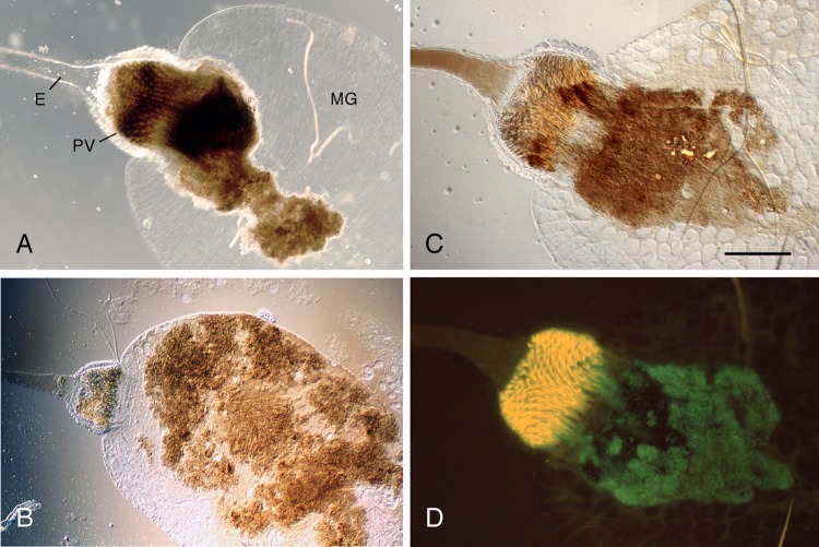
To compare the infectivity of the different Y. pestis strains for X. cheopis, samples of 20 female fleas were collected at 0 and 28 days after the infectious blood meal and used to determine Y. pestis CFU per flea. Neither the infection rate nor the average bacterial load differed significantly for fleas infected with the Y. pestis KIM6+, KIM6+ ΔphoP, or the complemented KIM6+ ΔphoP strains (Fig. 1B and C). Thus, the limited ability of the Y. pestis KIM6+ ΔphoP strain to block fleas was not due to decreased ability to establish a stable infection of the flea digestive tract. However, microscopic examination of the midguts dissected from infected fleas revealed a difference in infection phenotype (Fig. 2). Both phoP+ and phoP strains grew as a biofilm in the flea gut, i.e., they formed large bacterial aggregates that were surrounded by a brown extracellular matrix, as described previously (32, 33, 44). However, the Y. pestis ΔphoP aggregates were less cohesive and more easily fragmented than the aggregates formed by the phoP+ parent strain, and they appeared to adhere to and colonize the proventricular spines to a much lesser extent.
Fig 2.
Fragile biofilm produced by PhoP− Y. pestis in the flea gut. Digestive tracts of X. cheopis fleas infected with Y. pestis KIM6+ (A), KIM6+ ΔphoP (B), or KIM6+ ΔphoP (pGFP) (C and D) were dissected and examined by light (A to C) and fluorescent (D) microscopy. The proventriculus (PV) of the flea (A) is filled and blocked with a dense cohesive biofilm that extends into the midgut (MG). The biofilm produced by the phoP mutant is less cohesive and is usually confined to the MG (B) or attached only peripherally to the posterior ends of the autofluorescent spines of the PV (C and D). The examples shown are representative of several flea dissections. Bar = 0.1 mm. E, esophagus.
PhoPQ- or PmrAB-regulated aminoarabinose modification of Y. pestis lipid A is not required to infect or block fleas.
Modification of the lipid A moiety of LPS is regulated by PhoP and has important effects on bacterium-host interactions. For example, loss of phoP does not change the acylation pattern of lipid A from Y. pestis grown in vitro (29, 48) but greatly reduces aminoarabinose addition (29). Aminoarabinose modification makes the LPS less negatively charged, decreasing the binding affinity to CAMPs and thereby conferring resistance to them. Since changes in the electrostatic properties of the bacterial surface also affect adherence and biofilm formation (49, 50), we hypothesized that the blockage-deficient phenotype of the Y. pestis phoP mutant in fleas was attributable to a lack of aminoarabinose modification. In Y. pestis, aminoarabinose modification can be regulated independently by both the PhoP-PhoQ and PmrA-PmrB two-component systems (30). Therefore, we constructed additional Y. pestis mutant strains, one with pmrA deleted and one with pbgP and ugd, which are required for the biosynthesis of aminoarabinose (51), deleted. As shown in Fig. 1, the Y. pestis ΔpmrA and ΔpbgP Δugd strains were no different than wild-type KIM6+ in their ability to infect and block fleas. Y. pestis ΔphoP ΔpmrA, like the ΔphoP single mutant, had a greatly reduced ability to block fleas but not to infect them. Thus, loss of PhoP-mediated aminoarabinose addition to Y. pestis lipid A is not responsible for the altered biofilm phenotype of the Y. pestis phoP mutant in the flea. Lastly, because fleas that become blocked are unable to feed, they starve to death within a few days, and excess mortality is thus a surrogate indicator of blockage (43). Fleas infected with the PhoP− strains experienced significantly lower mortality during the 4-week experiments than PhoP+ strains (data not shown); the mortality rates mirrored the blockage rates shown in Fig. 1A.
Effect of phoP mutation on in vitro biofilm formation on a glass surface.
Colonization and blockage of the flea proventriculus by Y. pestis is essentially a biofilm phenomenon, and the ability of Y. pestis to block X. cheopis fleas correlates with the ability to produce a biofilm on the surface of a glass flow cell at low temperatures (32). Therefore, we examined the phenotype of in vitro biofilms produced by the Y. pestis phoP mutant. Both Y. pestis KIM6+ and KIM6+ ΔphoP produced confluent biofilm in flow cells at 21°C (Fig. 3A and B). At 25°C, the PhoP+ parent strain still produced a dense, confluent biofilm, but the phoP mutant strain did not (Fig. 3E, F). The flow-cell biofilm phenotypes of the wild-type and complemented phoP mutant strains were identical at 25°C (Fig. 3C and G).
Fig 3.
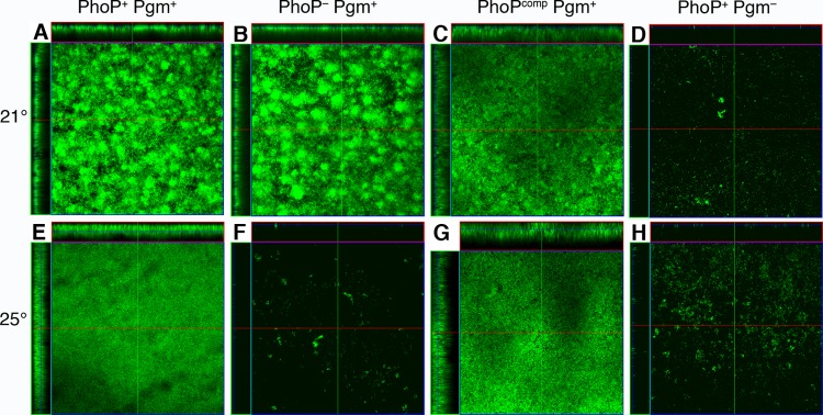
Effect of PhoP on Pgm-dependent biofilm formation in Y. pestis. In vitro biofilms produced by Y. pestis KIM6+ (PhoP+ Pgm+) (A and E), KIM6+ ΔphoP (pLG338) (PhoP− Pgm+) (B and F), KIM6+ ΔphoP (pLGphoP) (PhoPcomp Pgm+) (C and G), and KIM6 (PhoP+ Pgm−) (D and H) at 21°C (A to D) and 25°C (E to H). The results are representative of at least three independent experiments.
The Y. pestis hms gene products, including the hmsHFRS operon within the chromosomal Pgm locus, regulate and catalyze the synthesis of a polysaccharide extracellular matrix (ECM) that is essential for biofilm formation both in vitro (e.g., Fig. 3A versus D) and in the flea and for an in vitro pigmentation (Pgm) phenotype based on binding of hemin or Congo red (32, 41, 52–55). As there is some evidence that one of the hms genes, hmsT, is PhoP regulated in Yersinia pseudotuberculosis (56), we quantitated Congo red binding to Y. pestis at 21, 25, and 37°C to determine if the Pgm phenotype is influenced by PhoP (Fig. 4). Deletion of phoP did not affect temperature-dependent binding of the dye, indicating that production of the ECM is independent of the PhoP-PhoQ system. In addition, the pigmentation phenotype of Y. pestis KIM6+ ΔphoP, ΔpmrA, ΔphoP ΔpmrA, and ΔpbgP Δugd colonies on Congo red agar was identical to that of the Y. pestis KIM6+ parent strain (data not shown).
Fig 4.
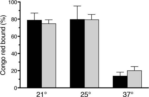
Loss of PhoP does not affect the Hms-dependent in vitro pigmentation phenotype of Y. pestis. The percentage of Congo red dye bound by Y. pestis KIM6+ (black bars) and KIM6+ ΔphoP (gray bars) after growth at different temperatures is shown (means and standard deviations [SD] from three experiments).
Induction of the PhoP-PhoQ system in the flea.
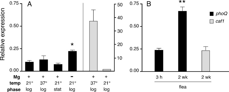
The preceding results indicated that the Y. pestis PhoP-PhoQ signal transduction system regulates genes that affect the ability to produce an adherent proventriculus-blocking biofilm in the flea. This finding is consistent with a microarray study in which transcription of the Y. pestis phoQ gene was reported to be upregulated ∼2-fold in the flea (36). Since the phoPQ operon is autogenously upregulated by PhoP, we quantified relative amounts of phoQ mRNA to assess activation of the Y. pestis system both in vitro and in the flea at different times after infection (Fig. 5). As a control, relative expression of the Y. pestis caf1 gene, which encodes the extracellular F1 capsular protein whose expression is known to be downregulated at 21°C and in the flea (57, 58), was also quantitated. As for other Gram-negative bacteria, growth in low Mg2+ activated the PhoP-PhoQ system of Y. pestis (Fig. 5A). However, PhoP-PhoQ induction was even higher in the flea gut 2 weeks after infection, a time corresponding to the peak incidence of proventricular blockage (Fig. 5B). The greater in vivo activation of the Y. pestis PhoP-PhoQ system occurred in spite of the fact that the concentration of Mg2+ in flea gut contents was 0.1 mM (Table 3), about 10-fold higher than in the low-Mg2+ medium used.
Fig 5.
Induction of the Y. pestis PhoP-PhoQ regulatory system during infection of X. cheopis fleas. Relative amounts of phoQ mRNA expressed by Y. pestis KIM6+ in logarithmic-phase (log) and stationary-phase (stat) cultures grown at 21 or 37°C in media containing high (+) or low (−) Mg2+ (A) and in fleas 3 h and 2 weeks after infection (B) were determined by quantitative RT-PCR. As a control, relative expression of caf1, a highly expressed gene which is known to be downregulated at 21°C and in the flea, was also determined. The means and SEM from three experiments performed in triplicate are shown. *, P < 0.05 compared to Mg+ cultures by one-way analysis of variance and Tukey's multiple comparison test; **, P = 0.0005 compared to the 3-h sample by unpaired t test (two tailed).
Table 3.
Cation concentrations in the midgut of X. cheopis fleas at different times after a blood meal
| Cation | Concentration (mM) at: |
|
|---|---|---|
| 1 day | 6 days | |
| Mg | 0.14 | 0.10 |
| Ca | 0.14 | 0.30 |
| Fe | 3.6 | 2.0 |
Role of PhoP in resistance to flea innate immunity.
A major biological role of the Salmonella PhoP-PhoQ system is to upregulate genes required for bacterial survival in macrophages and for resistance to host CAMPs. The PhoP-PhoQ system is likewise responsible for resistance to polymyxin B and cecropin, an insect-derived CAMP, in Y. pestis (29, 30, 48) and Y. pseudotuberculosis (28). To demonstrate that fleas, like other insects, mount an antibacterial immune response, a mixture of Gram-positive and -negative bacteria was introduced into the hemocoel (body cavity) of fleas. Six hours after challenge, flea extracts were screened in a bioassay for antibacterial activity. The results showed that bacterial challenge induced the expression of antibacterial components in the flea (Fig. 6).
Fig 6.
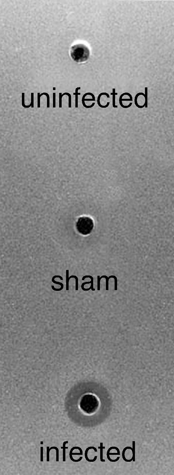
Antibacterial immune response of X. cheopis fleas induced by bacterial challenge. Zone of inhibition assay of extracts collected from fleas 6 h after challenge by piercing the exoskeleton with either a needle contaminated with E. coli and M. luteus (infected) or a sterile needle (sham) and from unchallenged fleas (uninfected).
We determined the susceptibility of PhoP+ and PhoP− Y. pestis to CAMPs, a major component of the insect antibacterial response (Table 4). As expected, the PhoP− strains as well as the Δugd and ΔpbgP Δugd mutants that are unable to synthesize or add aminoarabinose to lipid A were highly susceptible. In contrast, the PhoP+ and the complemented ΔphoP and Δugd Y. pestis strains were highly resistant. Because the Y. pestis ΔpbgP Δugd mutant had a wild-type phenotype in the flea (Fig. 1), these results suggest that any CAMP response of the flea to oral infection is not sufficient to control CAMP-sensitive, aminoarabinose-negative Y. pestis in the flea gut.
Table 4.
MICs of cationic antimicrobial peptides to Y. pestis KIM6+ strains
| Y. pestis strain or genotype | MIC (μg/ml) of: |
|
|---|---|---|
| Polymyxin B | Cecropin A | |
| KIM6+ (wild type) | 50 | >100 |
| ΔphoP (pLG338) | <0.1 | 1.6 |
| ΔphoP (pLGphoP) | 25 | >100 |
| ΔpmrA | 50 | >100 |
| ΔphoP ΔpmrA | 0.8 | 1.6 |
| ΔpbgP Δugd | <0.1 | 1.6 |
| Δugd (pLG338-30) | <0.1 | 1.6 |
| Δugd (pLGugd) | 50 | >100 |
DISCUSSION
Y. pestis colonizes its flea vector by growing as a biofilm in the digestive tract (32). The bacteria enter the flea midgut in a blood meal as individual cells but then reproduce and grow in the form of dense multicellular aggregates that are embedded within an extracellular matrix. Adherent biofilm in the flea proventriculus is crucial for the regurgitative transmission mechanism. When blocked or partially blocked fleas attempt to feed, the bacterial biofilm attached to the spines of the proventricular valve impedes the normal flow of blood into the midgut. This potentiates transmission, which occurs when blood containing Y. pestis released from the periphery of the biofilm is regurgitated into the bite site (33, 34).
The ability of Y. pestis to form a biofilm, both in the flea and in vitro, depends on the hms gene products encoded in the Pgm locus, which synthesize the β-1,6 N-acetyl-d-glucosamine polysaccharide component of the extracellular matrix of the biofilm (32, 41, 43, 54, 59, 60). The Pgm-dependent biofilm is produced only at temperatures below 28°C. In this study, we found that the Y. pestis PhoP-PhoQ system influences biofilm development and localization in the flea.
The biofilms formed by the Pgm+ PhoP− Y. pestis strains in the flea were less cohesive and less adherent to the proventricular spines than those formed by wild-type strains (Fig. 2). This phenotype resulted in a greatly decreased incidence of proventricular blockage in fleas infected with PhoP− Y. pestis (Fig. 1). It is likely that the biofilm produced by PhoP− Y. pestis is less adherent to the acellular, hydrophobic surface of the proventricular spines and too fragile to withstand the threshing action generated by the rapid contractions of the proventricular valve that occur when a flea feeds and is swept back into the midgut along with the incoming blood (Fig. 2C and D). The effects of phoP mutation on in vitro biofilm formation are consistent with this interpretation. Sun et al. reported that, although a Y. pestis phoP mutant produced thicker biofilms than the wild type in the microtiter plate assay, the biofilms made by the phoP mutant were more loosely adherent, and special care had to be taken to prevent dislodging them during washing and staining steps (56). In our flow cell system, an effect of phoP mutation on biofilm formation was evident at the intermediate temperature of 25°C (Fig. 3). At this temperature, the phoP mutant usually failed to produce an adherent biofilm, whereas the wild-type parent strain always produced one. In Y. pseudotuberculosis, loss of phoP correlates with increased biofilm formation on the surface of Caenorhabditis elegans nematodes, but neither PhoP+ nor PhoP− Y. pseudotuberculosis strains are able to block fleas (56, 61, 62). Thus, PhoP affects the biofilm phenotype of the two species differently under different environmental conditions. This is not surprising, because biofilm development depends on complex regulatory pathways and other physiologic factors that are sensitive to environmental conditions, as well as on the surface characteristics of the bacteria and the substrate to which they adhere (49, 63).
The results indicate that, in addition to the hms genes, PhoP-regulated genes of Y. pestis are required for the production of a stable, adherent biofilm in the flea proventriculus. We do not know which PhoP-regulated genes are responsible for this, but obvious candidates are those that affect cell surface characteristics. Changes in LPS structure, cell surface charge, and adhesion expression affect the biofilm phenotype of other bacteria (49, 64–67). Y. pestis undergoes temperature-dependent phase variation in the acylation pattern of its lipid A—during growth at 21°C (the temperature typical of the flea gut environment), the lipid A is hexa-acylated, whereas at 37°C the lipid A is primarily tetra-acylated (29, 68). We have previously shown that a Y. pestis mutant lacking msbB and lpxP, which encode the two acyltransferases required to produce hexa-acylated lipid A, constitutively makes the tetra-acylated form at both low and high temperatures but that this mutant is still able to infect and block fleas normally (69). The Y. pestis PhoP-PhoQ system does not appear to regulate lipid A acylation (29, 48) but greatly reduces aminoarabinose addition (29). We ruled out our hypothesis that PhoP-dependent addition of aminoarabinose to lipid A was required for normal biofilm formation in the flea; however, PhoP has been implicated in another modification of LPS—the addition of galactose to the oligosaccharide core (48). Alteration of LPS has previously been shown to affect biofilm formation in Y. pestis: gmhA, which is responsible for synthesis of the heptose component of the oligosaccharide core, is required for proventricular blockage of fleas (70), and yrbH and waaA, which are involved in the addition of Kdo (3-deoxy-D-manno-octulosonic acid) monosaccharide to the inner core of LPS, are required for normal biofilm formation in vitro (71). The Y. pestis hms genes themselves do not appear to be PhoP regulated, at least in vitro, because the prototypical Hms-dependent phenotype (pigmentation due to Congo red binding) was not affected by phoP mutation (Fig. 4).
The environmental factors in the flea gut that induce the Y. pestis PhoP-PhoQ system are also unknown. In Gram-negative bacteria, low Mg2+and Ca2+ stimulate PhoQ, and high Fe3+ can activate a subset of PhoP-regulated genes by stimulating the PmrA-PmrB two-component regulatory system (9, 30, 72). As in Salmonella, the Y. pestis PhoP-PhoQ system was induced by low Mg2+ concentration in vitro, but induction was even higher during infection of the flea digestive tract (Fig. 5). Mg2+ and Ca2+ levels in the flea midgut would be expected to be high immediately after a blood meal, reflecting mammalian plasma levels (0.8 to 1.0 mM and 2.0 to 2.5 mM, respectively). However, the concentration of these cations is much higher in insect hemolymph, so it is likely that they are actively transported by the midgut epithelium (73). We detected 0.1 to 0.3 mM Mg2+ and Ca2+ in flea midgut contents (Table 3). These levels are intermediate between the micromolar concentrations that maximally stimulate Salmonella PhoP activation and the millimolar concentrations that do not stimulate (9); nevertheless, Y. pestis PhoP activation was greater in fleas than in low-Mg2+ medium (Fig. 5). One possible explanation is that bacteria within the dense biofilm are not exposed to the level of Mg2+ present in the lumen of the flea gut. In contrast, pmrA expression was lower in the flea than in N-minimal medium containing 10 μM MgCl2 with or without 0.1 mM FeSO4 (data not shown). These results are consistent with a recent microarray study in which pmrA expression was lower in the flea than in vitro and no pmrB expression was detected in the flea (36) as well as with the lack of a phenotype of the Y. pestis pmrA mutant in the flea (Fig. 1). Although Y. pestis has a Fe3+-responsive PmrA-PmrB system (30) and the Fe concentration in the flea midgut was high (Table 3), as might be expected following digestion of red blood cells and hemoglobin), the ionic and chemical state (e.g., Fe2+ or Fe3+; free or bound to heme) of this metal in the flea gut is unknown. The microarray study indicated that the pbgP and ugd genes were not upregulated in the flea compared to in vitro growth conditions (36).
Other environmental signals in the flea gut may contribute to PhoQ stimulation. For example, the Salmonella PhoP-PhoQ system responds to mildly acidic pH and to CAMPs (13, 14, 74). The flea midgut pH is reportedly acidic (75), and the presence of bacteria in the blood meal induces the production and secretion of CAMPs into the midgut in other blood-feeding insects (76, 77). Fleas, like other insects, mount an immune response against bacterial challenge (Fig. 6), and PhoP mutation in Y. pestis and in the insect pathogen Photorhabdus luminescens results in an inability to infect the hemocoel and loss of virulence to insect (order Lepidoptera) larvae (7, 78). The PhoP-PhoQ system of Sodalis glossinidius, a Gram-negative endosymbiont of the tsetse fly, is also required for infectivity in the insect host (79). Consistent with previous results (29, 48) and the importance of aminoarabinose-modified lipid A for resistance to CAMPs (16), the Y. pestis phoP, ugd, and pbgP mutants were highly sensitive to both polymyxin and cecropin, a type of antibacterial peptide commonly produced by insects (Table 4). In spite of this, however, the Y. pestis ΔphoP and ΔpbgP Δugd strains multiplied normally in the flea midgut (Fig. 1). This suggests that the presence of Y. pestis in the blood meal does not induce a strong antimicrobial response into the midgut. Alternatively, since bacteria in a biofilm are more resistant to many antimicrobials (80), the biofilm ECM or other PhoP-independent, midgut-specific phenotype may have protected the Y. pestis ΔphoP and Δugd strains from flea CAMPs in the midgut.
Our results provide independent proof that the Y. pestis PhoP-PhoQ regulatory system is induced in the flea and that this induction is actually required to produce a normal transmissible infection. The specific inducing stress that stimulates PhoQ can result in the activation of different subsets of the PhoP-regulated genes (12, 31), and we are currently identifying the Y. pestis genes that are PhoP regulated in the flea gut environment. Certain known PhoP-regulated virulence factors have been shown to be upregulated in the flea, however, and their induction prior to transmission may enhance infectivity in the mammal by conferring resistance to the initial innate immune response encountered at the flea bite site (36). In addition to resistance to CAMPs, the PhoP-PhoQ two-component regulatory system is required for Y. pestis resistance to killing by neutrophils (81), which the bacteria encounter very soon after transmission. In this regard, upregulation of the PhoP-PhoQ system prior to transmission in the flea may be especially important because the major antiphagocytic virulence factors encoded by the type III secretion system are not expressed at a low temperature in the flea and are not fully functional until a few hours after a shift to 37°C (57, 75, 82).
ACKNOWLEDGMENTS
This work was supported by the Division of Intramural Research, NIAID, NIH, and by the Ellison Medical Foundation (New Scholars in Global Infectious Diseases award to B.J.H.).
We thank Eszter Deak for molecular biology assistance, Frank DeLeo for help with confocal microscopy, and C. Bosio, I. Chouikha, J. Shannon, and J. Spinner for review of the manuscript.
Footnotes
Published ahead of print 22 February 2013
REFERENCES
- 1. Miller JF, Mekalanos JJ, Falkow S. 1989. Coordinate regulation and sensory transduction in the control of bacterial virulence. Science 243:916–922 [DOI] [PubMed] [Google Scholar]
- 2. Miller SI, Kukral AM, Mekalanos JJ. 1989. A two-component regulatory system (phoP phoQ) controls Salmonella typhimurium virulence. Proc. Natl. Acad. Sci. U. S. A. 86:5054–5058 [DOI] [PMC free article] [PubMed] [Google Scholar]
- 3. Groisman EA, Chiao E, Lipps CJ, Heffron F. 1989. Salmonella typhimurium phoP virulence gene is a transcriptional regulator. Proc. Natl. Acad. Sci. U. S. A. 86:7077–7081 [DOI] [PMC free article] [PubMed] [Google Scholar]
- 4. Groisman EA. 2001. The pleiotropic two-component regulatory system PhoP-PhoQ. J. Bacteriol. 183:1835–1842 [DOI] [PMC free article] [PubMed] [Google Scholar]
- 5. Moss JE, Fisher PE, Vick B, Groisman EA, Zychlinsky A. 2000. The regulatory protein PhoP controls susceptibility to the host inflammatory response in Shigella flexneri. Cell. Microbiol. 2:443–452 [DOI] [PubMed] [Google Scholar]
- 6. Llama-Palacios A, Lopez-Solanilla E, Poza-Carrion C, Garcia-Olmedo F, Rodriguez-Palenzuela P. 2003. The Erwinia chrysanthemi phoP-phoQ operon plays an important role in growth at low pH, virulence and bacterial survival in plant tissue. Mol. Microbiol. 49:347–357 [DOI] [PubMed] [Google Scholar]
- 7. Derzelle S, Turlin E, Duchaud E, Pagges S, Kunst F, Givaudan A, Danchin A. 2004. The PhoP-PhoQ two-component regulatory system of Photorhabdus luminescens is essential for virulence in insects. J. Bacteriol. 186:1270–1279 [DOI] [PMC free article] [PubMed] [Google Scholar]
- 8. Grabenstein JP, Marceau M, Pujol C, Simonet M, Bliska JB. 2004. The response regulator PhoP of Yersinia pseudotuberculosis is important for replication in macrophages and for virulence. Infect. Immun. 72:4973–4984 [DOI] [PMC free article] [PubMed] [Google Scholar]
- 9. Garcia-Vescovi E, Soncini FC, Groisman AA. 1996. Mg2+ as an extracellular signal: environmental regulation of Salmonella virulence. Cell 84:165–174 [DOI] [PubMed] [Google Scholar]
- 10. Miller SI, Mekalanos JJ. 1990. Constitutive expression of the PhoP regulon attenuates Salmonella virulence and survival within macrophages. J. Bacteriol. 172:2485–2490 [DOI] [PMC free article] [PubMed] [Google Scholar]
- 11. Monsieurs P, De Keersmaecker S, Navarre WW, Bader MW, De Smet F, McClelland M, Fang FC, De Moor B, Vanderleyden J, Marchal K. 2005. Comparison of the PhoPQ regulon in Escherichia coli and Salmonella typhimurium. J. Mol. Evol. 60:462–474 [DOI] [PubMed] [Google Scholar]
- 12. Perez JC, Shin D, Zwir I, Latifi T, Hadley TJ, Groisman EA. 2009. Evolution of a bacterial regulon controlling virulence and Mg2+ homeostasis. PLoS Genet. 5:e1000428 doi:10.1371/journal.pgen.1000428 [DOI] [PMC free article] [PubMed] [Google Scholar]
- 13. Bader MW, Sanowar S, Daley ME, Schneider AR, Cho U, Xu W, Klevit RE, Le Moual H, Miller SI. 2005. Recognition of antimicrobial peptides by a bacterial sensor kinase. Cell 122:461–472 [DOI] [PubMed] [Google Scholar]
- 14. Prost LR, Daley ME, Le Sage V, Bader MW, Le Moual H, Klevit RE, Miller SI. 2007. Activation of the bacterial sensor kinase PhoQ by acidic pH. Mol. Cell 26:165–174 [DOI] [PubMed] [Google Scholar]
- 15. Fields PI, Groisman EA, Heffron F. 1989. A Salmonella locus that controls resistance to microbicidal proteins from phagocytic cells. Science 243:1059–1062 [DOI] [PubMed] [Google Scholar]
- 16. Gunn JS, Lim KB, Krueger J, Kim K, Guo L, Hackett M, Miller SI. 1998. PmrA-PmrB-regulated genes necessary for 4-aminoarabinose lipid A modification and polymyxin resistance. Mol. Microbiol. 27:1171–1182 [DOI] [PubMed] [Google Scholar]
- 17. Guo L, Lim KB, Gunn JS, Bainbridge B, Darveau RP, Hackett M, Miller SI. 1997. Regulation of lipid A modifications by Salmonella typhimurium virulence genes phoP-phoQ. Science 276:250–253 [DOI] [PubMed] [Google Scholar]
- 18. Guo L, Lim KB, Poduje CM, Daniel M, Gunn JS, Hackett M, Miller SI. 1998. Lipid A acylation and bacterial resistance against vertebrate antimicrobial peptides. Cell 95:189–198 [DOI] [PubMed] [Google Scholar]
- 19. Kato A, Groisman EA. 2008. The PhoP/PhoQ regulatory network of Salmonella enterica, p 7–21 In Utsumi R. (ed), Bacterial signal transduction: networks and drug targets. Springer Science+Business Media and Landes Bioscience, New York, NY [Google Scholar]
- 20. Zwir I, Shin D, Kato A, Nishino K, Latifi T, Solomon F, Hare JM, Huang H, Groisman EA. 2005. Dissecting the PhoP regulatory network of Escherichia coli and Salmonella enterica. Proc. Natl. Acad. Sci. U. S. A. 102:2862–2867 [DOI] [PMC free article] [PubMed] [Google Scholar]
- 21. Parkhill J, Wren BW, Thomson NR, Titball RW, Holden MTG, Prentice MB, Sebhaihia M, James KD, Churcher C, Mungall KL, Baker S, Basham D, Bentley SD, Brooks K, Cerdeño-Tárraga AM, Chillingworth T, Cronin A, Davies RM, Davis P, Dougan G, Feltwell T, Hamlin N, Holroyd S, Jagels K, Karlyshev AV, Leather S, Moule S, Oyston PCF, Quail M, Rutherford K, Simmonds M, Skelton J, Stevens K, Whitehead S, Barrell BG. 2001. Genome sequence of Yersinia pestis, the causative agent of plague. Nature 413:523–527 [DOI] [PubMed] [Google Scholar]
- 22. Oyston PCF, Dorrell N, Williams K, Li S-R, Green M, Titball RW, Wren BW. 2000. The response regulator PhoP is important for survival under conditions of macrophage-induced stress and virulence in Yersinia pestis. Infect. Immun. 68:3419–3425 [DOI] [PMC free article] [PubMed] [Google Scholar]
- 23. Grabenstein JP, Fukuto HS, Palmer LE, Bliska JB. 2006. Characterization of phagosome trafficking and identification of PhoP-regulated genes important for survival of Yersinia pestis in macrophages. Infect. Immun. 74:3727–3741 [DOI] [PMC free article] [PubMed] [Google Scholar]
- 24. Li Y, Gao H, Qin L, Li B, Han Y, Guo Z, Song Y, Zhai J, Du Z, Wang X, Zhou D, Yang R. 2008. Identification and characterization of PhoP regulon members in Yersinia pestis biovar microtus. BMC Genomics 9:143. [DOI] [PMC free article] [PubMed] [Google Scholar]
- 25. Zhou D, Han Y, Qin L, Chen Z, Qiu J, Song Y, Li B, Wang J, Guo Z, Du Z, Wang X, Yang R. 2005. Transcriptome analysis of the Mg2+-responsive PhoP regulator in Yersinia pestis. FEMS Microbiol. Lett. 250:85–95 [DOI] [PubMed] [Google Scholar]
- 26. Perez JC, Groisman EA. 2009. Evolution of transcriptional regulatory circuits in bacteria. Cell 138:233–244 [DOI] [PMC free article] [PubMed] [Google Scholar]
- 27. Perez JC, Groisman EA. 2009. Transcription factor function and promoter architecture govern the evolution of bacterial regulons. Proc. Natl. Acad. Sci. U. S. A. 106:4319–4324 [DOI] [PMC free article] [PubMed] [Google Scholar]
- 28. Marceau M, Sebbane F, Ewann F, Collyn F, Lindner B, Campos MA, Bengoechea J-A, Simonet M. 2004. The pmrF polymyxin-resistance operon of Yersinia pseudotuberculosis is upregulated by the PhoP-PhoQ two-component system but not by PmrA-PmrB, and is not required for virulence. Microbiology 150:3947–3957 [DOI] [PubMed] [Google Scholar]
- 29. Rebeil R, Ernst RK, Gowen BB, Miller SI, Hinnebusch BJ. 2004. Variation in lipid A structure in the pathogenic yersiniae. Mol. Microbiol. 52:1363–1373 [DOI] [PubMed] [Google Scholar]
- 30. Winfield MD, Latifi T, Groisman EA. 2005. Transcriptional regulation of the 4-amino-4-deoxy-l-arabinose biosynthetic genes in Yersinia pestis. J. Biol. Chem. 280:14765–14772 [DOI] [PubMed] [Google Scholar]
- 31. Miyashiro T, Goulian M. 2007. Stimulus-dependent differential regulation in the Escherichia coli PhoQ PhoP system. Proc. Natl. Acad. Sci. U. S. A. 104:16305–16310 [DOI] [PMC free article] [PubMed] [Google Scholar]
- 32. Jarrett CO, Deak E, Isherwood KE, Oyston PC, Fischer ER, Whitney AR, Kobayashi SD, DeLeo FR, Hinnebusch BJ. 2004. Transmission of Yersinia pestis from an infectious biofilm in the flea vector. J. Infect. Dis. 190:783–792 [DOI] [PubMed] [Google Scholar]
- 33. Bacot AW, Martin CJ. 1914. Observations on the mechanism of the transmission of plague by fleas. J. Hygiene 13:423–439 [PMC free article] [PubMed] [Google Scholar]
- 34. Bacot AW. 1915. Further notes on the mechanism of the transmission of plague by fleas. J. Hygiene 14:774–776 [PMC free article] [PubMed] [Google Scholar]
- 35. Pollitzer R. 1954. Plague. World Health Organization, Geneva, Switzerland [Google Scholar]
- 36. Vadyvaloo V, Jarrett C, Sturdevant DE, Sebbane F, Hinnebusch BJ. 2010. Transit through the flea vector induces a pretransmission innate immunity resistance phenotype in Yersinia pestis. PLoS Pathog. 6:e1000783 doi:10.1371/journal.ppat.1000783 [DOI] [PMC free article] [PubMed] [Google Scholar]
- 37. Ling MM, Robinson BH. 1997. Approaches to DNA mutagenesis: an overview. Anal. Biochem. 254:157–178 [DOI] [PubMed] [Google Scholar]
- 38. Donnenberg MS, Kaper JB. 1991. Construction of an eae deletion mutant of enteropathogenic Escherichia coli by using a positive-selection suicide vector. Infect. Immun. 59:4310–4317 [DOI] [PMC free article] [PubMed] [Google Scholar]
- 39. Wang J, Wilkinson MF. 2001. Deletion mutagenesis of large (12-kb) plasmids by a one-step PCR protocol. Biotechniques 31:722–724 [DOI] [PubMed] [Google Scholar]
- 40. Derbise A, Lesic B, Dacheux D, Ghigo JM, Carniel E. 2003. A rapid and simple method for inactivating chromosomal genes in Yersinia. FEMS Immunol. Med. Microbiol. 38:113–116 [DOI] [PubMed] [Google Scholar]
- 41. Kirillina O, Fetherston JD, Bobrov AG, Abney J, Perry RD. 2004. HmsP, a putative phosphodiesterase, and HmsT, a putative diguanylate cyclase, control Hms-dependent biofilm formation in Yersinia pestis. Mol. Microbiol. 54:75–88 [DOI] [PubMed] [Google Scholar]
- 42. Chaussee MS, Watson RO, Smoot JC, Musser JM. 2001. Identification of Rgg-regulated exoproteins of Streptococcus pyogenes. Infect. Immun. 69:822–831 [DOI] [PMC free article] [PubMed] [Google Scholar]
- 43. Hinnebusch BJ, Perry RD, Schwan TG. 1996. Role of the Yersinia pestis hemin storage (hms) locus in the transmission of plague by fleas. Science 273:367–370 [DOI] [PubMed] [Google Scholar]
- 44. Hinnebusch BJ, Fischer ER, Schwan TG. 1998. Evaluation of the role of the Yersinia pestis plasminogen activator and other plasmid-encoded factors in temperature-dependent blockage of the flea. J. Infect. Dis. 178:1406–1415 [DOI] [PubMed] [Google Scholar]
- 45. Chalk R, Townson H, Natori S, Desmond H, Ham PJ. 1994. Purification of an insect defensin from the mosquito, Aedes aegypti. Insect Biochem. Mol. Biol. 24:403–410 [DOI] [PubMed] [Google Scholar]
- 46. Castelli ME, Garcia Vescovi E, Soncini FC. 2000. The phosphatase activity is the target for Mg2+ regulation of the sensor protein PhoQ in Salmonella. J. Biol. Chem. 275:22948–22954 [DOI] [PubMed] [Google Scholar]
- 47. Sun Y-C, Koumoutsi A, Jarrett C, Lawrence K, Gherardini FC, Darby C, Hinnebusch BJ. 2011. Differential control of Yersinia pestis biofilm formation in vitro and in the flea vector by two c-di-GMP diguanylate cyclases. PLoS One 6:e19267 doi:10.1371/journal.pone.0019267 [DOI] [PMC free article] [PubMed] [Google Scholar]
- 48. Hitchen PG, Prior JL, Oyston PCF, Panico M, Wren BW, Titball RW, Morris HR, Dell A. 2002. Structural characterization of lipo-oligosaccharide (LOS) from Yersinia pestis: regulation of LOS structure by the PhoPQ system. Mol. Microbiol. 44:1637–1650 [DOI] [PubMed] [Google Scholar]
- 49. Beloin C, Da Re S, Ghigo J-M. 29 August 2005, posting date Chapter 8.3.1.3, Colonization of abiotic surfaces. In Böck A, Curtis R, III, Kaper JB, Neidhardt FC, Nyström T, Rudd KE, Squires CL. (ed), EcoSal—Escherichia coli and Salmonella: cellular and molecular biology. ASM Press, Washington, DC: doi:10.1128/ecosal.8.3.1.3 [Google Scholar]
- 50. Mayer C, Moritz R, Kirschner C, Borchard W, Maibaum R, Wingender J, Flemming H-C. 1999. The role of intermolecular interactions: studies on model systems for bacterial biofilms. Int. J. Biol. Macromol. 26:3–16 [DOI] [PubMed] [Google Scholar]
- 51. Raetz CRH, Reynolds CM, Trent MS, Bishop RE. 2007. Lipid A modification systems in gram-negative bacteria. Annu. Rev. Biochem. 76:295–329 [DOI] [PMC free article] [PubMed] [Google Scholar]
- 52. Jackson S, Burrows TW. 1956. The pigmentation of Pasteurella pestis on a defined medium containing haemin. Br. J. Exp. Pathol. 37:570–576 [PMC free article] [PubMed] [Google Scholar]
- 53. Pendrak ML, Perry RD. 1993. Proteins essential for expression of the Hms+ phenotype of Yersinia pestis. Mol. Microbiol. 8:857–864 [DOI] [PubMed] [Google Scholar]
- 54. Darby C, Hsu JW, Ghori N, Falkow S. 2002. Caenorhabditis elegans: plague bacteria biofilm blocks food intake. Nature 417:243–244 [DOI] [PubMed] [Google Scholar]
- 55. Bobrov AG, Kirillina O, Ryjenkov DA, Waters CM, Price PA, Fetherston JD, Mack D, Goldman WE, Gomelsky M, Perry RD. 2011. Systematic analysis of cyclic di-GMP signalling enzymes and their role in biofilm formation and virulence in Yersinia pestis. Mol. Microbiol. 79:533–551 [DOI] [PMC free article] [PubMed] [Google Scholar]
- 56. Sun Y-C, Koumoutsi A, Darby C. 2009. The response regulator PhoP negatively regulates Yersinia pseudotuberculosis and Yersinia pestis biofilms. FEMS Microbiol. Lett. 290:85–90 [DOI] [PMC free article] [PubMed] [Google Scholar]
- 57. Cavanaugh DC, Randall R. 1959. The role of multiplication of Pasteurella pestis in mononuclear phagocytes in the pathogenesis of flea-borne plague. J. Immunol. 83:348–363 [PubMed] [Google Scholar]
- 58. Hudson BW, Kartman L, Prince FM. 1966. Pasteurella pestis detection in fleas by fluorescent antibody staining. Bull. World Health Organ. 34:709–714 [PMC free article] [PubMed] [Google Scholar]
- 59. Bobrov AG, Kirillina O, Forman S, Mack D, Perry RD. 2008. Insights into Yersinia pestis biofilm development: topology and co-interaction of Hms inner membrane proteins involved in exopolysaccharide production. Environ. Microbiol. 10:1419–1432 [DOI] [PubMed] [Google Scholar]
- 60. Erickson DL, Jarrett CO, Callison JA, Fischer ER, Hinnebusch BJ. 2008. Loss of a biofilm-inhibiting glycosyl hydrolase during the emergence of Yersinia pestis. J. Bacteriol. 190:8163–8170 [DOI] [PMC free article] [PubMed] [Google Scholar]
- 61. Joshua GWP, Karlyshev AV, Smith MP, Isherwood KE, Titball RW, Wren BW. 2003. A Caenorhabditis elegans model of Yersinia infection: biofilm formation on a biotic surface. Microbiology 149:3221–3229 [DOI] [PubMed] [Google Scholar]
- 62. Erickson DL, Jarrett CO, Wren BW, Hinnebusch BJ. 2006. Serotype differences and lack of biofilm formation characterize Yersinia pseudotuberculosis infection of the Xenopsylla cheopis flea vector of Yersinia pestis. J. Bacteriol. 188:1113–1119 [DOI] [PMC free article] [PubMed] [Google Scholar]
- 63. O'Toole G, Kaplan HB, Kolter R. 2000. Biofilm formation as microbial development. Annu. Rev. Microbiol. 54:49–79 [DOI] [PubMed] [Google Scholar]
- 64. Beveridge TJ, Makin SA, Kadurugamuwa JL, Li Z. 1997. Interactions between biofilms and the environment. FEMS Microbiol. Rev. 20:291–303 [DOI] [PubMed] [Google Scholar]
- 65. Nesper J, Lauriano CM, Klose K, Kapfhammer D, Kraiss A, Reidl J. 2001. Characterization of Vibrio cholerae O1 El Tor galU and galE mutants: influence on lipopolysaccharide structure, colonization, and biofilm formation. Infect. Immun. 69:435–445 [DOI] [PMC free article] [PubMed] [Google Scholar]
- 66. Prouty AM, Gunn JS. 2003. Comparative analysis of Salmonella enterica serovar Typhimurium biofilm formation on gallstones and on glass. Infect. Immun. 71:7154–7158 [DOI] [PMC free article] [PubMed] [Google Scholar]
- 67. Swords WE, Moore ML, Godzicki L, Bukofzer G, Mitten MJ, VonCannon J. 2004. Sialylation of lipooligosaccharides promotes biofilm formation by nontypeable Haemophilus influenzae. Infect. Immun. 72:106–113 [DOI] [PMC free article] [PubMed] [Google Scholar]
- 68. Kawahara K, Tsukano H, Watanabe H, Lindner B, Matsuura M. 2002. Modification of the structure and activity of lipid A in Yersinia pestis lipopolysaccharide by growth temperature. Infect. Immun. 70:4092–4098 [DOI] [PMC free article] [PubMed] [Google Scholar]
- 69. Rebeil R, Ernst RK, Jarrett CO, Adams KN, Miller SI, Hinnebusch BJ. 2006. Characterization of late acyltransferase genes of Yersinia pestis and their role in temperature-dependent lipid A variation. J. Bacteriol. 188:1381–1388 [DOI] [PMC free article] [PubMed] [Google Scholar]
- 70. Darby C, Ananth SL, Tan L, Hinnebusch BJ. 2005. Identification of gmhA, a Yersinia pestis gene required for flea blockage, by using a Caenorhabditis elegans biofilm system. Infect. Immun. 73:7236–7242 [DOI] [PMC free article] [PubMed] [Google Scholar]
- 71. Tan L, Darby C. 2006. Yersinia pestis YrbH is a multifunctional protein required for both 3-deoxy-d-manno-oct-2-ulosonic acid biosynthesis and biofilm formation. Mol. Microbiol. 61:861–870 [DOI] [PubMed] [Google Scholar]
- 72. Wösten MMSM, Kox LFF, Chamnongpol S, Soncini FC, Groisman EA. 2000. A signal transduction system that responds to extracellular iron. Cell 103:113–125 [DOI] [PubMed] [Google Scholar]
- 73. Mullins D. 1985. Chemistry and physiology of the hemolymph, p 355–400 In Kerkut GA, Gilbert LI. (ed), Comprehensive insect physiology, biochemistry, and pharmacology, vol. 3: integument, respiration, and circulation. Pergamon Press, Oxford, United Kingdom [Google Scholar]
- 74. Alpuche Aranda CM, Swanson JA, Loomis WP, Miller SI. 1992. Salmonella typhimurium activates virulence gene transcription within acidified macrophage phagosomes. Proc. Natl. Acad. Sci. U. S. A. 89:10079–10083 [DOI] [PMC free article] [PubMed] [Google Scholar]
- 75. Perry RD, Fetherston JD. 1997. Yersinia pestis—etiologic agent of plague. Clin. Microbiol. Rev. 10:35–66 [DOI] [PMC free article] [PubMed] [Google Scholar]
- 76. Dimopoulos G, Richman A, Müller H-M, Kafatos FC. 1997. Molecular immune responses of the mosquito Anopheles gambiae to bacteria and malaria parasites. Proc. Natl. Acad. Sci. U. S. A. 94:11508–11513 [DOI] [PMC free article] [PubMed] [Google Scholar]
- 77. Lehane MJ, Wu D, Lehane SM. 1997. Midgut-specific immune molecules are produced by the blood-sucking insect Stomoxys calcitrans. Proc. Natl. Acad. Sci. U. S. A. 94:11502–11507 [DOI] [PMC free article] [PubMed] [Google Scholar]
- 78. Erickson DL, Russell CW, Johnson KL, Hileman T, Stewart RM. 2011. PhoP and OxyR transcriptional regulators contribute to Yersinia pestis virulence and survival within Galleria mellonella. Microb. Pathog. 51:389–395 [DOI] [PubMed] [Google Scholar]
- 79. Pontes MH, Smith KL, De Vooght L, Van Den Abbeele J, Dale C. 2011. Attenuation of the sensing capabilities of PhoQ in transition to obligate insect-bacterial association. PLoS Genet. 7:e1002349 doi:10.1371/journal.pgen.1002349 [DOI] [PMC free article] [PubMed] [Google Scholar]
- 80. Mah TF, O'Toole GA. 2001. Mechanisms of biofilm resistance to antimicrobial agents. Trends Microbiol. 9:34–39 [DOI] [PubMed] [Google Scholar]
- 81. O'Loughlin JL, Spinner JL, Minnich SA, Kobayashi SD. 2010. Yersinia pestis two-component gene regulatory systems promote survival in human neutrophils. Infect. Immun. 78:773–782 [DOI] [PMC free article] [PubMed] [Google Scholar]
- 82. Burrows TW, Bacon GA. 1956. The basis of virulence in Pasteurella pestis: the development of resistance to phagocytosis in vitro. Br. J. Exp. Pathol. 37:286–299 [PMC free article] [PubMed] [Google Scholar]
- 83. Une T, Brubaker RR. 1984. In vivo comparison of avirulent Vwa− and Pgm− or Pstr phenotypes of yersiniae. Infect. Immun. 43:895–900 [DOI] [PMC free article] [PubMed] [Google Scholar]
- 84. Sikkema DJ, Brubaker RR. 1987. Resistance to pesticin, storage of iron, and invasion of HeLa cells by yersiniae. Infect. Immun. 55:572–578 [DOI] [PMC free article] [PubMed] [Google Scholar]
- 85. Simon R, Priefer U, Pühler A. 1983. A broad host range mobilization system for in vivo genetic engineering: transposon mutagenesis in gram negative bacteria. Biotechnology 1:784–791 [Google Scholar]
- 86. Boman HG, Nilsson-Faye I, Paul K, Rasmuson T. 1974. Insect immunity. 1. Characteristics of an inducible cell-free antibacterial reaction in hemolymph of Samia cynthia pupae. Infect. Immun. 10:136–145 [DOI] [PMC free article] [PubMed] [Google Scholar]
- 87. Stoker NG, Fairweather NF, Spratt BG. 1982. Versatile low-copy-number plasmid vectors for cloning in Escherichia coli. Gene 18:335–341 [DOI] [PubMed] [Google Scholar]
- 88. Cunningham TP, Montelaro RC, Rushlow KE. 1993. Lentivirus envelope sequences and proviral genomes are stabilized in Escherichia coli when cloned in low-copy-number plasmid vectors. Gene 124:93–98 [DOI] [PubMed] [Google Scholar]


