The regulation of cerebral blood flow (CBF) is critical for the maintenance of oxygen and nutrient supply to the metabolically active brain. The control of CBF is multifactorial, influenced largely by the partial pressure of arterial carbon dioxide ( ), mean arterial pressure (MAP) and cerebral metabolism. Exercise-induced elevations in cerebral neuronal activity and metabolism increase CBF by approximately 10–30%. Increases in exercise intensity up to approximately 60–70% of maximal oxygen uptake (
 ) increase CBF whereas a reduction towards baseline values due to hyperventilation-induced hypocapnia and subsequent cerebral vasoconstriction are observed at higher exercise intensities. With healthy ageing, CBF is reduced at rest and during exercise by 28–50% from the age of 30 to 70 years likely mediated via brain atrophy and/or reduction in neuronal activity (reviewed in: Ogoh & Ainslie, 2009).
In this issue, Fisher and co-workers examined changes in blood flow velocity in the middle cerebral artery, cerebral oxygenation and metabolism in young (∼22 years) and old (∼66 years) participants at rest and during cycling exercise to exhaustion (Fisher et al. 2013). At each workload, MAP and  were monitored and arterial–jugular venous differences assessed to determine the transcerebral metabolic exchange of oxygen, glucose and lactate. Their findings confirm that the middle cerebral artery was indeed reduced at rest and during exercise in older participants. However, in spite of the clear reduction in cerebral perfusion, exercise-induced changes in (estimated) the cerebral metabolic rate of oxygen, mitochondrial oxygen tension, and the cerebral uptake of oxygen, glucose and lactate were remarkably similar in both groups.
Why are such findings noteworthy? This study is the first to employ a transcerebral exchange approach to determine if older participants are indeed characterized by a more pronounced reduction in CBF compared to their younger counterparts during exercise. This hypothesis is eminently justified based on previous reports of a lower CBF at rest and during exercise. The similarities between age groups emphasize two important points: (1) substrate delivery (i.e. the net arterial inflow) of oxygen, glucose and lactate is a key determinate of cerebral metabolism. However, despite arterial inflow of oxygen, lactate and glucose being lower in the aged, the net cerebral uptake was preserved. Thus, normal reductions in CBF with healthy ageing do not adversely influence the brain's capacity to use essential nutrients, even at maximal exercise. (2) The age-induced reductions in mitochondrial PO2 were similar at exhaustion despite lower absolute workloads in the older group. It would seem unlikely that maximal exercise is limited ‘centrally’ in the aged, given lower workloads in the face of preserved mitochondrial oxygenation.
Partitioning the mechanisms
As reflected in the study, with ageing,  is lower. Normally this would be reflected by small elevations in blood [H+] levels and reductions in bicarbonate (i.e. mild metabolic acidosis) resulting in a slight increase in ventilation. In addition, at maximal exercise, the older group were comparatively less acidotic. Despite these clear differences, temporal changes in the middle cerebral artery were similar, suggesting that other factors (e.g. neuronal activity) are important in provoking elevations in CBF, at least until moderate intensity exercise. After this point hypocapnia seems to dominate the relative decline in CBF. Nevertheless, given the powerful influence of
  , it is likely that the lower
  in the elderly individuals contributed to the blunted cerebrovascular conductance response observed in this group. Additionally, an inverse relationship exists between CBF and haemoglobin (Hb) to offset the reduction in
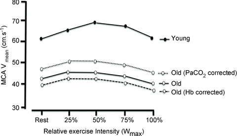
  (e.g. Ibaraki et al. 2010). Therefore, the lower perfusion in the aged group is likely ‘underestimated’ given their lower Hb and, conversely, ‘overestimated’ due to hypocapnia. Thus, as illustrated in Fig. 1, the age-related changes in
  and Hb, respectively, account for approximately 13% of the reduction in CBF and 6% of the elevation in CBF.
Figure 1. Influence of ageing on MCAV responses during exercise.

(Modified from Fisher et al. 2013.) Dashed lines indicate the ‘theoretical’ changes in MCAV that would have occurred in the absence of age-related changes in  and haemoglobin. See text for details. MCAV, middle cerebral artery.
Future research directions
It is clear that many aspects of CBF measurement in ageing warrant further investigation. For example, what are the underlying mechanisms driving an increase in CBF during exercise, and how are these altered with age-related disease processes? Are there age, training status and/or sex differences in ‘regional’ (as opposed to global) CBF during exercise? This is particularly important given recent findings that demonstrate – using positron emission tomography – an age-related decline in CBF and the cerebral metabolic rate of oxygen in the cerebral cortex whereas the CBF to the primary motor and sensory areas appear to be maintained (Aanerud et al. 2012). Such findings indicate that ‘regional’ reductions in oxygen and nutrient supply colocalize with those (i.e. cortex) that are the most vulnerable to neurodegeneration. Addressing some of these intriguing questions will not only provide new information concerning the mechanisms by which CBF is regulated during healthy ageing, but will also provide insight into the pathophysiology of many age-related diseases.
References
- Aanerud J, Borghammer P, Chakravarty MM, Vang K, Rodell AB, Jonsdottir KY, Moller A, Ashkanian M, Vafaee MS, Iversen P, Johannsen P, Gjedde A. J Cereb Blood Flow Metab. 2012;32:1177–1187. doi: 10.1038/jcbfm.2012.18. [DOI] [PMC free article] [PubMed] [Google Scholar]
- Fisher JP, Hartwich D, Seifert T, Olesen ND, McNulty CL, Nielsen HB, van Lieshout JJ, Secher NH. J Physiol. 2013;591:1859–1870. doi: 10.1113/jphysiol.2012.244905. [DOI] [PMC free article] [PubMed] [Google Scholar]
- Ibaraki M, Shinohara Y, Nakamura K, Miura S, Kinoshita F, Kinoshita T. J Cereb Blood Flow Metab. 2010;30:1296–1305. doi: 10.1038/jcbfm.2010.13. [DOI] [PMC free article] [PubMed] [Google Scholar]
- Ogoh S, Ainslie PN. J Appl Physiol. 2009;107:1370–80. doi: 10.1152/japplphysiol.00573.2009. [DOI] [PubMed] [Google Scholar]


