Abstract
Objectives
The aim of this study was to determine whether there is any significant difference in temporal measurements of pain, function and rates of re-tear for arthroscopic rotator cuff repair (RCR) patients compared with those patients undergoing open RCR.
Methods
This study compared questionnaire- and clinical examination-based outcomes over two years or longer for two series of patients who met the inclusion criteria: 200 open RCR and 200 arthroscopic RCR patients. All surgery was performed by a single surgeon.
Results
Most pain measurements were similar for both groups. However, the arthroscopic RCR group reported less night pain severity at six months, less extreme pain and greater satisfaction with their overall shoulder condition than the open RCR group. The arthroscopic RCR patients also had earlier recovery of strength and range of motion, achieving near maximal recovery by six months post-operatively whereas the open RCR patients took longer to reach the same recovery level. The median operative times were 40 minutes (20 to 90) for arthroscopic RCR and 60 minutes (35 to 120) for open RCR. Arthroscopic RCR had a 29% re-tear rate compared with 52% for the open RCR group (p < 0.001).
Conclusions
Arthroscopic RCR involved less extreme pain than open RCR, earlier functional recovery, a shorter operative time and better repair integrity.
Keywords: Rotator cuff repair, Arthroscopy, Open surgery, Clinical outcomes, Pain assessment, Strength measurements, Range-of-motion measurements
Article focus
We aimed to determine whether there is any significant difference in temporal measurements of pain and function, and/or re-tear rates for arthroscopic rotator cuff repair (RCR) patients compared with open RCR patients
Key messages
Arthroscopic RCR provides earlier recovery of strength and passive range of motion than open RCR
The two groups reported similar pain levels for most types of pain considered. The exceptions were that night pain was significantly less severe at six months and extreme pain occurred significantly less frequently from six weeks to six months post-surgery in arthroscopic RCR patients
Arthroscopic RCR patients rated their overall shoulder condition significantly better than open RCR patients.
Re-tear rates were significantly less and operative times significantly shorter in the arthroscopic RCR group than in the open RCR group.
Strengths and limitations
A limitation of this study is that the open RCR group preceded the arthroscopic RCR group in time rather than as contemporary randomised cohorts
Other potential limitations were that the examiners and ultrasonographers were unblinded as to the identity of the operative groups at the two-year visit and the sling of the arthroscopic RCR group differed by having a small abduction pillow
A strength of this study is that all surgical repairs were performed by a single surgeon who used a standardised assessment system
Another strength is that this is the largest study that has compared open RCR with arthroscopic RCR as of this date
Introduction
Tear of the rotator cuff is a common incapacitating condition of the adult shoulder that is often treated with surgical repair. The goals of rotator cuff repair (RCR) are to restore the normal anatomy of the affected shoulder, improve its strength and range-of-motion, and decrease pain. Surgery for RCR has been used since 1911,1 evolving from the all open technique, to the arthroscopy-assisted (mini-open) RCR and, more recently, to an all-arthroscopic RCR.
Concerns have been raised about arthroscopic RCR since its introduction. These include: 1) doubts about its efficacy compared with the proven efficacy of open RCR2,3; 2) its potentials for requiring a longer operative time4; 3) the production of a biomechanically weaker construct2,3,5,6; and 4) its steep learning curve.7 However, improvements continue to be made in suture anchors, arthroscopic instruments, suturing techniques and knot-tying as well as intra-articular visualisation and tendon quality assessment.8-10 Surgeons have also developed expertise in preparing bone for soft-tissue arthroscopic attachment.
There are perceptions among some surgeons who use arthroscopic RCR that their patients have less post-operative pain and more rapid functional recovery, such as Gartsman, Khan and Hammerman,9 who stated ‘Although we cannot document our impressions statistically, we believe that arthroscopic repair results in an improved cosmetic appearance, decreased pain post-operatively, and more rapid gains in motion compared with open operative treatment of similar lesions.’ Yamaguchi et al8 similarly report that complete arthroscopic repair ‘appears to offer less pain and morbidity as well as quicker recovery than do alternative techniques such as open or mini-open repair’. However, objective data that could aid in the assessment of these perceptions are lacking. To our knowledge, only a few published articles based on relatively small subject numbers have compared outcomes of patients with open RCR and arthroscopic RCR.7,11-13
The aim of the present study was to analyse and compare outcomes of a relatively large series of patients treated by a single surgeon: some with open RCR and others with arthroscopic RCR. We assessed rotator cuff integrity by rate of re-tear, operating time, patient-determined assessments of pain, function, and overall shoulder condition and clinician-determined assessments of range of movement (ROM), dynamometer-based strength and specific shoulder signs. This analysis was intended to clarify whether there is any basis for the perception that arthroscopic RCR patients have less post-operative pain and more rapid gains in movement and strength than open RCR patients.
Patients and Methods
Study subjects
This study was based on prospectively collected data from two non-randomised cohorts of consecutivepatients with symptomatic rotator cuff tears who underwent either open RCR (n = 200) or arthroscopic RCR (n = 200) and who satisfied the eligibility requirements. Ethical approval for this study was given by the South East Health Human Research Ethics Committee (Sydney, Australia).
There were two inclusion criteria for this study: 1) each patient had to undergo an open RCR or arthroscopic RCR by the senior author (GACM), and 2) attend a follow-up visit for clinical examination and in-house ultrasound investigation at two years post-operatively. Exclusion criteria included: revision surgery, severe glenohumeral arthritis, fracture or osteonecrosis, tears with a pre-operative size larger than 16 cm2 (to increase uniformity between groups), partially repairable or irreparable tears and patients whose surgery fell within the first ten weeks after the surgeon switched to arthroscopic RCR in 2004. This last criterion was in acknowledgement of the steep learning curve associated with arthroscopic RCR. With regard to the open group, 66 cases were excluded because the tears exceeded 16 cm2, 78 were either partially repairable or irreparable and 30 were excluded because they had moderate to severe osteoarthritis. For the arthroscopic group, 43 cases were excluded because the tears exceeded 16 cm2, 76 were irreparable or partially repairable and 22 had moderate to severe osteoarthritis.
These exclusions left a total of 200 patients in each group who satisfied the eligibility requirements. The open RCR group had a mean follow-up period of 34 months (24 to 81) compared with 31 months (24 to 72) for the arthroscopic group. Open surgery was performed between 2001 and 2004 whereas arthroscopic surgery was performed from 2004 to 2007. Follow-up visits continued until 2010.
Diagnostic arthroscopy
Both groups of patients underwent arthroscopic assessment prior to RCR through a standard three-portal technique.14 Diagnostic arthroscopy was used to confirm the presence or absence of an RCR tear in patients’ shoulders, to estimate the size of the tear, and to assess for other shoulder conditions that, if appropriate, were addressed at the same time. The method and its reproducibility for measuring the size of the rotator cuff tear during surgery are described in another paper.15 The estimated size of the tear, additional findings and operative details were recorded on a surgery form. This part of the procedure usually took less than 15 minutes. Operative time was defined as the number of minutes elapsing between the first incision until wound closure.
Operative techniques
All procedures were performed as day cases with the patient in the upright beach chair position under interscalene block. A pre-operative dose of 1 g cefazolin was given intravenously and a post-operative dose was given 4 hours after completing the procedure. The surgical techniques used for open RCR13 and arthroscopic RCR16,17 have been previously described.
Briefly, for open RCR, the deltoid is split in line with its fibres. The coracoacromial ligament is detached and subsequently reattached to the anterior acromion. The greater tuberosity is visualised and gently roughened with a rasp, and the edges of the torn tendon debrided prior to repair. Anterior acromioplasty and bursectomy are performed along with appropriate soft-tissue releases (subacromial and extra-articular adhesions, coracohumeral ligament and rotator cuff interval). Metallic suture anchors (Quickanchor; DePuy, Warsaw, Indiana) are tapped directly into the proximal humerus without pre-drilling. Suture anchors are placed in the rotator cuff footprint and sutures are passed through the tendon edges: the tendons are repaired using a horizontal mattress stitch configuration. A two-row anchor technique was used for fixation when sufficient excursion of the torn tendon was available.
For arthroscopic RCR, the three-portal technique is used.16 The edge of the rotator cuff tear and the landing site at the greater tuberosity are gently debrided, smoothing the tuberosity with an arthroscopic burr. Acromioplasty was performed in 73% of the arthroscopic patients (n = 146). The repair was undertaken using a single-row technique.9 Double suture-loaded 5 mm metal corkscrew anchors (Mitek Fastin; DePuy) were inserted through the lateral accessory portal, anteriorly to posteriorly in a single row in the rotator cuff footprint in 58 patients (29%) of the arthroscopic group. After August 2005, Opus metallic knotless suture anchors (Arthrocase, Austin, Texas) were used in a single inverted mattress (tension band) configuration, accounting for the remaining 142 patients (71%).
Ultrasound investigations
All patients were given an ultrasound at their final follow-up visit, except for one patient in the arthroscopic group, who attended clinic for the two-year follow-up, filled in a questionnaire and had a clinical exam and then left without having an ultrasound. Ultrasonography was carried out on their operated shoulders using a standardised procedure15 to determine whether the repair was still intact or whether the cuff had re-torn. Two specialists highly-experienced in musculoskeletal ultrasonography, each of them with over 15 years of ultrasound experience, and who routinely ultrasound 40 shoulders per week, used either a General Electric Logiq 9 (GE Corp., Fairfield, Connecticut) or a Logiq E9 (GE Corp.) with a linear ML6-15-D transducer set at 12 MHz to assess the rotator cuff. Both ultrasonographers scanned patients for this study. One sonographer scanned approximately two-thirds of patients and the other, one-third. The ultrasonographers could not be blinded to the surgical procedure because of their extensive experience in evaluating the post-operative appearance of various RCRs. However, they were unaware that the patients were participants of any study.
Post-operative care
Immediately after surgery, the shoulder was immobilised to protect the repair. This involved placing the patient’s arm in a sling for up to six weeks, supported by a small abduction pillow for arthroscopic RCR patients only. An ice pack was provided for use on the affected shoulder for 20 minutes at two-hour intervals before going to sleep.
All patients were instructed to follow the same rehabilitation protocol, regardless of the type of RCR. This involved immediate gentle passive ROM exercises followed by active ROM exercises at six weeks and strengthening exercises with Thera-Band (The Hygenic Corp., Akron, Ohio) activities starting at the 12th post-operative week. The exercises continued until the six-month follow-up, at which time full activity was allowed. The patients’ return-to-work dates were based on their individual requirements as tolerated.
Data and statistical analysis
The surgeon’s routine practice is for patients to attend clinic pre-operatively, at six weeks, three months and six months. At each clinical visit, the patients complete the pain-and-function questionnaire based on the L’Insalata questionnaire,18 and patients are given a standardised clinical examination by fellows and medical students working in our Sports Medicine and Shoulder Service.The examiners were not blinded to the surgical procedure.
The standardised clinical shoulder examination included tests for shoulder strength, passive ROM and special signs, including the drop arm sign and impingement both in internal rotation and external rotation. The ROM and strength tests have been validated by studies that compared the reliability of methods for making such measurements.19,20
The statistical analyses were performed with SigmaStat (Systat Software Inc., Point Richmond, California). Outcomes data were graphed using SigmaPlot (Systat Software Inc.). Mean scores of the open RCR and arthroscopic RCR groups were compared using Student’s t-tests and Mann-Whitney rank sum tests at specific time-points.
Chi-squared tests were used to evaluate for differences in the proportions of re-tear between the open and the arthroscopic RCR groups as an indicator of repair integrity. In the arthroscopic RCR patients, the incidences of re-tear were also compared between those that had double-row and those that had single-row repair. In order to evaluate the effect of the learning curve, we sorted the patients’ data records according to their date of surgery and used chi-squared testing to compare the proportion of re-tears in the first 100 patients of each operative group with that of the second 100 patients.
A multiple logistic regression analysis was used to predict factors important to the integrity of RCR, ultimately using re-tear rate as the dependent variable with pre-operative tear-size and operative technique as independent variables. Re-tear was defined as a full-thickness defect that could be smaller or larger than the original tear. A p-value < 0.05 was considered statistically significant.
Results
Table I describes the demographics of the two cohorts, which were largely similar. Median operative times were 40 minutes (20 to 90) for arthroscopic RCR and 60 minutes (35 to 120) for open RCR (Mann-Whitney rank sum test, p < 0.001). The median number of anchors required for tendon repair was two (1 to 5) for the arthroscopic RCR patients and four (1 to 12) for the open RCR patients (p < 0.001). Pre-operative tear-sizes were similar between the two cohorts (p = 0.083) (Table I). Most pre-operative tears of this study fell into the medium size-category, showing comparable distribution between the groups. There were no infections or other surgical complications.
Table I.
Demographics of the arthroscopic rotator cuff repair (RCR) and open RCR groups
| Arthroscopic RCR (n = 200) | Open RCR (n = 200) | p-value* | |
|---|---|---|---|
| Male (n, %) | 99 (49) | 102 (51) | 0.765 |
| Mean age at surgery (yrs) (range) | 61 (34 to 90) | 61 (26 to 87) | 0.965† |
| Estimated duration of symptoms‡ (days) (range) | 310 (18 to 2920) | 356 (6 to 2903) | 0.801† |
| Patient awareness of precipitating event (n, %) | 186 (93) | 184 (92) | 0.849 |
| Dominant/affected shoulder (%) | |||
| R/R | 127/137 (93) | 100/118 (85) | 0.065 |
| L/L | 12/63 (19) | 33/82 (40) | 0.011 |
| R/L | 10/137 (7) | 18/118 (15) | 0.068 |
| L/R | 51/63 (81) | 49/82 (60) | 0.011 |
| Patients with shoulder related insurance claims (n, %) | 46 (23) | 45 (23) | 0.981 |
| Pre-operative tear size (cm2) (range) | 4.4 (0.25 to 16) | 3.8 (0.50 to 16) | 0.083† |
* chi-squared test, unless otherwise stated † Mann-Whitney rank sum test ‡ approximately 36% of these patients named a specific date of their injury, 42% named the month and year and 22% estimated the number of years they had their shoulder problem
The open RCR group had 13 patients who also presented with adhesive capsulitis, 15 with mild osteoarthritis, three with superior labral tear from anterior to posterior (SLAP) lesions and two with calcific tendinosis. In the arthroscopic group, nine patients had adhesive capsulitis, 12 had mild osteoarthritis and three had calcific tendinosis.
Pain-and-function questionnaire
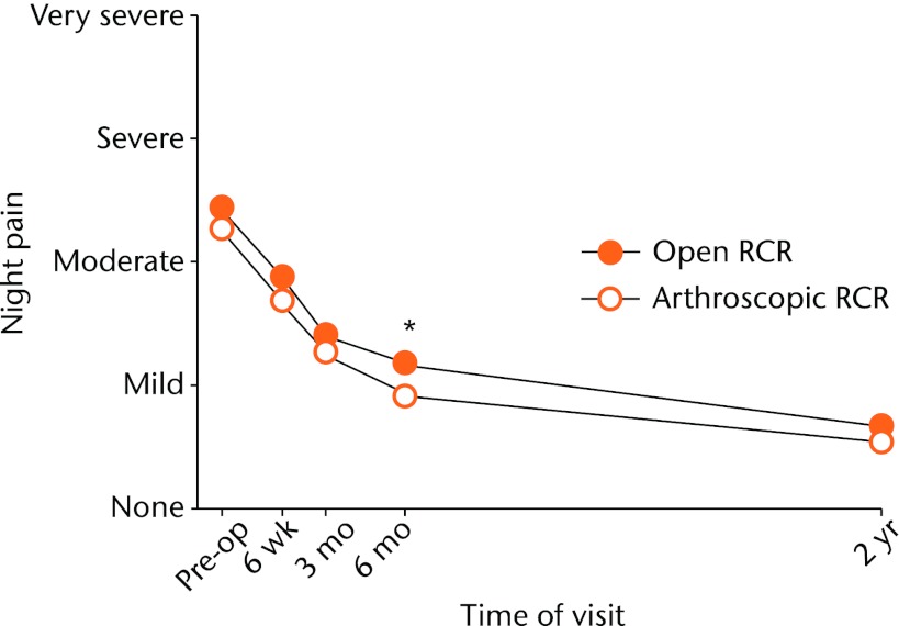
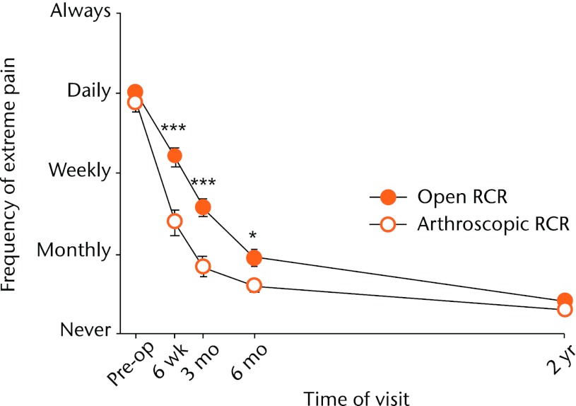
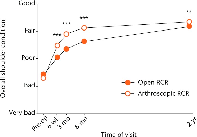
Pre-operative baseline data were similar for the two RCR groups for all patient-assessed questionnaire outcomes. Post-operative severity of pain during rest and activity, and frequency of pain at night and during activity, showed no significant difference between the RCR groups at any time-point. The only statistically significant differences in pain between the two RCR groups were for severity of night pain at six months (p = 0.012; Fig. 1) and frequency of extreme pain at three time-points (six weeks, p < 0.001; three months, p < 0.001; and six months, p = 0.011), all of which showed significantly more pain with open RCR (Fig. 2). The arthroscopic RCR patients rated the overall condition of their operated shoulder as significantly better than the open RCR patients at six weeks, three months and six months (all p < 0.001, Fig. 3), and also at the two-year follow-up visit (p = 0.004). Patients of both RCR groups assessed post-operative stiffness of their affected shoulder almost the same throughout their two-year follow-up period, the overall value declining from ‘moderate’ before surgery to less than ‘a little’ by their final visit.
Fig. 1.
Graph showing the mean patient-assessed severity of night pain for the open and arthroscopic rotator cuff repair (RCR) groups pre-operatively and at different post-operative time-points. There was a statistically significant difference between the groups at six months (* p = 0.012).
Fig. 2.
Graph showing the mean patient-assessed frequency of extreme pain for the open and arthroscopic rotator cuff repair (RCR) groups pre-operatively and at different post-operative time-points. Extreme pain was encountered significantly more frequently in the open RCR group at six weeks (*** p < 0.001), three months (*** p < 0.001) and six months (* p = 0.011).
Fig. 3.
Graph showing the mean patient-assessed overall shoulder condition for the open and arthroscopic rotator cuff repair (RCR) groups pre-operatively and at different post-operative time-points. The arthroscopic RCR cohort reported a significantly better overall condition at six weeks and three and six months (*** all p < 0.001), and also at two years (** p = 0.004).
Clinical examination
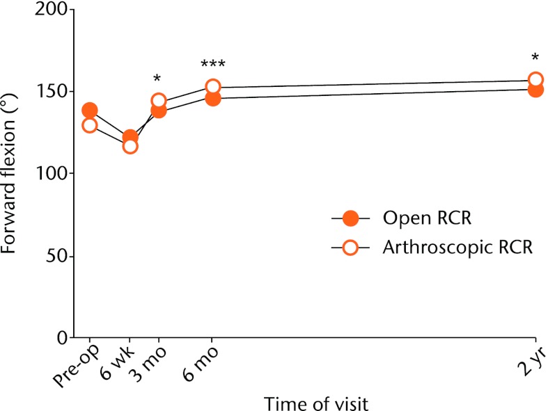
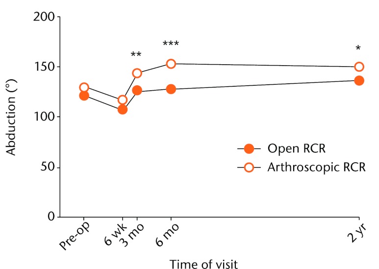
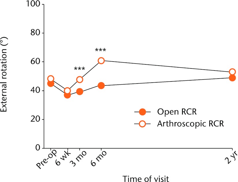
Pre-operative baseline values for the clinical measurements were similar for both RCR groups. The arthroscopic RCR group had significantly greater post-operative ROM in forward flexion, abduction and external rotation than the open RCR group (Figs 4 to 6). This was first apparent at three months (p = 0.020, p = 0.005 and p < 0.001, respectively) and the difference was larger at six months (all p < 0.001). At six months post-operatively, the arthroscopic RCR patients had, on average, 10° more forward flexion, 25° more abduction, and an additional 20° of external rotation than the open RCR patients. ROM measurements were still converging for the two groups by the time of the two-year follow-up. ROM measurements for internal rotation were almost the same for both groups at all time points.
Fig. 4.
Graph showing the mean forward flexion range of movement (ROM) for the open and arthroscopic rotator cuff repair (RCR) groups pre-operatively and at different post-operative time-points. The arthroscopic RCR group had a significantly greater forward flexion ROM at three months (* p = 0.020), six months (*** p < 0.001) and two years (* p = 0.046).
Fig. 5.
Graph showing the mean abduction range of movement (ROM) for the open and arthroscopic rotator cuff repair (RCR) groups pre-operatively and at different post-operative time-points. The arthroscopic RCR group had a significantly greater abduction at three months (** p = 0.005), six months (*** p < 0.001) and two years (* p = 0.010).
Fig. 6.
Graph showing the mean external rotation range of movement (ROM) for the open and arthroscopic rotator cuff repair (RCR) groups pre-operatively and at different post-operative time-points. The arthroscopic RCR group had a significantly greater external rotation ROM at three months and six months (*** both p < 0.001).
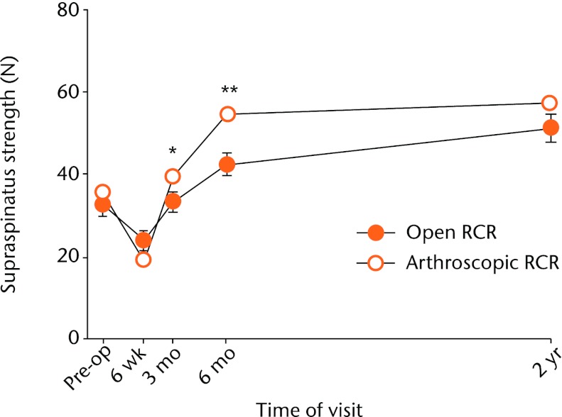
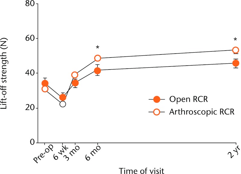
Dynamometer-assessed supraspinatus strength was significantly greater in the arthroscopic RCR group by three months (p = 0.045) and this difference increased further by the six-month follow-up (p = 0.005) (Fig. 7). At two years the mean supraspinatus strength remained comparatively higher in the arthroscopic RCR group, but this difference was no longer statistically significant (p = 0.145). The mean lift-off strength was similar in both groups until six months post-operatively, at which point the arthroscopic RCR group had significantly greater strength (p = 0.040). This difference was still significant at two years (p = 0.011) (Fig. 8). Adduction strength was higher at all time-points for the arthroscopic RCR group, but without reaching statistical significance (e.g., p = 0.201 at six months post-operatively).
Fig. 7.
Graph showing the mean supraspinatus strength for the open and arthroscopic rotator cuff repair (RCR) groups pre-operatively and at different post-operative time-points. The arthroscopic group had significantly greater strength at three (* p = 0.045) and six months (** p = 0.005). The error bars show the standard error of the mean.
Fig. 8.
Graph showing the mean lift-off strength for the open and arthroscopic rotator cuff repair (RCR) groups pre-operatively and at different post-operative time-points. The arthroscopic group had significantly greater strength at six months (* p = 0.040) and at two years (* p = 0.011). The error bars show the standard error of the mean.
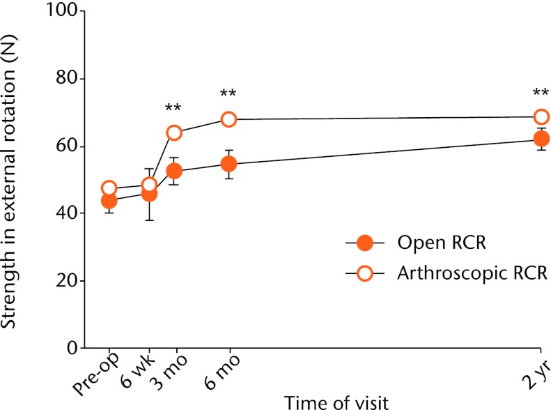
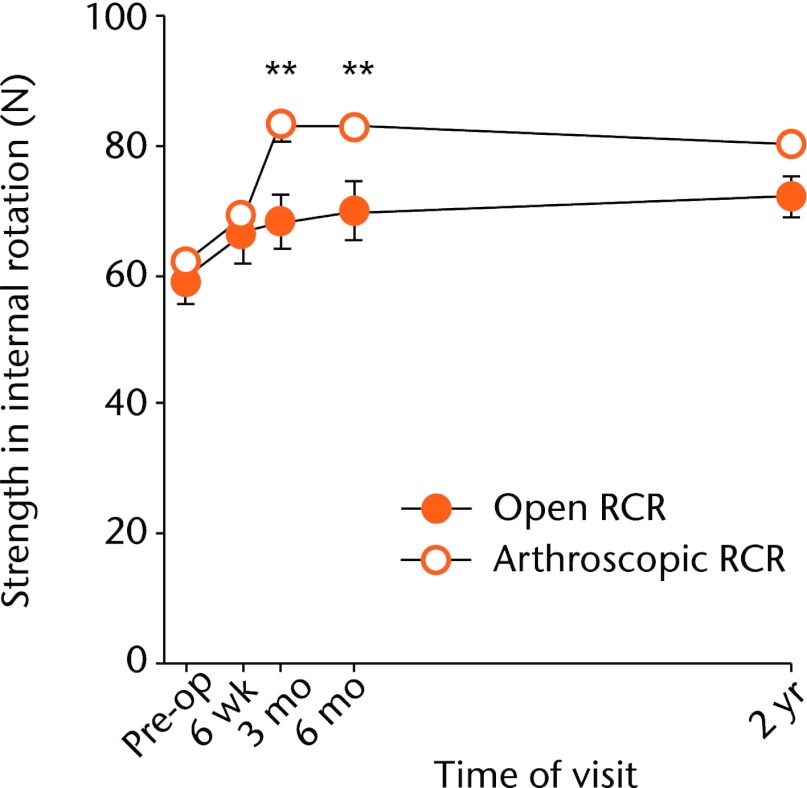
Measurements for strength in external rotation and internal rotation showed significantly greater increase for the arthroscopic RCR group at three and six months (Figs 9 and 10). At six months, the differences between the groups for mean strengths in external and internal rotation were 6 N (p = 0.008) and 12 N (p = 0.003), respectively. The difference for the two-year strength measurements remained higher for the arthroscopic RCR group but were only significant for strength in external rotation (p = 0.009).
Fig. 9.
Graph showing the mean strength in external rotation for the open and arthroscopic rotator cuff repair (RCR) groups pre-operatively and at different post-operative time-points. There was a significant difference between the groups at three months (** p = 0.008), at six months (** p = 0.006) and at two years (** p = 0.009).
Fig. 10.
Graph showing the mean strength in internal rotation for the open and arthroscopic rotator cuff repair (RCR) groups pre-operatively and at different post-operative time-points. There was a significant difference between the groups at three months (** p = 0.003) and six months (** p = 0.004).
Pre-operatively, 26 patients (13%) in the open RCR group and 20 (10%) in the arthroscopic RCR group demonstrated the drop-arm sign, which was not a statistically significantly difference (p = 0.433). At the two-year follow-up visit these numbers had decreased to ten patients (5%) in the open and four (2%) in the arthroscopic RCR groups, which again were not significantly different (p = 0.174). Approximately 150 patients (75%) of the open RCR group and 174 (87%) of the arthroscopic RCR group exhibited impingement pre-operatively, in either or both directions (p = 0.003). These proportions decreased to 46 (23%) for both the open RCR patients and the arthroscopic RCR patients at two years (p = 0.905).
The proportions of re-tear were 53% (n = 105 of 200) for the open RCR patients and 28% (n = 55 of 199) for the arthroscopic RCR patients (p < 0.001). The proportion of patients who re-tore with single row RCR did not significantly differ from those with double row repair (p = 0.262). The effect of the surgeon’s experience and overall learning curve approached but did not reach significance for either the open RCR group (p = 0.064) or the arthroscopic RCR group (p = 0.098).
A total of 14 patients (7%) in the open group underwent revision surgery for re-tear, compared with eight patients (4%) in the arthroscopy group. There were no other complications.
A multiple logistic regression analysis showed factors important to the integrity of the rotator cuff repair. When the program was run using re-tear as the dependent variable, and size of the original tear, age, gender, operative technique (open versus arthroscopic RCR), duration of symptoms and operative time as dependent variables, the rate of re-tear was found to mainly depend on the pre-surgical tear size (p < 0.001) and the operative technique (p = 0.007). Age (p = 0.055), gender (p = 0.377), duration of symptoms (p = 0.579) and operating time (p = 0.231) were less relevant to the re-tear rate. With open RCR assigned the value “1” and arthroscopic RCR the value “0”, the final multiple logistic regression equation is:
Logit P = -0.263 + (0.248 × tear-size) - (0.774 × operative technique)
Discussion
Some shoulder surgeons with extensive experience in arthroscopic RCR have stated their impression that arthroscopy provides less pain and earlier recovery of strength and ROM.9 The present study found that extreme pain was less frequent in arthroscopic RCR patients than open RCR patients and night pain was significantly greater in the open group at six months post-surgery. However, both groups exhibited a similar time course for resolution of all other pain types studied. Patient-based assessment of their overall shoulder condition was also significantly better in the arthroscopic RCR group.
Decreased post-operative stiffness is reported as an advantage of arthroscopic RCR.8 In the two patient groups, perceptions of shoulder stiffness were similar at all time points yet the clinical examinations revealed that shoulders of arthroscopic RCR patients had significantly greater passive ROM than those of open RCR patients at three and six months post-surgery. The arthroscopic group also showed earlier improvements in strength, achieving near maximal recovery by six months whereas the open RCR group continued to show some strength deficits at the two-year follow-up, confirming impressions that arthroscopic RCR allows earlier recovery of strength and ROM.
Buess et al7 estimated the time needed to be pain-free took “roughly” three months in both of their arthroscopic and open RCR patient groups (based on mailed questionnaires at between 15 and 40 months post-operatively) and surmised that three months is probably the time required for tendon repair to bone. Our results, however, indicate that pain is still diminishing and strength and movement are continuing to improve at three months.
The present study has potential advantages over others, such as the large group sizes with all surgery performed by the same surgeon. Two highly experienced sonographers performed all of the ultrasound assessments at two years. The patients had a wide range of pre-operative tear sizes, measuring up to 16 cm2. Many surgical practices have a significant proportion of patients with insurance claims related to their shoulder damage so this category of patients was also included in both RCR groups. Most other published comparisons of open and arthroscopic RCR techniques relate the pre-operative status of patients against their post-operative status at a single post-operative time point.7,11,12 A theoretical advantage of the present study is that the senior author’s practice systematically collects the same questionnaire and clinical data at set intervals of follow-up, thus documenting patient progression towards pain alleviation and functional recovery.
The difference in median operating times required for arthroscopic versus open RCR (40 minutes versus 60 minutes) was surprising in view of the greater technical difficulty of arthroscopic repair. The present study suggests that greater arthroscopic experience can reduce the operating time for arthroscopic RCR to a significantly shorter time than that required for open RCR.
A limitation of the present study is that the open RCR group preceded the arthroscopic RCR group in time, rather than as contemporary randomised cohorts. As a result, the first 130 open RCR patients were strength-tested with manual muscle tests before the surgeon’s institution converted to routine use of dynamometer-based strength measurements in order to provide more quantitative and objective strength measurements. Therefore, the 70 open RCR patients whose strength measurements were performed with hand-held dynamometry were compared with the 200 arthroscopic RCR patients that all had dynamometry data. Nevertheless, both group sizes were large and the numbers were sufficient to reveal significant differences between the two RCR groups. The increases in strength measurements are relatively small, in the order of 0.7 kg (1.5 lbs), but we regard this as clinically significant.
Another potential limitation concerns post-operative care for the two groups. The arthroscopic RCR group wore a small abduction pillow with their sling whereas the open RCR group did not. In other respects, post-operative conditions were the same for both RCR groups, including their exercise regimes. A number of authors have evaluated the benefit or otherwise of acromioplasty during RCR, and have concluded it provides no additional benefit over RCR alone.21,22
Results from the present study indicate that arthroscopic RCR used by an experienced surgeon can provide more secure repairs that are less prone to re-tear. Despite the greater technical difficulty of arthroscopic RCR, its results are equal to or better than those of the open procedure, even relatively early in the learning curve.7
Conclusions
The findings presented here show that arthroscopic RCR is associated with less extreme post-operative pain as well as earlier return of strength and ROM, thus providing quicker recovery and rehabilitation than that offered by the open procedure. Integrity of repair, as assessed by re-tear rates, was found to depend on both the pre-surgical tear size and the operative technique, in that order of importance. The operating time was significantly longer for open RCR than for arthroscopic RCR. Moreover, patients who had arthroscopic RCR rated their overall shoulder condition significantly better than those repaired with open RCR throughout the two-year follow-up period.
Funding Statement
No outside funding was received for this study.
Footnotes
The authors would like to thank L. Briggs and R. Tantau for their expert musculoskeletal ultrasonography.
Author contributions:J. R. Walton: Study design, Data analysis, Writing the paper
G. A. C. Murrell: Study design, Performed all surgeries, Data collection
ICMJE Conflict of Interest:None declared
References
- 1.Codman EA. Complete rupture of the supraspinatus tendon: operative treatment with report of two successful cases. Boston Med Surg J 1911;164:708–710 [DOI] [PubMed] [Google Scholar]
- 2.Snyder SJ. Arthroscopic evaluation and treatment of the rotator cuff. In:. Shoulder arthroscopy . New York: McGraw-Hill, 1994:133–178 [Google Scholar]
- 3.Norberg FB, Field LD, Savoie FH. Repair of the rotator cuff: mini-open and arthroscopic repairs. Clin Sports Med 2000;19:77–99 [DOI] [PubMed] [Google Scholar]
- 4.Churchill RS, Ghorai JK. Total cost and operating room time comparison of rotator cuff repair techniques at low, intermediate, and high volume centers: miniopen versus all-arthroscopic. J Shoulder Elbow Surg 2010;19:716–721 [DOI] [PubMed] [Google Scholar]
- 5.Schneeberger AG, von Roll A, Kalberer F, Jacob HA, Gerber C. Mechanical strength of arthroscopic rotator cuff repair techniques: an in vitro study. J Bone Joint Surg [Am] 2002;84-A:2152–2160 [DOI] [PubMed] [Google Scholar]
- 6.Sauerbrey AM, Getz CL, Piancastelli M, et al. Arthroscopic versus mini-open rotator cuff repair: a comparison of clinical outcome. Arthroscopy 2005;21:1415–1420 [DOI] [PubMed] [Google Scholar]
- 7.Buess E, Steuber KU, Waibl B. Open versus arthroscopic rotator cuff repair: a comparative view of 96 cases. Arthroscopy 2005;21:597–604 [DOI] [PubMed] [Google Scholar]
- 8.Yamaguchi K, Levine WN, Marra G, et al. Transitioning to arthroscopic rotator cuff repair: the pros and cons. Instr Course Lect 2003;52:81–92 [PubMed] [Google Scholar]
- 9.Gartsman GM, Khan M, Hammerman SM. Arthroscopic repair of full-thickness tears of the rotator cuff. J Bone Joint Surg [Am] 1998;80-A:832–840 [DOI] [PubMed] [Google Scholar]
- 10.Tauro JC. Arthroscopic rotator cuff repair: analysis of technique and results at 2- and 3-year follow-up. Arthroscopy 1998;14:45–51 [DOI] [PubMed] [Google Scholar]
- 11.Ide J, Maeda S, Takagi K. A comparison of arthroscopic and open rotator cuff repair. Arthroscopy 2005;21:1090–1098 [DOI] [PubMed] [Google Scholar]
- 12.Bishop J, Klepps S, Lo IK, et al. Cuff integrity after arthroscopic versus open rotator cuff repair: a prospective study. J Shoulder Elbow Surg 2006;15:290–299 [DOI] [PubMed] [Google Scholar]
- 13.Millar NL, Wu X, Tantau R, Silverstone E, Murrell GA. Open versus two forms of arthroscopic rotator cuff repair. Clin Orthop Relat Res 2009;467:966–978 [DOI] [PMC free article] [PubMed] [Google Scholar]
- 14.Gartsman GM, Hammerman SM. Full-thickness tears: arthroscopic repair. Orthop Clin North Am 1997;28:83–98 [DOI] [PubMed] [Google Scholar]
- 15.Bryant L, Shnier R, Bryant C, Murrell GA. A comparison of clinical estimation, sonography, magnetic resonance imaging, and arthroscopy in determining the size of rotator cuff tears. J Shoulder Elbow Surg 2002;11:219–224 [DOI] [PubMed] [Google Scholar]
- 16.Cummins CA, Strickland S, Appleyard RC, et al. Rotator cuff repair with bioabsorbable screws: an in vivo and ex vivo investigation. Arthroscopy 2009;19:239–248 [DOI] [PubMed] [Google Scholar]
- 17.Wu XL, Baldwick C, Briggs L, Murrell GA. Arthroscopic undersurface rotator cuff repair. Techn Shoulder Elbow Surg 2009;10:112–118 [Google Scholar]
- 18.L’Insalata JC, Warren RF, Cohen SB, Altchek DW, Peterson MG. A self-administered questionnaire for assessment of symptoms and function of the shoulder. J Bone Joint Surg [Am] 1997;79-A:738–748 [PubMed] [Google Scholar]
- 19.Hayes K, Walton JR, Szomor ZR, Murrell GA. Reliability of five methods for assessing shoulder range of motion. Aust J Physiother 2001;47:289–294 [DOI] [PubMed] [Google Scholar]
- 20.Hayes K, Walton JR, Szomor ZL, Murrell GA. Reliability of 3 methods for assessing shoulder strength. J Shoulder Elbow Surg 2002;11:33–39 [DOI] [PubMed] [Google Scholar]
- 21.Gartsman GM. Arthroscopic acromioplasty for lesions of the rotator cuff. J Bone Joint Surg [Am] 1990;72-A:169–180 [PubMed] [Google Scholar]
- 22.MacDonald P, McRae S, Leiter J, Mascarenhas R, Lapner P. Arthroscopic rotator cuff repair with and without acromioplasty in the treatment of full-thickness rotator cuff tears: a multicenter, randomized controlled trial. J Bone Joint Surg [Am] 2011;93-A:1953–1960 [DOI] [PubMed] [Google Scholar]


