Abstract
It can be difficult to appreciate just how small bacteria and phages are or how large, in comparison, the volumes that they occupy. A single milliliter, for example, can represent to a phage what would be, with proper scaling, an “ocean” to you and me. Here I illustrate, using more easily visualized macroscopic examples, the difficulties that a phage, as a randomly diffusing particle, can have in locating bacteria to infect. I conclude by restating the truism that the rate of phage adsorption to a given target bacterium is a function of phage density, that is, titer, in combination with the degree of bacterial susceptibility to adsorption by an encountering phage.
Keywords: mean free path, phage adsorption, phage therapy
Bacteria and phages are sufficiently small that their sizes effectively are abstractions for those of us more familiar with the “macro” world that we experience immediately around us, that is, rather than the “micro” world found under a light or electron microscope. Thus, if a dimension of a bacterium is 1 μm, then what, intuitively, does that mean? What about a phage with a dimension, for example, of 100 nm? A simplifying trick used by physicists is to assume that objects are spherical, even if they really are not. This is done, for example, when making calculations of the mean free path of molecules making up an ideal gas. Key especially is to calculate the collision cross section, here of area π(D/2),2 where D is the diameter of the sphere in question.1 D for a typical bacterial cell is 1 μm while D for a typical tailed phage is 100 nm. In meters, these are 10−6 and 10−7, respectively. A standard means of presenting phage adsorption rate constants is in terms of the likelihood of collision of one phage with one bacterium when both are suspended in 1 ml of fluid. A milliliter fills a volume that is a cube of the dimension, 1 cm, where 1 cm is 10−2 meters. The diameter of a bacterium is 1/10,000 that of 1 cm while phages are a further 10-fold smaller.
Hagens and Loesner2 provide a striking visualization of the relative sizes of phages and bacteria by scaling these organisms up to more familiar sizes, particularly considering phages to be the size of apples and bacteria the size of humans. Here I revisit these ideas. For the sake of simplicity, I base my calculations on linear dimensions, 10−2, 10−6 and 10−7 meters, i.e., as just indicated.
The typical human is approximately 1 m in diameter, if visualized as a sphere or about the size of a large beach ball. That provides a volume that is in the range of 10-fold too great: ~500 L vs. a more reasonable 50 to 100 L. However, in terms of cross section, which is a better measure of target size than volume, a one-meter-diameter sphere is less erroneous. One meter, as indicated, is approximately one-million-fold larger than the size of a typical bacterium.
Similarly scaled up, a typical phage would have a diameter of approximately 10 cm, or one-tenth the human diameter. Ten centimeters is about the diameter of a softball, which are approximately 30 cm in circumference depending on league, or the diameter of a rather large apple. This is one-million-fold larger than the actual phage diameter that we are using of 100 nm. One milliliter, in turn, would therefore occupy a cube that was one-million times longer in one dimension than 1 cm, with one million centimeters equal to 10,000 m or 10 km. The volume of 1 ml that has been expanded to 10 km on a side is 103 km3. These various diameters and volumes are summarized in Table 1.
Table 1. Scaling of diameters (D) and volumes (proportional to D3) of typical phage, typical bacterium, and one milliliter.
| Actual Measures | 106-fold Scaled-Up Measures | ||||||||
|---|---|---|---|---|---|---|---|---|---|
| |
D (nm) |
D (mm) |
D (cm) |
D (m) |
D3 (cm3) |
D (cm) |
D (m) |
D (km) |
D3 (m3) |
| Phage |
102 |
10−1 |
10−5 |
10−7 |
10−15 |
101 |
10−1 |
10−4 |
10−3 |
| Bacterium |
103 |
100 |
10−4 |
10−6 |
10−12 |
102 |
100 |
10−3 |
100 |
| Milliliter* | 107 | 104 | 100 | 10−2 | 100 | 106 | 104 | 101 | 1012 |
As measured assuming cubic in shape.
How large is 1,000 km3? Given that I am writing from a location that is roughly 100 km south of Cleveland, Ohio, I am compelled to answer that it is about twice the volume of Lake Erie (480 km3), and over 100-times the volume of Loch Ness (7.4 km3). Note that closer in volume to 1,000 km3 are Lake Ladoga in Russia, which at 837 km3 is the largest lake in Europe, and Lake Titicaca, which is shared by Bolivia and Peru and is the largest lake in South America (893 km3).
If we were to scale up that phage another 10-fold, to the size of you and me, then instead of a 1,000 km3 volume, our milliliter would be a cube that is 100 km on a side, or one million km3. This is not quite an ocean at less than one hundredth of the volume of the Arctic Ocean, Earth’s smallest (~107 km3). Still, it is rather substantial at about ten times the volume of the world’s largest inland body of water, the Caspian Sea (6.9 × 10⁴ km3).
For phages seeking out bacteria, all movement would be done under water and the totally of the volume is explored via the equivalent of a drunkard’s “walk.” In fact, this exploration is accomplished, essentially, by searchers that are “blindfolded” since phages have available to them only a sense of touch to detect the presence of other objects (or, more correctly stated, phages have a sense of taste since it is direct interactions within an aqueous environment between chemicals and phage proteins that is being perceived). Then again, fluid flow on microscales can be quite a bit faster than one normally observes if scaling up to kilometers. This means that mixing should speed up phage movement through 1 ml quite a bit more than is conventionally possible through the volume of a very large lake.
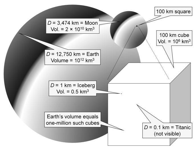
As an alternative analogy to that of apples, large beach balls and Lake Titicaca, note that the RMS Titanic could be viewed as a roughly 100 m-diameter sphere (102 m). If said sphere were to strike a kilometer-wide iceberg (103 m), which is large but certainly not unprecedented, then how big would a similarly scaled up ocean be? One kilometer is 109-fold longer than 1 μm (iceberg vs. bacterial cell). This implies a cube that is 1013 μm on each side (ocean vs. ml), or 10,000 km. (1013 is calculated as follows: 109 is the ratio of 1 km to the diameter of a typical bacterium, i.e., 103 m vs. 10−6 m; 10−2 is the ratio of the size of a meter to the size of 1 cm, with the latter defining the volume of 1 ml; 10-6 refers to the diameter of a bacterium in meters, that is, which is being scaled up to a kilometer-sized iceberg, where 10−6 m is 1/10,000th = 10−4 the length of 1 cm; thus, 109 × 10−2/10−6 = 109 × 104 = 1013 μm = 104 km). 104 km squared is 108 km2, which is roughly the surface area of the Atlantic Ocean. Of course relative motion in this example, as depicted so far, occurs in only two rather than three dimensions, meaning that the likelihood of contact between Titanic and iceberg would be grossly overestimated. Indeed, if the Titanic were a submarine, and ice had the same density as water, then the Atlantic Ocean would have to be ~10,000 km deep on average for the analogy of a phage and bacterium in 1 ml of fluid to hold, whereas the real Atlantic Ocean is shallower by 3–4 orders of magnitude (average depth ~3 km). Remarkably, 10,0003 = 1012, which in cubic kilometers is approximately the volume of the entire Earth (1.1 × 10¹² km3). See Figure 1 for summary.
Figure 1. Bacteria and phages as very small things. Shown is a profile of planet Earth as well as that of the moon, with diameters of about 1.3 × 10⁴ km and 3.5 × 10³ km, respectively (both are drawn to dimensional scale, that is, rather than in terms of linear perspective). Upon the moon is a 100 km-wide cube presented as a square. Below the moon sits a blow up representation of that cube, within which is a circle representing a 1 km diameter sphere, as equivalent to a moderately large iceberg. Also within this cube, but not visible, is a 0.1 km sphere (as equivalent to the Titanic). In terms of relative sizes, the Earth represents 1 ml of fluid, the larger sphere within the cube a bacterium (seen as the small dot), and the invisible sphere also found within the cube is a phage.
For a T-even phage, which adsorbs to E. coli B with a rate constant of approximately 2.5 × 10⁻⁹ ml/min,3 it would take on average the inverse of that number, i.e., 1/(2.5 × 10−9), before collision followed by infection would be expected to occur. This assumes that bacteria are non-motile, that phage movement occurs by diffusion only, and further that only a single phage along with one bacterium are present within 1 ml of fluid. The calculation involved is equivalent to that of the phage mean free time, 1/kN, where k is the phage adsorption rate constant and N is the bacterial concentration, except that with bacterial density equal to 1 per ml then N = 1.4 This mean free time thus works out to 4.0 × 10⁸ min, 6.7 × 10⁶ hours, 2.8 × 10⁵ days, 4.0 × 10⁴ weeks, 9.3 × 10³ 30-d months or 761 y before one adsorption would, on average, be expected to occur. In other words, approximately three adsorptions would be predicted to have taken place—adding phages as they are lost, disregarding phage as well as bacterial replication, and assuming multiple adsorption to that single bacterium—since the time of Aristotle (considered by many to be the first biologist). About 3 × 10⁵ adsorptions will occur for every trip the Sun makes around the Milky Way (one galactic or cosmic year, is equal to about 225 to 250 million years) while animals have been present on Earth for only about the last three such trips.5 Saltzman6 similarly calculates that the protein albumin, which has a diameter in the range of 6 nm, vs. 10-fold larger or more for most tailed phages, could take, on average, 800 y to traverse two meters by diffusion alone. See the appendix to Goodridge7 for comparable considerations as applied to phages.
Bottom line: The collision of individual phages with individual bacteria is not a trivial process, unless the volumes within which phages are diffusing are greatly constrained. The latter, though, is just another way of stating that, for a given density of bacteria, the greater the density of phages present then the faster that bacteria will be adsorbed and, given infection by obligately lytic phages, subsequently die. The progress of bactericidal activity during phage therapy8-10 thus is limited explicitly by the density that phages are able to achieve within the immediate vicinity of target bacteria in combination with the susceptibility of target bacteria to phage adsorption and subsequent phage-mediated killing. Again, if a phage had the cross section of the RMS Titanic and a bacterium that of a moderately large iceberg, then the likelihood of phage collision with bacterium, per unit time, would occur as though an ml filled the volume of our entire planet.
Acknowledgments
Thank you to Cameron Thomas-Abedon and Rodney Michael who read and commented on the manuscript.
Footnotes
Previously published online: www.landesbioscience.com/journals/bacteriophage/article/17281
References
- 1.Noggle JH. Physical Chemistry. Glenview, Illinois: Scott, Foresman and Co., 1989 [Google Scholar]
- 2.Hagens S, Loessner MJ. Bacteriophage for biocontrol of foodborne pathogens: calculations and considerations. Curr Pharm Biotechnol. 2010;11:58–68. doi: 10.2174/138920110790725429. [DOI] [PubMed] [Google Scholar]
- 3.Stent GS. Molecular Biology of Bacterial Viruses. San Francisco, CA: WH Freeman and Co., 1963 [Google Scholar]
- 4.Abedon ST, Herschler TD, Stopar D. Bacteriophage latent-period evolution as a response to resource availability. Appl Environ Microbiol. 2001;67:4233–41. doi: 10.1128/AEM.67.9.4233-4241.2001. [DOI] [PMC free article] [PubMed] [Google Scholar]
- 5.Merril CR. Interaction of bacteriophages with animals. In: Abedon ST, ed. Bacteriophage Ecology. Cambridge, UK: Cambridge University Press, 2008:332-52 [Google Scholar]
- 6.Saltzman WM. Tissue Engineering: Principles for the Design of Replacement Organs and Tissues. Oxford: Oxford University Press, 2004 [Google Scholar]
- 7.Goodridge LD. Phages, bacteria, and food. In: Abedon ST, ed. Bacteriophage Ecology. Cambridge, UK: Cambridge University Press, 2008:302-31 [Google Scholar]
- 8.Abedon ST. The ‘nuts and bolts’ of phage therapy. Curr Pharm Biotechnol. 2010;11:1. doi: 10.2174/138920110790725438. [DOI] [PubMed] [Google Scholar]
- 9.Abedon ST, Kuhl SJ, Blasdel BG, Kutter EM. Phage treatment of human infections. Bacteriophage. 2011;1:66–85. doi: 10.4161/bact.1.2.15845. [DOI] [PMC free article] [PubMed] [Google Scholar]
- 10.Loc-Carrillo C, Abedon ST. Pros and cons of phage therapy. Bacteriophage. 2011;1:111–4. doi: 10.4161/bact.1.2.14590. [DOI] [PMC free article] [PubMed] [Google Scholar]


