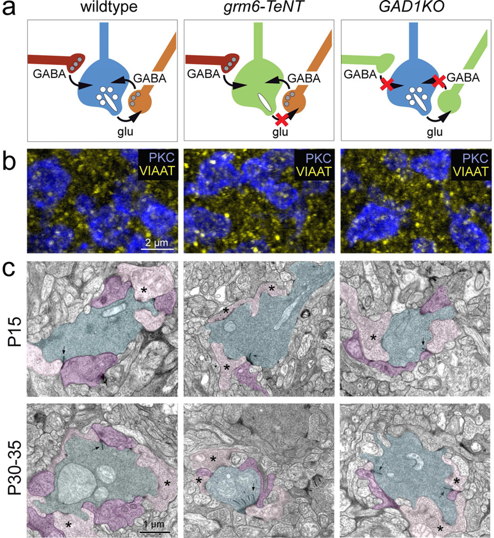
Figure 2. Inhibitory synapses onto RBC axons in the grm6-TeNT and GAD1KO retina.
(a) Schematic showing RBC axon terminal circuitry in wildtype and transgenic lines with suppressed glutamatergic transmission from ON-bipolar cells (grm6-TeNT) and reduced GABAergic transmission from amacrine cells (GAD1KO). (b) Immunolabeling for VIAAT (yellow) at the level of PKC positive RBC boutons (blue) in P30 wildtype, grm6-TeNT and GAD1KO retina. (c) Electron micrographs showing dyad synapses between RBC boutons (cyan) and AII (purple) and A17 (pink) amacrine cell processes in the wildtype (WT), grm6-TeNT and GAD1KO retinae at P15 and P30–35. Presynaptic ribbons are indicated by arrows. Asterisks mark examples of A17 contact. Note that RBCs in the P30 grm6-TeNT retina have multiple ribbons at a single dyad synapse.

