
Figure 7.
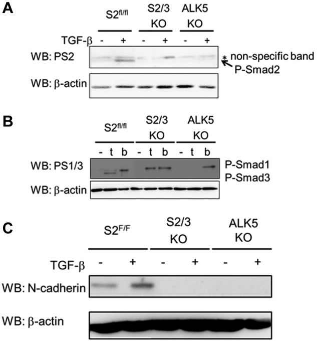
TGF-β/Smad2/3 signaling induces N-cadherin expression. (A) TGF-β–induced Smad2 phosphorylation in ECs. MEECs were stimulated with TGF-β for 1 hour. The filters were incubated with anti-phosphorylated Smad2 (PS2; top panel) and anti–β-actin antibodies (bottom panel). P-Smad2, phosphorylated Smad2. (B) Detection of phosphorylated Smad1 and phosphorylated Smad3 on TGF-β (t) or BMP (b) stimulation. MEECs were stimulated with TGF-β or BMP6 for 1 hour. The filters were incubated with anti-phosphorylated Smad1/3 (PS1/3; top panel), which specifically recognize phosphorylated Smad1 and Smad3, and anti–β-actin antibodies (bottom panel). P-Smad1 indicates phosphorylated Smad1; and P-Smad3, phosphorylated Smad3. (C) TGF-β–induced N-cadherin expression in MEECs. MEECs were stimulated with TGF-β for 48 hour. The filters were incubated with anti–N-cadherin (top panel) and anti–β-actin antibodies (bottom panel).