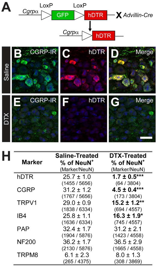
Figure 1. Conditional ablation of peptidergic DRG neurons in adult CGRPα-DTR+/− mice.
(A) Advillin-Cre was used to remove floxed GFP and drive selective expression of hDTR in CGRPα-expressing sensory neurons. (B–G) Lumbar DRG from CGRPα-DTR+/− mice treated with saline or DTX were stained with antibodies to (B,E) CGRP, (C,F) hDTR and (B–G) NeuN (blue). Images were acquired by confocal microscopy. Scale bar in (G) is 50 μm. See Figure S1 for additional markers. (H) Sensory neuron marker quantification, as percentage relative to total number of NeuN+ neurons. Cell counts are from representative sections of L3–L6 ganglia (8 representative sections per animal, with n=3 animals per treatment group). The number of neurons expressing each marker is shown in parentheses. All values are means ± SEM. *p<0.05, **p<0.005, ***p<0.0005. (B–H) Tissue was collected 7 days after second saline/DTX injection.

