Abstract
Adult neurogenesis in the hippocampal subgranular zone (SGZ) and the anterior subventricular zone (SVZ) is regulated by multiple factors, including neurotransmitters, hormones, stress, aging, voluntary exercise, environmental enrichment, learning, and ischemia. Chronic treatment with selective serotonin reuptake inhibitors (SSRIs) modulates adult neurogenesis in the SGZ, the neuronal area that is hypothesized to mediate the antidepressant effects of these substances. Layer 1 inhibitory neuron progenitor cells (L1-INP cells) were recently identified in the adult cortex, but it remains unclear what factors other than ischemia affect the neurogenesis of L1-INP cells. Here, we show that chronic treatment with an SSRI, fluoxetine (FLX), stimulated the neurogenesis of γ-aminobutyric acid (GABA)ergic interneurons from L1-INP cells in the cortex of adult mice. Immunofluorescence and genetic analyses revealed that FLX treatment increased the number of L1-INP cells in all examined cortical regions in a dose-dependent manner. Furthermore, enhanced Venus reporter expression driven by the synapsin I promoter demonstrated that GABAergic interneurons were derived from retrovirally labeled L1-INP cells. In order to assess if these new GABAergic interneurons possess physiological function, we examined their effect on apoptosis surrounding areas following ischemia. Intriguingly, the number of neurons expressing the apoptotic marker, active caspase-3, was significantly lower in adult mice pretreated with FLX. Our findings indicate that FLX stimulates the neurogenesis of cortical GABAergic interneurons, which might have, at least, some functions, including a suppressive effect on apoptosis induced by ischemia.
Keywords: adult neurogenesis, antidepressant, interneuron, neocortex, neural progenitor, stroke
INTRODUCTION
γ-Aminobutyric acid (GABA) is a widespread inhibitory neurotransmitter in the brain. Excitation in the brain must be balanced with inhibition, which allows fine-tuning of our moods, thoughts, and actions. An imbalance between excitation and inhibition may be involved in the onset of ischemic excitotoxicity (Blaesse et al, 2009) as well as psychiatric disorders, such as schizophrenia, bipolar disorder, major depressive disorder, and anxiety disorder (Luscher et al, 2011). One subset of interneurons, generated from Layer 1 inhibitory neuron progenitor cells (L1-INP cells) after transient global forebrain ischemia, are GABAergic (Ohira et al, 2010), and therapies using L1-INP cell-derived GABAergic interneurons may be useful in the treatment of cortical GABA-related diseases. However, it is currently unclear what additional factors regulate the proliferation and neurogenesis of L1-INP cells, and the identification and characterization of these factors could be clinically important.
Selective serotonin reuptake inhibitors (SSRIs) are possible regulators of L1-INP cell neurogenesis. SSRIs, which are widely used to treat depression, mood, and anxiety disorders, have been extensively studied in the regulation of subgranular zone (SGZ) and subventricular zone (SVZ) neurogenesis. Chronic administration of the SSRI fluoxetine (FLX) increases neurogenesis in the hippocampal dentate gyrus (DG) (Kodama et al, 2004; Malberg et al, 2000; Santarelli et al, 2003). In contrast, neurogenesis in the SVZ of adult rodents is not measurably affected by FLX treatments for 3–4 weeks (Kodama et al, 2004; Malberg et al, 2000; Santarelli et al, 2003), but administration of FLX for >6 weeks adversely affects neurogenesis (Ohira and Miyakawa, 2011). Additionally, FLX reverses the established mature state of granule cells, designated as ‘dematuration,' in the adult hippocampus (Kobayashi et al, 2010, 2012; Ohira and Miyakawa, 2011). These findings led us to examine whether SSRI treatments affect the proliferation and neurogenesis of L1-INP cells.
MATERIALS AND METHODS
Animals and Antidepressant Treatment
Adult male C57BL/6J mice (Charles River Laboratories Japan, Yokohama, Japan) (3 months old at the start of our experiments) were used. Fluorescent ubiquitination-based cell-cycle indicator (Fucci) mice (line #596/#504) (Sakaue-Sawano et al, 2008a, 2008b) were purchased from Amalgaam (Tokyo, Japan) and maintained using standard husbandry procedures. All animal experiments were approved by the Institutional Animal Care and Use Committee of Fujita Health University, based on the Law for the Humane Treatment and Management of Animals (2005) and the Standards Relating to the Care and Management of Laboratory Animals and Relief of Pain (2006). Every effort was made to minimize the number of animals used.
FLX solution was intraperitoneally injected into mice every day for 3 weeks, and the appropriate FLX concentration was determined for each body weight. FLX pellet treatment was performed as described previously (Ohira and Miyakawa, 2011). The drug pellets contained 7.245 mg or 20.7 mg FLX; these dosages were calculated so that a mouse with a body weight of 23 g received FLX at 15 mg/kg/day for 21 days or 60 days, respectively (Innovative Research of America, Sarasota, FL). Pellets without FLX were administered to control mice. FLX treatments of 15 mg/kg/day for 21 days inhibited the decrease in latency to fall in the rotarod test, suggesting that our FLX treatment protocol may lead to a behavioral recovery (Supplementary Figure S1).
BrdU Labeling
BrdU (Sigma-Aldrich, St Louis, MO) stock solution was prepared in phosphate-buffered saline (PBS; pH 7.2, 0.1 M) with 0.007 N NaOH at 20 mg/ml. The mice were injected intraperitoneally with BrdU (100 mg/kg body weight) every 24 h for the last 3 days of the FLX injection period to label L1-INP cells. Mice were then perfused for fixation 3 weeks after the last injection.
Retrovirus-Mediated Venus Labeling of Newly Generated Neurons
The retrovirus vector was derived from murine leukemia virus and constructed as follows. The pBluescript II SK (+) (BSII SK)-Esyn-GFP-WPRE construct (a gift from Dr Kaneko) (Hioki et al, 2007) was digested with HindIII and EcoRI. The Venus/pCS2 construct (a gift from Dr Miyawaki) was digested with the same set of restriction enzymes. The resulting Venus fragment was inserted into the same site of the pBSII SK-Esyn-WPRE fragment. The Esyn-Venus-WPRE sequence was amplified by PCR with the following primers: 5′-ACGTTAACTGCTCGAGGTTTTCCCAGTCACGACGTT-3′ and 5′-TTATTTTATCCTCGAGTGTGGAATTGTGAGCGGATA-3′ (complementary sequences of pDON-5 underlined). The PCR product of Esyn-Venus-WPRE with the pDON-5 complementary sequences in both the 5′ and 3′ termini was ligated using the In-Fusion HD Cloning Kit (Clontech-Takara Bio, Mountain View, CA) into pDON-5 that was previously cut with XhoI. The pDON-5 Esyn-Venus-WPRE construct was used for retrovirus production.
The retrovirus was produced according to the manufacturer's instructions accompanying the retrovirus packaging kit (Takara Bio, Otsu, Japan). The resulting viral particles in the culture supernatant were collected and adjusted to 1.0 × 10⁶ transducing units/ml.
The virus injection was performed as described previously (Ohira et al, 2010). Briefly, the virus solution (0.2 μl per site) was stereotaxically injected by air pressure through a glass micropipette attached to Picospritzer III (Parker, Cleveland, OH) into the mouse frontal (1.8–2.8 mm anterior to bregma, 1–2 mm lateral, and 0.2 mm depth below the cortical surface) or somatosensory region (1–2 mm anterior to bregma, 2–3 mm lateral, and 0.2 mm depth below the cortical surface). One hemisphere received a total of 5 μl of the virus solution (25 injection sites in 1.0 mm × 1.0 mm square). Two days after injection, the mice were treated with FLX and subsequently perfused after 1, 2, 4, or 8 weeks.
Arabinosylcytosine (Ara-C) Infusion and Ischemia
Mice were deeply anesthetized with chloral hydrate. Brain infusion cannulae (Brain Infusion Kit 3, Alzet, Cupertino, CA) were stereotaxically placed into the frontoparietal region of both hemispheres (1.5–2.0 mm anterior to bregma, 1.5 mm lateral, and 0.5 mm depth below the skull surface). Each cannula was connected to a micro-osmotic pump (1004, Alzet) that was filled with an Ara-C solution (2% w/v) containing Fast Green FCF (2% w/v, Sigma-Aldrich) or PBS (vehicle). Thus, one hemisphere received Ara-C solution for 6 weeks, and the other received PBS. FLX (15 mg/kg/day) was simultaneously administered at the onset of Ara-C treatment and continued for 2 weeks. After completing FLX treatment, mice were housed for 4 weeks to allow for the differentiation of new neurons from L1-INP cells. Global ischemia was induced as described previously (Ohira et al, 2010). Briefly, both common carotid arteries were transiently occluded with clamps for 10 min. The mice were killed 48 h later and then perfused.
Immunohistological Analysis
Fixation and immunofluorescent staining were performed as described previously (Ohira et al, 2010). A confocal laser-scanning microscope (LSM 700; Carl Zeiss, Oberkochen, Germany) was used to obtain images of the stained sections.
For BrdU staining, sections were incubated at 4 °C for 10 min in 0.1 N HCl and then at 37 °C for 30 min in 2 N HCl. Sections were washed twice for 5 min in PBS and then blocked in 0.2 M glycine in PBS at room temperature for at least 2 h.
To count the immunostained cells, the brains were coronally and sequentially cut from the olfactory bulb to the posterior region of the midbrain. After all sections were co-stained with the indicated antibodies, the total number of cells in each section was counted.
Sholl-like analysis was used to quantify the number of active caspase-3+ cells. First, the xy plane in the middle of each cell was defined. Next, the active caspase-3+ cell bodies that intersected with the concentric circles drawn around each Venus-expressing or control neuron were counted. The number of cells that were stained for active caspase-3 was then normalized by dividing the number of active caspase-3+ cell bodies by the circumference of each concentric circle. The same analysis was performed on cell nucleus images in the same regions to examine the uniform distribution of cells in the regions measured. We selected control cells that showed no active casapse-3 staining and were distributed in the same layers, areas, and locations in one hemisphere as Venus+ neurons in the other hemisphere. In addition, glial cells were omitted using both the morphology of their nuclei and staining of their cell bodies with anti-active-caspase-3 antibody and Hoechst 33258 dye in this quantification.
Antibodies
The following primary antibodies were used: mouse monoclonal antibodies for calbindin (1 : 2000, Sigma-Aldrich), calretinin (1 : 10000, Millipore, Billerica, MA), GAD67 (1 : 10000, Millipore), parvalbumin (1 : 2000, Sigma-Aldrich), and S100β (1 : 1000, Sigma-Aldrich); rat monoclonal antibody for BrdU (1 : 100, Abcam, Cambridge, MA); rabbit polyclonal antibodies for active caspase-3 (1 : 100, BD Pharmingen, San Jose, CA), GABA (1 : 1000, Sigma-Aldrich), Iba1 (1 : 200, Wako, Osaka, Japan), Ki67 (1 : 10, Ylem, Rome, Italy), Olig2 (1 : 200, Immuno-Biological Laboratories, Fujioka, Japan), NeuN (1 : 200, Millipore), neuropeptide Y (1 : 2000, Sigma-Aldrich), and somatostatin (1 : 2000, Bachem, Bubendorf, Switzerland); and guinea pig polyclonal antibodies for vesicular GABA transporter 1 (VGAT1, 1 : 200, Synaptic Systems, Göttingen, Germany), vesicular glutamate transporter 1 (VGLUT1, 1 : 200, Synaptic Systems), and VGLUT2 (1 : 200, Synaptic Systems). The following secondary antibodies were used: anti-mouse IgG Cy3 (1 : 200, Millipore), anti-mouse IgG Alexa Fluor 405 (1 : 200, Life Technology, Carlsbad, CA), anti-mouse IgG Alexa Fluor 488 (1 : 200, Life Technology), anti-mouse IgG Alexa Fluor 594 (1 : 200, Life Technology), anti-mouse IgG Alexa Fluor 647 (1 : 200, Life Technology), anti-rat IgG Alexa Fluor 594 (1 : 200, Life Technology), anti-rabbit IgG Alexa Fluor 350 (1 : 200, Life Technology), anti-rabbit IgG Alexa Fluor 488 (1 : 200, Life Technology), anti-rabbit IgG Alexa Fluor 594 (1 : 200, Life Technology), and anti-rabbit IgG Alexa Fluor 647 (1 : 200, Life Technology).
Quantification of Labeled Cells
A quantification analysis was performed as reported previously (Ohira et al, 2010). Slides were coded and quantified by blinded, independent observers.
Rotarod Test
Motor coordination and balance were assessed with the rotarod test using an accelerating rotarod (Accelerating Rotarod, UGO Basile, Comerio, Italy). Mice were placed on rotating drums (3 cm diameter), and the time (s) that each animal maintained its balance on the rod was measured. The speed of the rotarod accelerated from 4–40 r.p.m. over a 5-min period.
Statistical Analysis
Data were analyzed by Student's or Welch's t-test and one- or two-way ANOVA followed by Tukey's or Scheffé's post hoc test. If the underlying distributions were normally distributed with equal population variances, Student's t test was used to determine whether two independent groups differed. If the distributions of data were not normal, Welch's t test was employed. Error bars indicate SEM.
RESULTS
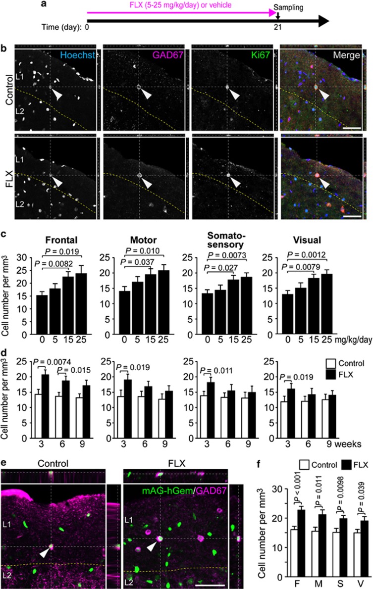
In the adult rat cortex, L1-INP cells are characterized by double staining for Ki67, which is expressed during late-G1/S/G2/M phases of the cell cycle (Scholzen and Gerdes, 2000), and glutamic acid decarboxylase 67 (GAD67), which is expressed in neural stem cells and neural progenitor cells (NPCs) of the SGZ and SVZ (Ohira et al, 2010; Stewart et al, 2002), as well as in GABAergic neurons. Because L1-INP cells have been reported in adult rats but not in other mammals, we first confirmed the presence of L1-INP cells in adult mice. Using anti-Ki67 and anti-GAD67 antibodies, we detected Ki67+/GAD67+ cells (L1-INP cells) in most cortical regions of adult mice (Figure 1b). Next, we examined the reactivity of L1-INP cells to FLX and found good linearity between the number of L1-INP cells and FLX concentration in the range of 5–25 mg/kg/day after 3 weeks of FLX treatment (Figure 1c: frontal, F(3,76)=3.06, p=0.033; motor, F(3,92)=5.34, p=0.019; somatosensory, F(3,92)=3.08, p=0.031; visual, F(3,100)=4.17, p=0.0079; n=5 each). We also performed a time-course analysis of the FLX responsiveness of L1-INP cells. In all the regions examined, the number of L1-INP cells was significantly increased after 3 weeks of FLX administration, and the rates of increase gradually declined at 6 and 9 weeks (Figure 1d, n=5 each).
Figure 1.
Increase in the number of L1-INP cells by FLX treatment in adult mice. (a) Summary of the time course of experiments. (b) The xy, xz, and yz planes showing co-localization of L1-INP cell markers, GAD67 and Ki67, in control and FLX-treated mice. The arrowheads indicate the same cells in each row. The cell images were taken from the frontal cortex of control (upper row) and FLX-treated mice (lower row) after 3 weeks of FLX treatment (15 mg/kg/day). L, layer. (c) Concentration dependence of L1-INP cells on 3-week FLX treatment (15 mg/kg/day; one-way ANOVA and Tukey's post hoc test, n=5 each). (d) Time-course analysis of the responsiveness of L1-INP cells to FLX (for each time point; Student's or Welch's t tests, n=5 each). Mice were treated with FLX for each period. (e, f) Detection of L1-INP cells in the cortex of adult Fucci transgenic mice and the increase in cell number induced by FLX (Welch's t test, n=6 each). Green signals, which indicate the expression of mAG-hGem, are derived from the Fucci-S/G2/M marker, while the magenta signals indicate GAD67+ structures. L1-INP cells are indicated by arrowheads. Mice were treated with FLX for 3 weeks. F, frontal; M, motor; S, somatosensory; V, visual. All data represent the mean±SEM. Scale bars, 50 μm.
Among cortical regions, the increase in the number of L1-INP cells was most prominent in the frontal cortex, with a significant increase at 6 weeks. In addition, we found no significant sex-specific differences in the number of L1-INP cells detected in any cortical region in mice treated with FLX for 3 weeks (Supplementary Figure S2). To examine the dose dependency and time course of the L1-INP cell response to FLX, we intraperitoneally injected FLX solutions or subcutaneously embedded FLX pellets into the mice and found no significant differences between treatment methods (FLX treatment at 15 mg/kg/day for 3 weeks in the frontal cortex is displayed in Figure 1c and d; p=0.50, n=5 each, Student's t test).
In the above experiments, Ki67 was used as a cell proliferation marker. Although the Ki67 antigen is strictly associated with cell proliferation, its functional role in cell proliferation is still unclear (Scholzen and Gerdes, 2000). To further examine the proliferation of L1-INP cells, we used another established method of cell-cycle detection (Sakaue-Sawano et al, 2008a, 2008b), the transgenic (Tg) mouse line expressing Fucci. In the Fucci mouse, monomeric Azami Green fluorescent protein is fused to a fragment of human Geminin (mAG-hGem) and labels the nuclei of S/G2/M phase cells green (Sakaue-Sawano et al, 2008a, 2008b). In addition, because we detected many mAG-hGem+ endothelial cells and glial progenitor cells (Ohira et al, 2010), we stained the sections with anti-GAD67 to identify L1-INP cells among mAG-hGem+ cells (proliferating cells). We found that the number of mAG-hGem+/GAD67+ cells (L1-INP cells) was significantly increased in all cortical areas of FLX-treated mice (Figure 1e and f). Additionally, the number of mAG-hGem+/GAD67+ cells was comparable to the number of Ki67+/GAD67+ cells (FLX treatment at 15 mg/kg/day for 3 weeks in the frontal cortex, Figure 1b and e; p=0.34, n=6 each, Student's t test), indicating a high reliability of both L1-INP cell detection methods.
In cortical layer 1 of mAG-hGem Tg mice not treated with FLX, ∼10% of total mAG-hGem+ cells, namely proliferating cells, were mAG-hGem+/GAD67+ and were considered L1-INP cells (mean±SEM of n=6 mice, 11.4±1.2% in frontal cortex; 11.8±1.9% in motor cortex; 10.8±1.6% in somatosensory cortex; 11.3±1.5% in visual cortex). By contrast, the percentage of L1-INP cells among total GAD67+ cells in cortical layer 1 was ∼2% in control mice (n=6 mice; 1.95±0.16% in frontal cortex; 2.01±0.20% in motor cortex; 1.92±0.18% in somatosensory cortex; 1.98±0.17% in visual cortex).
GABA is used as a feedback inhibition signal to control cell proliferation in the SVZ (Ge et al, 2007). L1-INP cells express GAD67, but it is not clear whether L1-INP cells express GABA or whether L1-INP cells generate only GABAergic cells. Therefore, we performed triple staining for either GAD67, Azami Green, and vesicular glutamate transporter 1 and 2 (VGLUTs) or GAD67, Azami Green, and vesicular GABA transporter (VGAT). Our data show that VGAT was expressed in L1-INP cells, while VGLUTs were not detected (Supplementary Figure S3). This result suggests that GABA is produced and packaged in L1-INP cells and that L1-INP cells may be committed to GABAergic interneuron progenitors.
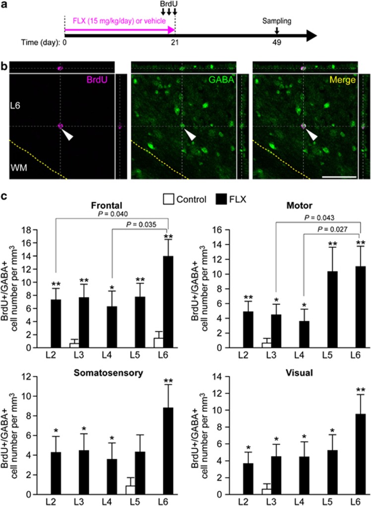
As shown in Figure 1, FLX treatments increased the number of L1-INP cells. To determine whether administration of FLX led to an increase in the number of GABAergic interneurons, we performed immunostaining of cortical sections for GABA. After 3 weeks of FLX administration, the number of GABAergic interneurons significantly increased in layer 6 of all cortical regions examined (Supplementary Figure S4). Layer-specific increases in GABAergic interneurons also occurred in layer 3 of the frontal cortex and layer 5 of the motor cortex. Moreover, we detected BrdU+/GABA+ cells in the cortex of mice at 4 weeks after the last FLX injection (Figure 2), but we found few BrdU+/GABA+ cells in control mice (frontal, F(9,230)=8.9, p<0.0001; motor, F(9,230)=7.01, p<0.0001; somatosensory, F(9,230)=4.9, p<0.0001; visual, F(9,230)=6.2, p<0.0001), indicating that at least a subset of detected GABAergic cells were newly generated.
Figure 2.
Increases in the number of GABAergic interneurons in the cortices of FLX-treated mice. (a) Time course of experiments. Mice were treated with FLX pellets (15 mg/kg/day) for 3 weeks. (b) Brain sections were stained with anti-GABA (green) and anti-BrdU (magenta). Scale bars, 50 μm. (c) In all layers of the cortical regions examined, FLX treatment increased the number of GABA+/BrdU+ interneurons (*p<0.05, **p<0.001; two-way ANOVA, n=8 each). In layer 6 particularly, the number of GABA+/BrdU+ interneurons was significantly elevated.
Next, we examined whether the production of GABAergic interneurons depended on the presence of L1-INP cells. To trace newly generated cells derived from L1-INP cells, we used a recombinant retroviral vector that infects only proliferating cells when its transgene is incorporated into the cell genome (Tashiro et al, 2006). In a previous study, we developed a tracing method for detecting the daughter cells of L1-INP cells using retroviral vectors (Ohira et al, 2010). To increase the rate of detection of L1-INP-derived daughter cells, we constructed a new retroviral vector that expressed the enhanced yellow-green fluorescence protein Venus driven by the neuron-specific synapsin I promoter (Hioki et al, 2007), which is activated during the differentiation of NPCs (Coufal et al, 2009), in the early developmental stage of neurons, and in adulthood (Melloni and DeGennaro, 1994). The retroviral vector was injected into the frontal regions because the reactivity of L1-INP cells to FLX was highest in the frontal cortex (Figure 1c–f). After 2 days, each mouse was injected intraperitoneally with FLX for 3 weeks and perfused at 1, 2, 4, or 8 weeks after the last FLX administration. In addition, we injected the viral vector into the somatosensory cortex to compare the results in this region with those in the frontal cortex and perfused mice at 4 and 8 weeks.
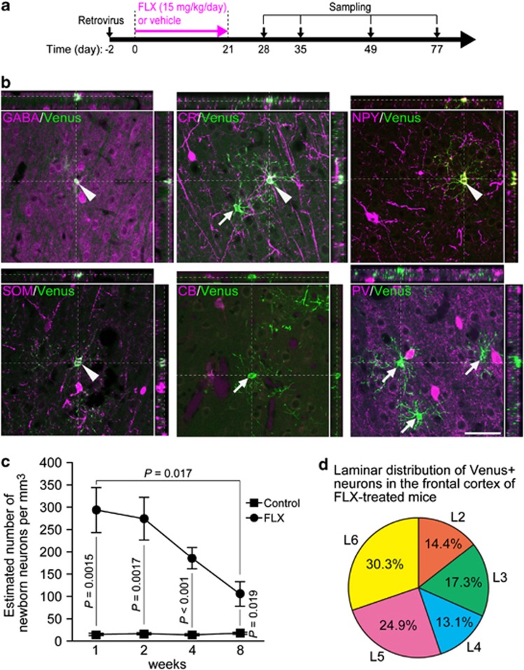
We examined the phenotypic differentiation of layer 2–6 of Venus+ neurons in FLX-treated mice using several neuronal markers. GABA, calretinin (CR), neuropeptide Y (NPY), and somatostatin (SOM) were expressed up to 11 weeks, but calbindin (CB) and parvalbumin (PV), which are expressed in a subpopulation of GABAergic neurons (Gonchar et al, 2008), and glial markers were undetectable in Venus+ neurons (Figure 3; Table 1; Supplementary Figure S5). Additionally, the labeling percentages of these markers in Venus+ cells increased up to 7 weeks (n=4 each, Table 1). In the somatosensory cortex, the differentiation rates of GABA, CR, NPY, and SOM cells at both 7 and 11 weeks were comparable to those in the frontal cortex (Supplementary Table S1). On the basis of these differentiation percentages, it seems that new neurons mature ∼7 weeks after their genesis.
Figure 3.
GABAergic phenotypes of new neurons derived from L1-INP cells by FLX treatment. (a) Representation of the experimental time course. (b) Expression of GABAergic markers in neurons derived from L1-INP cells as labeled by Venus-expressing retroviral vectors. Immunofluorescent analysis was performed using antibodies against molecular markers (magenta) expressed by GABAergic interneurons: GABA, calretinin (CR), neuropeptide Y (NPY), somatostatin (SOM), calbindin (CB), and parvalbumin (PV). All cell images were taken from the frontal cortex 4 weeks after FLX treatments (15 mg/kg/day) and are presented in the xy, xz, and yz planes. Arrowheads and arrows indicate immunopositive and immunonegative cells, respectively, for each marker. Scale bar, 50 μm. (c) Time course of the estimated number of layer 2–6 Venus+ cells in the frontal cortex of FLX-treated mice (15 mg/kg/day; two-way ANOVA followed by Scheffé's post hoc test, n=4 each). All data represent the mean±SEM. (d) Laminar distribution of Venus+ neurons in the frontal cortex of FLX-treated mice. Data represent the mean of four mice. L, layer.
Table 1. Phenotypes of Venus+ Neurons in the Frontal Cortex.
|
Labeling percentages of the markers expressed in Venus+
cells |
||||
|---|---|---|---|---|
| Period after FLX treatments | 1 week | 2 weeks | 4 weeks | 8 weeks |
| GABA | 39±4.6 (27/70) | 53±5.7 (32/59) | 77±3.9 (34/47)*1 | 84±3.7 (23/29)*1 |
| CR | 19±4.0 (14/72) | 24±3.8 (14/59) | 43±6.2 (19/49)*2 | 44±4.3 (13/28)*3 |
| NPY | 7.3±3.1 (5/66) | 13±4.0 (7/58) | 22±5.7 (10/41)*4 | 27±4.7 (7/25)*5 |
| SOM | 2.5±1.6 (2/69) | 2.1±1.4 (2/73) | 5.8±3.0 (3/44) | 6.1±3.1 (2/24) |
| CB | 0 (0/68) | 0 (0/64) | 0 (0/49) | 0 (0/30) |
| PV | 0 (0/68) | 0 (0/56) | 0 (0/39) | 0 (0/27) |
| S100β | 0 (0/76) | 0 (0/68) | 0 (0/39) | 0 (0/25) |
| Olig2 | 0 (0/80) | 0 (0/66) | 0 (0/40) | 0 (0/27) |
| Iba1 | 0 (0/81) | 0 (0/75) | 0 (0/35) | 0 (0/23) |
Abbreviations: CB, calbindin; CR, calretinin; Iba1, ionized calcium-binding adapter molecule 1; NPY, neuropeptide Y; Olig2, oligodendrocyte transcription factor 2; PV, parvalbumin; SOM, somatostatin; S100β, S100 calcium-binding protein β. Data are presented as the mean values±SEM from 4–5 mice at each time point. The denominator and numerator of the fraction in parentheses represent the total number of Venus+ cells and the number of marker+/Venus+ cells in the frontal cortex, respectively. One-way ANOVA and Scheffé's post hoc test were used in the statistical analysis of the time course of each marker. All P values are vs. 1-week samples for each marker. *1: p<0.001. *2: p=0.0063. *3: p=0.0011. *4: p=0.030. *5: p=0.0037.
To estimate the number of new neurons derived from L1-INP cells in the whole cortex, we calculated the labeling efficiency r (the number of Venus+ L1-INP cells [Venus+/Ki67+/GAD67+ cells] divided by the total number of L1-INP cells [Ki67+/GAD67+ cells] Supplementary Figures S6 and S7; Supplementary Table S2) and estimated the number of new neurons produced (the number of L2–6 total Venus+ neurons divided by r for each mouse).
At 1 week after the last FLX treatment, the estimated number of layer 2–6 Venus+ neurons was 19-fold greater than control mice (p=0.015, n=4 mice, two-way ANOVA followed by Tukey's post hoc test), corresponding to 0.32% of total neurons (Schüz and Palm, 1989) and 3–6% of GABAergic interneurons (Beaulieu, 1993; Gabbott et al, 1997) in the frontal cortex of adult rodents (Figure 3c: control vs FLX, F(7,21)=10.2, p<0.0001; time, F(3,21)=3.14, p=0.047). The estimated number of Venus+ neurons declined thereafter. In the somatosensory cortex, the estimated number of Venus+ neurons was lower than in the frontal cortex (Supplementary Figure S7), which may reflect the lower number of L1-INP cells in the somatosensory cortex (Figure 1). In control mice, we found a small number of layer 2–6 Venus+ neurons prior to 8 weeks, although this number might have been overestimated because brain damage caused by viral injection could have enhanced L1-INP cell proliferation.
At 4 weeks after the last FLX treatment, Venus+ cells were found in all layers and most frequently in the deep layers of the frontal and somatosensory cortices (Figure 3d; Supplementary Figure S7). This is consistent with the distribution of the BrdU+/GABA+ cells (Figure 2). Furthermore, many synaptophysin+ presynaptic boutons were in contact with Venus+ neurons (Supplementary Figure S8), and synaptophysin+ presynaptic boutons were found not only on cell bodies but also on dendrites.
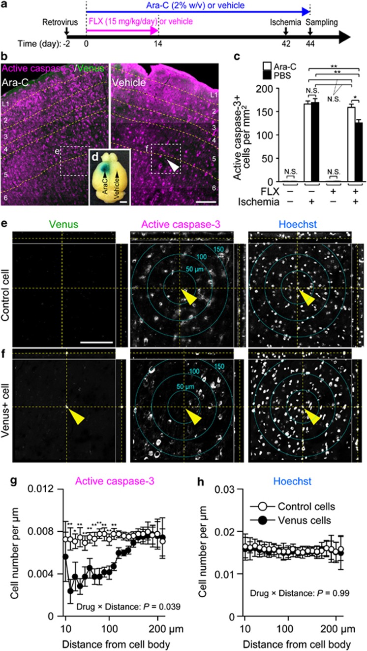
In ischemic brains, excitotoxicity is induced by enhanced excitatory neurotransmission (Blaesse et al, 2009), and upregulation of inhibitory mechanisms limits neuronal hyperexcitability in mammalian models of ischemic damage (Ovbiagele et al, 2003). In this study, FLX-induced neurons were GABAergic interneurons (Figures 2 and 3; Table 1; Supplementary Figure S7; Supplementary Table S1). Because the functional role of new neurons derived from L1-INP cells is unknown, we hypothesized that these newly generated GABAergic interneurons suppress apoptosis induced by ischemia. To test this hypothesis, we infused mice with PBS or Ara-C solution in each hemisphere during the experimental period. After 2 weeks of FLX treatment, mice were housed in cages for 4 weeks to allow sufficient neuronal differentiation. Thereafter, global ischemia was induced for 10 min, and cell death was evaluated according to the expression of active caspase-3 after 48 h. Apoptotic cells express active-casapse-3 for 1–2 days after ischemia and the expression of active-casapse-3 becomes weaker 3 days after ischemia in the cortex (Namura et al, 1998; Chen et al, 1998; Hu et al, 2000). Moreover, in our preliminary study, the expression of active caspase-3 reached the maximum level 2 days after ischemia. Thus, we chose this time point. Given that the new neurons derived from L1-INP cells coincided with a decrease in the expression of active caspase-3 following ischemia, then the generation of new neurons would be expected to reduce overall apoptosis in the PBS-treated hemispheres. As shown in Figure 4 and Supplementary Figures S9 and S10, neurogenesis of L1-INP cells was significantly suppressed by Ara-C treatments (p<0.001, n=4 each, two-way ANOVA) and also significantly increased after 2 weeks of FLX treatment (control vs Ara-C, F(1,42)=1.55, p=0.22; treatments, F(3,42)=600, p<0.0001). Interestingly, in FLX-treated, ischemia-induced mice, the number of active caspase-3+ cells in the PBS-infused hemispheres was significantly decreased compared with Ara-C-infused hemispheres (p<0.05, n=4 each, two-way ANOVA, Figure 4c). The number of active caspase-3+ cells in the Ara-C-infused hemispheres of FLX-treated and ischemia-induced mice was similar to the Ara-C- or PBS-infused hemispheres of ischemia-induced mice not treated with FLX, thus suggesting that newly generated neurons may have suppressed the apoptotic effect of ischemia at this time point. Although Ara-C treatment might affect active caspase-3 expression, there are several reasons it is unlikely here. In the ischemia-induced mice lacking FLX treatments, there was no significant difference between Ara-C- and PBS-treated hemispheres in the number of active caspase-3+ cells (Figure 4c). In the brains without ischemia, we observed few active caspase-3+ cells in either Ara-C or PBS hemispheres regardless of FLX treatment. Additionally, there were no significant differences between hemispheres of non-ischemic brains. These results indicate that Ara-C treatment alone had no effect on the expression of active caspase-3 observed in this study. Moreover, the number of active caspase-3+ cells within 20–110 μm of a Venus-expressing cell soma was significantly less than the number within that range of control cells (Figure 4e and g; control cells vs Venus+ cells, F(1,819)=56.3, p<0.0001; distance, F(20,819)=1.74, p=0.023). Hoechst staining showed that there are many surviving neurons, whose nuclei are much larger than glial nuclei and can be easily distinguished from those of glial cells or endothelial cells, around Venus+ cells. The control cells were defined as active-casapse-3-negative and distributed in the same layers, areas, and locations in one hemisphere as Venus+ neurons were in the other hemisphere (Figure 4f and h; control cells vs Venus+ cells, F(1,819)<0.0001, p=0.99; distance, F(20,819)=0.114, p=0.99). In addition, because glial cells, whose nuclei were smaller and denser than neurons' nuclei, were easily discerned, they were omitted in this quantification.
Figure 4.
New neurons derived from L1-INP cells coincide with a suppression of ischemia-induced active caspase-3 expression. (a) Representative time course of experiments. (b–d) The number of active caspase-3+ cells decreased in the vehicle-treated hemisphere, where new neurons were generated from L1-INP cells, compared with the Ara-C-treated hemisphere, where neurogenesis was inhibited (two-way ANOVA followed by Scheffé's post hoc test, n=4 each). Black arrowheads indicate the positions of the cannulae that were connected to micro-osmotic pumps filled with an Ara-C solution containing Fast Green FCF, or PBS (vehicle). NS, not significant. (e–h) The number of active caspase-3+ cells within 20–110 μm of a Venus-expressing cell soma was significantly less than the number within that range of the control cells (*p<0.05, **p<0.01, repeated-measures ANOVA, n=17 Venus-expressing cells and 23 control cells). The xy plane in the middle of each cell is shown. Scale bars, 200 μm (b), 2 mm (d), 100 μm (e, f).
Astrocyte activation, which was not accompanied by cell proliferation (Supplementary Figure S11) (Panickar and Norenberg, 2005), was induced by ischemia in both Ara-C- and PBS-treated hemispheres 48 h after ischemia. Activated astrocytes have beneficial effects on ischemic injury as well. However, because astrocyte activation was induced by ischemia regardless of the Ara-C treatment, the decrease in the number of active caspase-3+ cells in the PBS-treated hemispheres cannot be explained by astrocyte activation alone. Considering that astrocytes were distributed throughout the cortex (Supplementary Figure S11), astrocyte activation cannot account for the marked reduction of the number of active caspase-3+ cells within 20–110 μm of Venus-expressing cell somas. There was no difference in the number of microglial cells in both hemispheres during the experimental periods (Supplementary Figure S11) (Denes et al, 2007). These results indicate that neurogenesis of L1-INP cells coincides with a decrease in ischemia-induced active caspase-3 expression, a concurrence that may highlight the potential physiological activity of these interneurons.
DISCUSSION
To date, three reports demonstrate that chronic FLX treatments increase cell proliferation in the frontal cortex of adult rodents (Czéh et al, 2007; Hodes et al, 2010; Kodama et al, 2004). In two of them, the authors claimed that newborn cells that incorporated BrdU were glial and endothelial cells, but not neurons, based on immunohistochemistry for neuronal (NeuN), glial (GFAP, NG2, and O4), and endothelial cell markers (RECA-1) (Czéh et al, 2007; Kodama et al, 2004). However, importantly, GABAergic neurons derived from L1-INP cells were NeuN-immunonegative when immunostaining with two monoclonal and polyclonal NeuN antibodies was performed (0 NeuN+/Venus+ cells of 39 Venus+ cells, n=8 mice; Supplementary Figure S12). The NeuN antibody cannot stain some neuronal classes (eg, olfactory mitral cells, Purkinje cells, and all cerebellar interneurons except for granule cells) (Rakic, 2002). Moreover, GABAergic interneurons are rather small, and new neurons make up only 0.005–1% of all existing neurons (Ohira, 2011). In previous reports, FLX was administered at 5 mg/kg/day (Hodes et al, 2010; Kodama et al, 2004) or 10 mg/kg/day (Czéh et al, 2007; Hodes et al, 2010). We showed that the proliferation of L1-INP cells was significantly upregulated at >15 mg/kg/day (see Figure 1), although we did not use FLX at 10 mg/kg/day. In our study, the estimated number of new neurons derived from L1-INP cells was 290 cells/mm3 1 week after 3 weeks of FLX treatment, which means that there were 116 GABA+ cells/mm3 (0.4 × 290=116 cells/mm3). On the other hand, Czéh et al, (2007) reported that ∼3500 cells were generated in one hemisphere of the medial prefrontal cortex in adult rats after 4 weeks of FLX treatment. Because the hemispheric volume of the medial prefrontal cortex is 5.9 mm3 in adult rats (Day-Wilson et al, 2006), ∼600 BrdU+ cells/mm3 should have survived for 3 weeks after the last BrdU injection. Czeh et al showed that 80% of FLX-generated BrdU+ cells were glial and endothelial. If 20% of newly generated cells are GABAergic interneurons, this means that 120 cells/mm3 (0.2 × 600=120 cells/mm3) might have been GABAergic interneurons. Therefore, our data show that the number of new neurons generated from L1-INP cells is comparable to the number of BrdU+ cells previously reported by Czéh et al. In summary, by staining for the GABAergic neuron markers GABA, CR, NPY, and SOM conjointly with retroviral labeling of L1-INP cells, along with treatment with a high dose of FLX, we detected cortical adult neurogenesis of L1-INP cells following treatment with SSRIs.
We used retrovirus vectors to trace neurogenesis of L1-INP cells. Under these conditions, it is possible that the observed neurogenesis was the result of inflammation caused by the virus injection rather than by FLX treatment. However, this seems unlikely for the following reasons: first, the number of L1-INP cells in the untreated control frontal cortex (14.3±1.65 cells/mm3, n=5) was comparable to that in the frontal cortex of mice injected with the retrovirus injection for 2 weeks (16.4±0.982 cells/mm3, n=4; p=0.29, compared with intact control). This result indicates that the virus injection alone did not induce neurogenesis of L1-INP cells. Second, to examine the effect of retrovirus injection on GABAergic neurogenesis of L1-INP cells, we made a comparison between the number of BrdU+/GABA+ cells in vehicle-treated control cortex without retrovirus injection (taken from Figure 2c) and that in vehicle-treated control cortex with retrovirus injection (taken from Supplementary Figure S9b). The number of BrdU+/GABA+ cells in the vehicle-treated control cortex without retrovirus injection is 0.303±0.136 cells per mm3 (n=6 mice), whereas that in vehicle-treated control cortex with retrovirus injection is 0.205±0.096 cells per mm3 [n=5; the number of BrdU+/GABA+ cells per hemisphere is divided by 3.895±0.37 mm3, the hemispheric volume of medial prefrontal cortex in adult mice (Spanswick and Dyck, 2012)]. There was no significant difference between them (p=0.713, Student's t-test).
It follows that, if any inflammation caused by the retrovirus injection led to neurogenesis of L1-INP cells, a concurrent increase in the number of newborn interneurons should have been detected. Thus, this result indicates that the retrovirus injection did not induce GABAergic neurogenesis of L1-INP cells, and also that any resultant inflammation caused by the injections was weak, if it existed at all, and was certainly not potent enough to activate L1-INP cells in this study.
In this study, because ischemia was induced 4 weeks after the last FLX treatment, we are inclined to think that any subsequent reduction in active caspase-3 expression was mediated by FLX-induced new GABAergic interneurons rather than residual FLX. These new neurons contained not only GABA but also NPY and SOM, both of which suppress neuronal activity (Richichi et al, 2004; Tallent and Siggins, 1999). Thus, it is possible that these neuropeptides, together with GABA, suppress apoptosis in existing neurons following ischemia. However, there are two other possible interpretations of this result. First, activation of caspase-3 degrades proteins inside cells, including Venus protein. If degradation of Venus by activated caspase-3 occurred in ischemic brains, then the number of Venus+ neurons should have decreased after ischemia in all brain regions. However, the numbers of Venus+ new neurons were similar before and after ischemia, indicating degradation of Venus is unlikely (Supplementary Figure S10). The second possibility is that FLX treatments induced local conditions giving rise to niches that promoted neurotrophic factor release and/or survival. This may be why surviving Venus+ cells and reduced active caspase-3+ cells were located in the same area, without any causal relationship between neurogenesis and suppression of apoptosis. However, if there were FLX-induced niches, the expression of active-caspase-3 would be inhibited by those niches in both hemispheres. In our immunostaining assays, such niches were not detected in the PBS-treated side, and as described above, the active-caspase-3+ cells seemed to be uniformly distributed in the PBS-treated side. Moreover, there were no differences in the number of active caspase-3+ cells between the Ara-C- and PBS-treated hemispheres. Taken together, these data imply that GABAergic neurogenesis of L1-INP cells may suppress apoptosis induced by ischemic stroke; at least 2 days post the ischemic event.
In this study, we used the apoptotic marker, active caspase-3, to demonstrate the suppressing effect of new GABAergic interneurons on apoptosis. However, we did not elucidate precisely how new GABAergic interneurons suppress apoptosis induced by ischemia. In ischemic brains, excitotoxicity is induced by enhanced excitatory neurotransmission and results in neuronal death (Blaesse et al, 2009). As shown in Figure 4, active caspase-3 expression was reduced in the areas surrounding the new GABAergic interneurons, thus highlighting the possibility that inhibitory mechanisms related to GABAergic interneurons may limit the apoptotic effect of ischemia. Future studies employing electrophysiology and connectome techniques are required to clarify the inhibitory properties underlying new GABAergic interneurons, such as their firing patterns and wiring with neighboring neurons. In addition, the present study only assessed apoptosis at 1 time point, that is, 2 days following ischemia, which was based on the finding that apoptotic cells express active-caspase-3 for 1–2 days following ischemia, with its expression becoming weaker 3 days post cortical ischemia (Chen et al, 1998; Hu et al, 2000; Namura et al, 1998). Thus, future experiments should assess the potential apoptosis-suppressing effect of new GABAergic interneurons over a longer period; this may give some insight into the potential neuroprotective proclivity of these new interneurons against ischemia.
Interneurons in the cortical deep layers migrate from the SVZ in the early postnatal mouse pups (Le Magueresse et al, 2011), and new interneurons are generated in the SVZ during ischemia (Kreuzberg et al, 2010), suggesting that some new neurons might have been derived from the SVZ in this study. However, we have previously established that our method of injection restricts the viral solution, and therefore virus-infected cells, to cortical layer 1 (Ohira and Kaneko, 2010; Ohira et al, 2010), and new neurons from the SVZ can be excluded. In addition, Ara-C-treated regions can be visualized by co-injection of the dye Fast Green. As shown in Supplementary Figure S13, the Ara-C solution did not reach the SVZ region. If progenitor/stem cells in the SVZ were infected with the Venus-expressing viral vectors, FLX treatments would produce new Venus+ neurons regardless of Ara-C treatments. However, we found no increases in new neurons in either Ara-C- or FLX-treated hemispheres (Figure 4; Supplementary Figures S9 and S10), implying that Venus+ new neurons were generated from L1-INP cells. However, we cannot exclude that some new neurons, which were Venus negative and were derived from the SVZ were added to the deep layers.
Although we only used FLX to examine whether neurogenesis of L1-INP cells was regulated by SSRIs, this does not suggest whether other antidepressants increase or decrease the neurogenesis of L1-INP cells. In hippocampal neurogenesis of adult rodents, tricyclic antidepressants (imipramine), monoamine oxidase inhibitors (tranylcypromine), and norepinephrine-selective reuptake inhibitors (reboxetine) increase neurogenesis (Malberg et al, 2000; Santarelli et al, 2003), possibly mediated through the 5-HT1A receptor (Santarelli et al, 2003). These results suggest that the increase in the concentration of 5-HT around the NSCs and NPCs may be important for upregulation of hippocampal neurogenesis. If 5-HT increases neurogenesis of L1-INP cells, neurogenesis of L1-INP cells may be enhanced by antidepressants other than FLX. It would be of interest to determine whether other antidepressants also control neurogenesis of L1-INP cells.
It is possible that cortical neurogenesis of L1-INP cells partly underlies the neuroprotective/therapeutic effects of FLX during ischemia in animals (Chang et al, 2006; Li et al, 2009; Lim et al, 2009) and human (Chollet et al, 2011; Dam et al, 1996; Pariente et al, 2001). Considering the possible role of GABAergic interneurons in depression (Luscher et al, 2011; Sanacora et al, 2002), the stimulation of cortical neurogenesis of L1-INP cells may account for the positive therapeutic effects of FLX on depression, in addition to or as an alternative to its effects of increasing neurogenesis (Sahay and Hen, 2007), dematuration in the DG (Kobayashi et al, 2010, 2012; Ohira and Miyakawa, 2011), survival (Cameron et al, 1998) and differentiation of new neurons (Bibel et al, 2004), and synaptic plasticity of neural circuits via increased signaling through brain-derived neurotrophic factor (Karpova et al, 2011).
In adult neurogenesis of the SGZ and SVZ, various factors that affect neurogenesis have been identified, for example, neurotransmitters, hormones, stress, aging, exercise, learning, and enriched environments (Abrous et al, 2005). Further investigations are needed to examine whether such factors can also control the adult neurogenesis of L1-INP cells.
Acknowledgments
We thank T Kaneko for the kind gift of the enhanced synapsin I promoter, TJ Hope for the kind gift of WPRE, and A Miyawaki for the kind gift of Venus/pCS2. We are also grateful to S Noma and T Ueno for assistance with FLX treatments, immunostaining, and quantification. This work was supported by the NEXT Program (LS-116) and CREST, JST.
The authors declare no conflict of interest.
Footnotes
Supplementary Information accompanies the paper on the Neuropsychopharmacology website (http://www.nature.com/npp)
Supplementary Material
References
- Abrous DN, Koehl M, Le Moal M. Adult neurogenesis: from precursors to network and physiology. Physiol Rev. 2005;85:523–569. doi: 10.1152/physrev.00055.2003. [DOI] [PubMed] [Google Scholar]
- Beaulieu C. Numerical data on neocortical neurons in adult rat, with special reference to the GABA population. Brain Res. 1993;609:284–292. doi: 10.1016/0006-8993(93)90884-p. [DOI] [PubMed] [Google Scholar]
- Bibel M, Richter J, Schrenk K, Tucker KL, Staiger V, Korte M, et al. Differentiation of mouse embryonic stem cells into a defined neuronal lineage. Nat Neurosci. 2004;7:1003–1009. doi: 10.1038/nn1301. [DOI] [PubMed] [Google Scholar]
- Blaesse P, Airaksinen MS, Rivera C, Kaila K. Cation-chloride cotransporters and neuronal function. Neuron. 2009;61:820–838. doi: 10.1016/j.neuron.2009.03.003. [DOI] [PubMed] [Google Scholar]
- Cameron HA, Hazel TG, McKay RD. Regulation of neurogenesis by growth factors and neurotransmitters. J Neurobiol. 1998;36:287–306. [PubMed] [Google Scholar]
- Chang Y-C, Tzeng S-F, Yu L, Huang A-M, Lee H-T, Huang C-C, et al. Early-life fluoxetine exposure reduced functional deficits after hypoxic-ischemia brain injury in rat pups. Neurobiol Dis. 2006;24:101–113. doi: 10.1016/j.nbd.2006.06.001. [DOI] [PubMed] [Google Scholar]
- Chen J, Nagayama T, Jin K, Stetler RA, Zhu RL, Graham SH, et al. Induction of caspase-3-like protease may mediate delayed neuronal death in the hippocampus after transient cerebral ischemia. J Neurosci. 1998;18:4914–4928. doi: 10.1523/JNEUROSCI.18-13-04914.1998. [DOI] [PMC free article] [PubMed] [Google Scholar]
- Chollet F, Tardy J, Albucher J-F, Thalamas C, Berard E, Lamy C, et al. Fluoxetine for motor recovery after acute ischaemic stroke (FLAME): a randomised placebo-controlled trial. Lancet Neurol. 2011;10:123–130. doi: 10.1016/S1474-4422(10)70314-8. [DOI] [PubMed] [Google Scholar]
- Coufal NG, Garcia-Perez JL, Peng GE, Yeo GW, Mu Y, Lovci MT, et al. L1 retrotransposition in human neural progenitor cells. Nature. 2009;460:1127–1131. doi: 10.1038/nature08248. [DOI] [PMC free article] [PubMed] [Google Scholar]
- Czéh B, Müller-Keuker JIH, Rygula R, Abumaria N, Hiemke C, Domenici E, et al. Chronic social stress inhibits cell proliferation in the adult medial prefrontal cortex: hemispheric asymmetry and reversal by fluoxetine treatment. Neuropsychopharmacology. 2007;32:1490–1503. doi: 10.1038/sj.npp.1301275. [DOI] [PubMed] [Google Scholar]
- Dam M, Tonin P, De Boni A, Pizzolato G, Casson S, Ermani M, et al. Effects of fluoxetine and maprotiline on functional recovery in poststroke hemiplegic patients undergoing rehabilitation therapy. Stroke. 1996;27:1211–1214. doi: 10.1161/01.str.27.7.1211. [DOI] [PubMed] [Google Scholar]
- Day-Wilson KM, Jones DNC, Southam E, Cilia J, Totterdell S. Medial prefrontal cortex volume loss in rats with isolation rearing-induced deficits in prepulse inhibition of acoustic startle. Neuroscience. 2006;141:1113–1121. doi: 10.1016/j.neuroscience.2006.04.048. [DOI] [PubMed] [Google Scholar]
- Denes A, Vidyasagar R, Feng J, Narvainen J, McColl BW, Kauppinen RA, et al. Proliferating resident microglia after focal cerebral ischaemia in mice. J Cereb Blood Flow Metab. 2007;27:1941–1953. doi: 10.1038/sj.jcbfm.9600495. [DOI] [PubMed] [Google Scholar]
- Gabbott PL, Dickie BG, Vaid RR, Headlam AJ, Bacon SJ. Local-circuit neurones in the medial prefrontal cortex (areas 25, 32 and 24b) in the rat: morphology and quantitative distribution. J Comp Neurol. 1997;377:465–499. doi: 10.1002/(sici)1096-9861(19970127)377:4<465::aid-cne1>3.0.co;2-0. [DOI] [PubMed] [Google Scholar]
- Ge S, Pradhan DA, Ming G-L, Song H. GABA sets the tempo for activity-dependent adult neurogenesis. Trends Neurosci. 2007;30:1–8. doi: 10.1016/j.tins.2006.11.001. [DOI] [PubMed] [Google Scholar]
- Gonchar Y, Wang Q, Burkhalter A. Multiple distinct subtypes of GABAergic neurons in mouse visual cortex identified by triple immunostaining. Front Neuroanat. 2008;1:3. doi: 10.3389/neuro.05.003.2007. [DOI] [PMC free article] [PubMed] [Google Scholar]
- Hioki H, Kameda H, Nakamura H, Okunomiya T, Ohira K, Nakamura K, et al. Efficient gene transduction of neurons by lentivirus with enhanced neuron-specific promoters. Gene Ther. 2007;14:872–882. doi: 10.1038/sj.gt.3302924. [DOI] [PubMed] [Google Scholar]
- Hodes GE, Hill-Smith TE, Suckow RF, Cooper TB, Lucki I. Sex-specific effects of chronic fluoxetine treatment on neuroplasticity and pharmacokinetics in mice. J Pharmacol Exp Ther. 2010;332:266–273. doi: 10.1124/jpet.109.158717. [DOI] [PMC free article] [PubMed] [Google Scholar]
- Hu BR, Liu CL, Ouyang Y, Blomgren K, Siesjö BK. Involvement of caspase-3 in cell death after hypoxia-ischemia declines during brain maturation. J Cereb Blood Flow Metab. 2000;20:1294–1300. doi: 10.1097/00004647-200009000-00003. [DOI] [PubMed] [Google Scholar]
- Karpova NN, Pickenhagen A, Lindholm J, Tiraboschi E, Kulesskaya N, Agústsdóttir A, et al. Fear erasure in mice requires synergy between antidepressant drugs and extinction training. Science. 2011;334:1731–1734. doi: 10.1126/science.1214592. [DOI] [PMC free article] [PubMed] [Google Scholar]
- Kobayashi K, Haneda E, Higuchi M, Suhara T, Suzuki H. Chronic fluoxetine selectively upregulates dopamine D1-like receptors in the hippocampus. Neuropsychopharmacology. 2012;37:1500–1508. doi: 10.1038/npp.2011.335. [DOI] [PMC free article] [PubMed] [Google Scholar]
- Kobayashi K, Ikeda Y, Sakai A, Yamasaki N, Haneda E, Miyakawa T, et al. Reversal of hippocampal neuronal maturation by serotonergic antidepressants. Proc Natl Acad Sci USA. 2010;107:8434–8439. doi: 10.1073/pnas.0912690107. [DOI] [PMC free article] [PubMed] [Google Scholar]
- Kodama M, Fujioka T, Duman RS. Chronic olanzapine or fluoxetine administration increases cell proliferation in hippocampus and prefrontal cortex of adult rat. Biol Psychiatry. 2004;56:570–580. doi: 10.1016/j.biopsych.2004.07.008. [DOI] [PubMed] [Google Scholar]
- Kreuzberg M, Kanov E, Timofeev O, Schwaninger M, Monyer H, Khodosevich K. Increased subventricular zone-derived cortical neurogenesis after ischemic lesion. Exp Neurol. 2010;226:90–99. doi: 10.1016/j.expneurol.2010.08.006. [DOI] [PubMed] [Google Scholar]
- Li W-L, Cai H-H, Wang B, Chen L, Zhou Q-G, Luo C-X, et al. Chronic fluoxetine treatment improves ischemia-induced spatial cognitive deficits through increasing hippocampal neurogenesis after stroke. J Neurosci Res. 2009;87:112–122. doi: 10.1002/jnr.21829. [DOI] [PubMed] [Google Scholar]
- Lim C-M, Kim S-W, Park J-Y, Kim C, Yoon SH, Lee J-K. Fluoxetine affords robust neuroprotection in the postischemic brain via its anti-inflammatory effect. J Neurosci Res. 2009;87:1037–1045. doi: 10.1002/jnr.21899. [DOI] [PubMed] [Google Scholar]
- Luscher B, Shen Q, Sahir N. The GABAergic deficit hypothesis of major depressive disorder. Mol Psychiatry. 2011;16:383–406. doi: 10.1038/mp.2010.120. [DOI] [PMC free article] [PubMed] [Google Scholar]
- LE Magueresse C, Alfonso J, Khodosevich K, Martín ÁAA, Bark C, Monyer H. ‘Small axonless neurons' : postnatally generated neocortical interneurons with delayed functional maturation. J Neurosci. 2011;31:16731–16747. doi: 10.1523/JNEUROSCI.4273-11.2011. [DOI] [PMC free article] [PubMed] [Google Scholar]
- Malberg JE, Eisch AJ, Nestler EJ, Duman RS. Chronic antidepressant treatment increases neurogenesis in adult rat hippocampus. J Neurosci. 2000;20:9104–9110. doi: 10.1523/JNEUROSCI.20-24-09104.2000. [DOI] [PMC free article] [PubMed] [Google Scholar]
- Melloni RH, DeGennaro LJ. Temporal onset of synapsin I gene expression coincides with neuronal differentiation during the development of the nervous system. J Comp Neurol. 1994;342:449–462. doi: 10.1002/cne.903420311. [DOI] [PubMed] [Google Scholar]
- Namura S, Zhu J, Fink K, Endres M, Srinivasan A, Tomaselli KJ, et al. Activation and cleavage of caspase-3 in apoptosis induced by experimental cerebral ischemia. J Neurosci. 1998;18:3659–3668. doi: 10.1523/JNEUROSCI.18-10-03659.1998. [DOI] [PMC free article] [PubMed] [Google Scholar]
- Ohira K. Injury-induced neurogenesis in the mammalian forebrain. Cell Mol Life Sci. 2011;68:1645–1656. doi: 10.1007/s00018-010-0552-y. [DOI] [PMC free article] [PubMed] [Google Scholar]
- Ohira K, Furuta T, Hioki H, Nakamura KC, Kuramoto E, Tanaka Y, et al. Ischemia-induced neurogenesis of neocortical layer 1 progenitor cells. Nat Neurosci. 2010;13:173–179. doi: 10.1038/nn.2473. [DOI] [PubMed] [Google Scholar]
- Ohira K, Kaneko T.2010Injection of virus vectors into the neocortical layer 1 Protocol Exchange 10.1038/nprot.2010.21 .
- Ohira K, Miyakawa T. Chronic treatment with fluoxetine for more than 6 weeks decreases neurogenesis in the subventricular zone of adult mice. Mol Brain. 2011;4:10. doi: 10.1186/1756-6606-4-10. [DOI] [PMC free article] [PubMed] [Google Scholar]
- Ovbiagele B, Kidwell CS, Starkman S, Saver JL. Neuroprotective agents for the treatment of acute ischemic stroke. Curr Neurol Neurosci Rep. 2003;3:9–20. doi: 10.1007/s11910-003-0031-z. [DOI] [PubMed] [Google Scholar]
- Panickar KS, Norenberg MD. Astrocytes in cerebral ischemic injury: morphological and general considerations. Glia. 2005;50:287–298. doi: 10.1002/glia.20181. [DOI] [PubMed] [Google Scholar]
- Pariente J, Loubinoux I, Carel C, Albucher JF, Leger A, Manelfe C, et al. Fluoxetine modulates motor performance and cerebral activation of patients recovering from stroke. Ann Neurol. 2001;50:718–729. doi: 10.1002/ana.1257. [DOI] [PubMed] [Google Scholar]
- Rakic P. Neurogenesis in adult primate neocortex: an evaluation of the evidence. Nat Rev Neurosci. 2002;3:65–71. doi: 10.1038/nrn700. [DOI] [PubMed] [Google Scholar]
- Richichi C, Lin E-JD, Stefanin D, Colella D, Ravizza T, Grignaschi G, et al. Anticonvulsant and antiepileptogenic effects mediated by adeno-associated virus vector neuropeptide Y expression in the rat hippocampus. J Neurosci. 2004;24:3051–3059. doi: 10.1523/JNEUROSCI.4056-03.2004. [DOI] [PMC free article] [PubMed] [Google Scholar]
- Sahay A, Hen R. Adult hippocampal neurogenesis in depression. Nat Neurosci. 2007;10:1110–1115. doi: 10.1038/nn1969. [DOI] [PubMed] [Google Scholar]
- Sakaue-Sawano A, Kurokawa H, Morimura T, Hanyu A, Hama H, Osawa H, et al. Visualizing spatiotemporal dynamics of multicellular cell-cycle progression. Cell. 2008a;132:487–498. doi: 10.1016/j.cell.2007.12.033. [DOI] [PubMed] [Google Scholar]
- Sakaue-Sawano A, Ohtawa K, Hama H, Kawano M, Ogawa M, Miyawaki A. Tracing the silhouette of individual cells in S/G2/M phases with fluorescence. Chem Biol. 2008b;15:1243–1248. doi: 10.1016/j.chembiol.2008.10.015. [DOI] [PubMed] [Google Scholar]
- Sanacora G, Mason GF, Rothman DL, Krystal JH. Increased occipital cortex GABA concentrations in depressed patients after therapy with selective serotonin reuptake inhibitors. Am J Psychiatry. 2002;159:663–665. doi: 10.1176/appi.ajp.159.4.663. [DOI] [PubMed] [Google Scholar]
- Santarelli L, Saxe M, Gross C, Surget A, Battaglia F, Dulawa S, et al. Requirement of hippocampal neurogenesis for the behavioral effects of antidepressants. Science. 2003;301:805–809. doi: 10.1126/science.1083328. [DOI] [PubMed] [Google Scholar]
- Scholzen T, Gerdes J. The Ki-67 protein: from the known and the unknown. J Cell Physiol. 2000;182:311–322. doi: 10.1002/(SICI)1097-4652(200003)182:3<311::AID-JCP1>3.0.CO;2-9. [DOI] [PubMed] [Google Scholar]
- Schüz A, Palm G. Density of neurons and synapses in the cerebral cortex of the mouse. J Comp Neurol. 1989;286:442–455. doi: 10.1002/cne.902860404. [DOI] [PubMed] [Google Scholar]
- Spanswick SC, Dyck RH. Object/Context Specific Memory Deficits following Medial Frontal Cortex Damage in Mice. PLoS One. 2012;7:e43698. doi: 10.1371/journal.pone.0043698. [DOI] [PMC free article] [PubMed] [Google Scholar]
- Stewart RR, Hoge GJ, Zigova T, Luskin MB. Neural progenitor cells of the neonatal rat anterior subventricular zone express functional GABA(A) receptors. J Neurobiol. 2002;50:305–322. doi: 10.1002/neu.10038. [DOI] [PubMed] [Google Scholar]
- Tallent MK, Siggins GR. Somatostatin acts in CA1 and CA3 to reduce hippocampal epileptiform activity. J Neurophysiol. 1999;81:1626–1635. doi: 10.1152/jn.1999.81.4.1626. [DOI] [PubMed] [Google Scholar]
- Tashiro A, Sandler VM, Toni N, Zhao C, Gage FH. NMDA-receptor-mediated, cell-specific integration of new neurons in adult dentate gyrus. Nature. 2006;442:929–933. doi: 10.1038/nature05028. [DOI] [PubMed] [Google Scholar]
Associated Data
This section collects any data citations, data availability statements, or supplementary materials included in this article.


