Abstract
Latent equine herpesvirus type 1 (EHV-1) infection is common in horse populations worldwide and estimated to reach a prevalence nearing 90% in some areas. The virus causes acute outbreaks of disease that are characterized by abortion and sporadic cases of myeloencephalopathy (EHM), both severe threats to equine facilities. Different strains vary in their abortigenic and neuropathogenic potential and the simultaneous occurrence of EHM and abortion is rare. In this report, we present clinical observations collected during an EHV-1 outbreak caused by a so-called “neuropathogenic” EHV-1 G2254/D752 polymerase (Pol) variant, which has become more prevalent in recent years and is less frequently associated with abortions. In this outbreak with 61 clinically affected horses, 6/7 pregnant mares aborted and 8 horses developed EHM. Three abortions occurred after development of EHM symptoms. Virus detection was performed by nested PCR targeting gB from nasal swabs (11 positive), blood serum (6 positive) and peripheral blood mononuclear cells (9 positive) of a total of 42 horses sampled. All 6 fetuses tested positive for EHV-1 by PCR and 4 by virus isolation. Paired serum neutralization test (SNT) on day 12 and 28 after the index case showed a significant (≥ 4-fold) increase in twelve horses (n = 42; 28.6%). This outbreak with abortions and EHM cases on a single equine facility provided a unique opportunity for the documentation of clinical disease progression as well as diagnostic procedures.
Keywords: Equine herpesvirus type 1, Neuropathogenicity, Stud, Treatment, Recovery rate, Pregnancy rate
Background
Equine herpesvirus type 1 (EHV-1) is ubiquitous in horse populations worldwide. Many horses are latently infected with EHV-1 and reactivation of the virus can occur under stress, upon which latently infected carriers start to shed infectious virus that may spread to in-contact horses [1,2]. Clinical EHV-1 infection can manifest itself in the form of three different clinical syndromes: respiratory disease, usually mild, in horses under 2 years of age; abortions, typically in the last trimester of pregnancy; and equine herpesvirus myeloencephalopathy (EHM) [1-3]. Different strains vary in their abortigenic potential [4,5] as well as in neuropathogenicity [6]. A single nucleotide polymorphism (SNP) in the viral DNA polymerase (Pol) gene (ORF30) is considered one major marker for the neuropathogenic potential of EHV-1 strains [7,8]. The A2254/N752 and G2254/D752 Pol variants apparently differ in their capability of causing EHM [6,8,9]. A2254/N752 (non-neuropathogenic) viruses are mainly isolated from cases of abortion and less frequently from horses with EHM. On the other hand, the G2254/D752 Pol variant (neuropathogenic) is found predominantly associated with EHM outbreaks [10,11]. The estimated prevalence of latent EHV-1 infection varies between 54% and 88%, depending on the population sampled and the method of virus detection used [3,12]. In the last decades, the prevalence of the G2254/D752 EHV-1 genotype in EHV-1 positive horses in the United States has apparently increased from 3.3% in the 1960s to 19.4% at the beginning of this century [13-15]. This may indicate a selective advantage of the neuropathogenic strain, which also could increase prevalence of EHM in the future.
Simultaneous latent infection in lymphoid tissues with both A2254/N752 and G2254/D752 EHV-1 genotypes was documented in some studies [12,16,17]. Parallel reactivation of both genotypes was not addressed in these studies. In contrast, no dual infection with A2254/N752 and G2254/D752 EHV-1 was detected in 419 fetal isolates [15]. However, 2 EDTA blood samples of febrile horses tested positive for both genotypes in another study on one farm (n = 23; 20 abortion outbreaks, 3 EHM outbreaks) with EHM cases [14].
Detection of the D752 Pol variant in outbreaks without EHM cases is rare and occurred in only 5% of the cases in a retrospective worldwide study [7]. In a 23-year retrospective analysis, 11% (19/176) of isolates carried the G2254/D752 allele, of which 84% (16/19) were collected from EHM cases and only 16% (3/19) from respiratory or abortion cases [14]. A combination of abortions with EHM was documented in 3 of the sampled farms [14]. In Argentina, 7% (4/54) of the abortion outbreaks were induced by the G2254/D752 variant, and in 2 of these simultaneous neurologic disease occurred [18]. Two outbreaks with abortion and EHM cases caused by the neuropathogenic strain were recently documented in Croatia with breed-dependent clinical signs: a high incidence of abortion (53.1%) was described in Lippizaner mares compared to a lower incidence (10%) in Quarter horses infected with the identical virus [6].
Various reports with clinical data are available for outbreaks either with neurological cases or abortion storms [19-22]. This case report documents for the first time the clinical data collected during an EHV-1 outbreak with an uncommon combination of abortion and EHM on a single facility in Germany.
Case presentation
This report is an account of an acute outbreak of EHV-1 in a mixed horse operation. Consequently, all clinical decisions and laboratory diagnostic procedures were initiated during the course of the outbreak. The documented outbreak occurred on an equine riding and breeding facility in southern Germany in early 2009. The stud includes different areas for sport and breeding horses. Broodmares and breeding stallions were kept separate from the sports horses. Breeding stallions were attended by grooms, which did not enter the broodmare or sport horse facilities as required for a European Union accredited breeding facility. In the sports horse section, horses were brought onto the premise without quarantine prior to entering the facility.
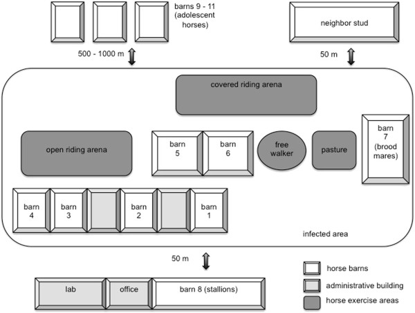
The clinical outbreak started in the broodmare section and spread rapidly to all other barns, except those for breeding stallions and adolescent horses. Affected horses were detected in seven of the eleven barns (Figure 1, Table 1). Adolescent horses (<2 years of age) were housed in groups of 20 in barns 9 to 11, where individual monitoring of body temperatures was not possible. In the affected areas, a total of 79 horses were housed. A stud in close proximity to the affected farm (50 m distance to broodmare barn 7) had no horses with fever, abortion or EHM, but no virus testing was performed. The vaccination status of the on-site animals was variable: All breeding stallions and broodmares were vaccinateda in accordance with the manufacturers recommendations, with boosters in the 5th, 7th and 9th month of gestation. In barns 1 to 6, only 21 (32%) of the horses were vaccinated. The group of adolescent horses (barns 9 to 11) was not vaccinated. We view it as likely that the poor overall vaccination status contributed to rapid virus spread on the premise. Another contributing factor for the severity of the outbreak may be the typical boarding facility situation that includes frequent horse traffic on and off the premise, which may have compromised overall herd immunity.
Figure 1.

Schematic presentation of the stud (not in scale). A total of 61 clinically affected (fever, abortion, EHM) horses were recorded. For distribution of clinical cases in the barns see Table 1.
Table 1.
Description of horse populations in the different barns on the stud
| Barn | No. of horses | Affected horses | Horse descriptiona | Same staff as horses of other barns |
|---|---|---|---|---|
| 1 |
6 |
5 |
SH; VH |
yes |
| 2 |
8 |
6 |
SH; VH |
yes |
| 3 |
12 |
8 |
SH; VH |
yes |
| 4 |
20 |
14 |
SH; VH |
yes |
| 5 |
12 |
10 |
SH; VH |
yes |
| 6 |
10 |
8 |
SH; VH |
yes |
| 7 |
11 |
10 |
BM; 1 foal |
yes |
| 8 |
10 |
0 |
BS |
no |
| 9-11 | 90 | 1 (?)b | AH | no |
aSH = Sport Horses; VH = Visiting Horses; BM = Brood Mares; BS = Breeding Stallions; AH = Adolescent Horses.
bClinical EHM signs, but no laboratory data available.
The index case was an abortion in barn 7. With the exception of the stallion barn and one questionable EHM case in the adolescent group (barns 9–11), all barns were affected. All affected barns were situated ≤ 50 meters next each other. The adolescent horses were stabled in barns 9–11, situated 500 to 1000 meters from the other barns. Horses from barns 1 – 7 used the same training facilities and paddocks.
The index case of the outbreak was an abortion in the broodmare barn 7. The day of the abortion is referred to as day 1 of the outbreak. Sixty-one clinical cases (77.2%) were detected during the outbreak and cases were identified over a period of 44 days. Onset, type and duration of clinical signs were documented for all individuals and are documented in the following sections.
Fever
In 71 horses, housed in barns 1 to 7, body temperatures were taken twice daily, starting on day 5 after the first abortion (index case). In the remaining 8 horses of the stabled population, control of body temperature as a routine procedure was practically impossible. Fever was defined as a body temperature of 38.1°C or higher (≥ 100.6°F).
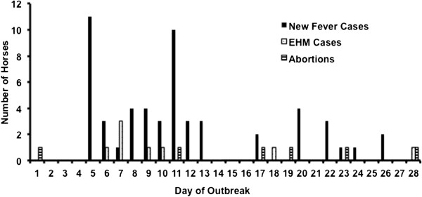
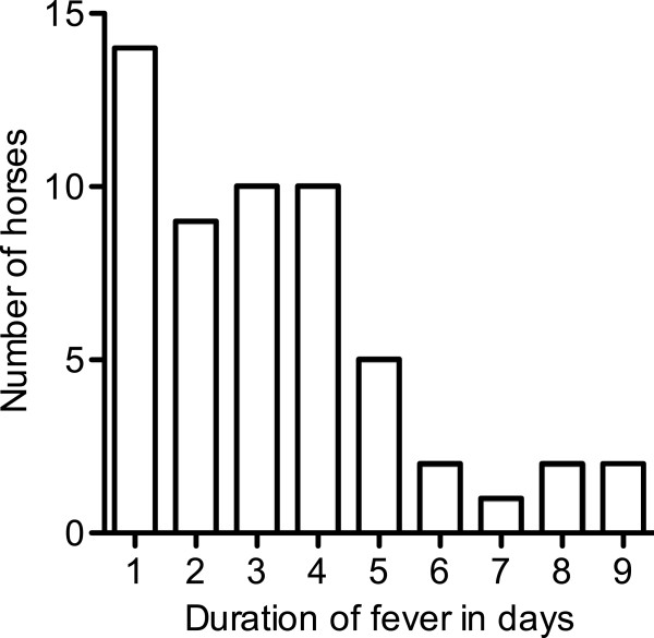
An increase of body temperature was detected in 55 of the regularly measured 71 horses (77.5%). Since body temperature measurements had not started before day 5 of the outbreak, we assume that fever was missed in several horses in the beginning. The maximum number of fever cases/day was documented on day 5 with 11 horses. On day 11, a second peak with 10 new cases was observed (Figure 2). The mean of the horses’ maximal body temperatures was 39.1 ± 0.7°C (minimum 38.1°C, maximum 40.8°C) and a mean duration of 3.2 ± 2.2 days of fever were recorded (minimum 1 day, maximum 9 days, Figure 3). Seven horses (12.7%) were febrile for more than 5 days. Only 3 (6.5%) of the 55 horses showed a second febrile episode. Five horses with fever > 38.7°C with a duration of 5 to 8 days showed mild lethargy. Body temperatures of individual horses peaked mainly in the evening with normal temperature readings in the morning. Without the twice-daily assessment of body temperatures, many of the infected horses would have been missed, as fever was only recorded once in eleven horses. Without the “index” abortion or the first EHM cases, spread of the disease could have remained undiscovered, as most of the horses only having fever and no other symptoms remained in excellent general condition and did not develop obvious clinical signs. The regular recording of rectal temperature can be an effective diagnostic tool during an EHV-1 outbreak [11] and seems especially important when no respiratory signs were detected like during this outbreak.
Figure 2.

Number of newly detected clinical cases per day during the outbreak.
Figure 3.
Duration of fever (≥38.1°C) in individual animals in days (n = 55/71).
Abortion
On day 1, 7 pregnant mares (8 to 11 months of gestation), including the index case mare, were housed in the broodmare barn 7. Six of the mares lost their foals (85.7%), one gave birth to a healthy foal. In four mares, the time between first fever and abortion ranged from 10 to 15 days (10, 10, 12 and 15 days) with fever durations of 3, 4, 2 and 4 days, respectively. In two mares that aborted on day 1 and 11, no fever was detected. Three of the mares developed EHM (Table 2) and abortions occurred 5, 7 and 10 days after the onset of neurological signs. One mare in the 8th month of pregnancy had a premature placental separation (red bag delivery). One of the mares was also diagnosed with dystocia following the incorrect position of the dead foal and absence of uterine contractions. One EHV-1-positive foal was delivered alive 19 days before term. However, due to prematurity and distress at birth, it had to be euthanized for humane reasons. Four mares retained the fetal membranes after abortion (66.6%).
Table 2.
Clinical and laboratory data in individual horses with EHM (n = 8) and/or abortion (n = 6)
| Animal | Ox | Ja | Ro | Wo | Le | Wa | Cl | Du | Ho | Da | Pu |
|---|---|---|---|---|---|---|---|---|---|---|---|
|
Age/Gendera |
10/F |
4/G |
4/F |
4/G |
16/F |
10/F |
4/F |
4/G |
5/F |
9/F |
11/F |
|
Vaccination |
+ |
- |
- |
- |
+ |
+ |
+ |
- |
+ |
+ |
+ |
|
Day of Onset of Fever/EHM/Abortionb (EHM grade)c |
-/-/1 |
-/9/- (2) |
-/7/-(2) |
-/7/-(2) |
-/6/11 (4) |
5/10/17 (4) |
9/-/19 |
-/7/-(4) |
13/-/23 |
13/18/28 (4) |
20/28/-(5) |
|
Fever Max/Durationd |
-/- |
-/- |
-/- |
-/- |
-/- |
39.2/2 |
39.4/3 |
38.9/3 |
39.1/4 |
39.8/4 |
39.4/8 |
|
PCRe D12 |
-/-/- |
-/-/- |
+/-/- |
-/-/- |
+/-/- |
+/-/- |
+/+/+ |
+/+/+ |
-/-/- |
-/-/- |
n.a. |
|
SNTf D12/D28 |
384/192 |
64/48 |
32/64 |
32/128 |
128/128 |
48/96 |
16/128 |
128/192 |
48/128 |
96/128 |
n.a./96 |
| Fetus Cell Culture/PCRg | +/+ | +/+ | -/+ | -/+ | +/+ | +/+ |
agender: (F = female; G = gelding).
bgiven is the day of the outbreak.
cEHM grade according to Reed and Andrews (2004).
dHighest measured body temperature and duration of fever.
ePCR results from nasal swabs, serum and PBMC, separated by the dash.
fSNT values for the animals sampled on day 12 and 28 of the outbreak.
gLaboratory results of the fetus samples of the abortion cases (cell culture and PCR).
Animals are titled with name abbreviations. Ox is the index case. (n.a. = not available).
EHM
Eight of the 61 clinically affected horses (13.1%) developed neurological signs, which we all attributed to EHV-1 infection. Neurological symptoms were graded according to Reed and Andrews [23]. Fever was detected before onset of clinical signs of EHM in 3 of the 8 cases. In 5 horses, EHM symptoms became evident between day 5 and 9 of the outbreak without a preceding fever period, which could be the result of the delayed implementation of regular body temperature measurements. The incubation period for EHM symptoms in experimental and natural infected horses was previously described to range between 6 and 8 days [24]. In this outbreak EHM symptoms developed 5 (two horses) or 9 (one horse) days after the first fever, and 1, 2 and 4 days after the last fever (Table 2). As reported previously by other authors, all horses developing EHM were non-febrile at the onset of neurologic disease [19].
In three horses, neurological signs were restricted to mild ataxia of the hind limbs (grade 2). Four other horses suffered from more severe ataxia and upper motor neuron (UMN) symptoms as evidenced by an inability to open the urethral sphincter for autonomous urination (grade 4). One horse became temporarily recumbent (grade 5) [23]. The “UMN bladder” was observed between 2 and 13 days after onset of ataxia (2, 4, 10, 12 and 13 days in grade 4 and 5 horses). The average age of the eight EHM cases was 7.8 ± 4.5 years with a range of 4 to 16 years. In other studies, horses under five years of age had a decreased risk to develop EHM [19,20,22]. This outbreak included 4 EHM horses (50%) that were only 4 years old, with 3 of these younger horses developing grade 2 neurological signs. Just one horse with grade 4 neurological signs was 4 years old and the 3 other horses suffering from neurological signs grade 4 were over 9 years of age. Other authors also described a more severe clinical presentation in older horses. It was recently shown that the amount of virus in biological samples was not systematically related to the intensity of clinical signs observed [11]. During the outbreak described here, we too were unable to correlate virus detection and development of EHM (Table 2).
The 13.1% incidence of EHM observed in this outbreak appears relatively low. A cumulative analysis of records from the United States showed a 26% incidence of EHM in 452 EHV-1 infected horses, out of which 29% required euthanasia [25]. A recent report described a case fatality rate for EHV-1 of 71.4% (5 out of 7) [11]. The high mortality associated with EHM is at odds with our observations, where all 8 EHM cases survived and returned to their former level of activities, regardless of the severity of the disease. How the low EHM incidence and mortality were influenced by the implemented heparin treatment or supportive veterinary care remains speculative. Other reports document factors beside the neuropathogenicity of the virus (breed, sex, age or vaccination) that may contribute to the risk to develop severe EHM after EHV-1 infection and may be also responsible for the good outcomes in this outbreak [6,26,27].
Laboratory data
The laboratory results confirmed the clinical diagnosis “EHV-1 infection” that was based on the typical clinical course with fever (n = 55), abortions (n = 6) and neurological deficits (n = 8).
Gross pathology and histopathology were performed on all abortions. An ORF30 (Pol)-specific polymerase chain reaction (PCR) was used for detection of viral nucleic acid in fetal lung, liver and fetal membranes. Typical lesions in spleen and liver, with necrosis and intranuclear inclusion bodies, were detected in all 6 aborted fetuses. In 4 of the aborted fetuses, cell culture isolation of the virus was successful and in all 6 fetuses EHV-1-specific PCR amplification was positive. Sequencing of the amplification product was performed after digestion with restriction enzyme SalI to identify the EHV-1 Pol D752 genotype [14].
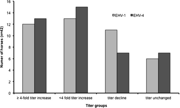
Due to practical reasons, on day 12 of the outbreak, nasal swabs, blood and sera were collected from 42 horses, irrespective of the development of clinical symptoms in individual horses. Antibody titer determinations specific for EHV-1 and EHV-4 were done using serum neutralization tests (SNT) and were repeated on day 28 of the outbreak [28]. A ≥ 4-fold titer increase was detected in 12 horses for EHV-1 (29%) and 13 horses for EHV-4 (31%). The highest EHV-1 antibody titer determined was 1:384 and was measured in the index mare on day 12. This low percentage of horses with seroconversion is probably the result of the delayed sampling, which was initiated only 12 days after the index case (Figure 4). Serum samples were analyzed in two batches on different days. Therefore, inter-test variability may have skewed the results. The rise in EHV-4 titers is likely due to the high cross-reactivity of antibodies against these closely related viruses [29]. These results confirm earlier reports suggesting SNT results are no reliable tool to diagnose acute EHV-1 infection, especially when the first sampling is not performed early in the beginning of disease in individual horses [30]. Virus detection in swabs, blood and peripheral blood mononuclear cells (PBMC) was performed by conventional nested PCR targeting glycoprotein B (gB) [31]. PCR diagnosed 11 EHV-1-positive nasal swabs, 6 positive sera and 9 positive PBMC samples; in total, 17 horses were positive for EHV-1 in at least one sample. All samples tested positive for EHV-1 in 3 horses. EHV-4 was not detectable in any of the samples. The results of our virological and serological tests confirmed that PCR of nasal swabs and PBMC samples should be initiated early in the febrile phase in suspect EHV-1 cases. Discrimination between EHV-1 and EHV-4 antibodies could have been achieved using an ELISA assay based on the C terminus of glycoprotein G [29]. But PCR and cell culture results from blood samples, nasal swabs and fetuses confirmed the clinical suspicion of an acute EHV-1 infection, which, in our opinion, decreased the importance of a differentiation of the antibody response. Additionally to routine diagnostic samples, semen samples of 3 infected non-breeding stallions, housed in the sport horse section, tested positive for EHV-1 by PCR, but not by virus isolation. Which role, if any, venereal shedding plays in EHV-1 spread requires further investigation [32].
Figure 4.

Changes in EHV-1 and EHV-4 antibody titers of serum samples collected on days 12 and 28 after the index case. A ≥ 4-fold increase in antibody titer is considered a significant rise. A simultaneous and significant increase of EHV-1 and EHV-4 antibodies was detected in 9 horses.
Disease management
From day 1, quarantine was imposed for broodmare barn 7. From day 5, different grooms attended separate barns. Facilities were entered through hygienic barriers in which personnel had to change clothes. Horse movements were prohibited and all horses remained indoors. However, new cases of fever were detected in nearly all barns during the outbreak. We concluded from this observation that quarantine measures were implemented too late and infection had already been fully established in all areas of the premise (except the breeding stallion part). Separation of horses, which were suspected to shed virus based on clinical symptoms such as fever, EHM and/or abortion in the affected barns, was deemed impossible. Isolation facilities were not available and barn construction could not be changed fast enough, so horses were able to maintain nose-to-nose contact. All abortions occurred in the broodmare stall and aborted fetuses and placentas were submitted to pathological examination and taken with precautions in order not to contaminate the environment. The facility imposed on itself a voluntary quarantine and all horse transports on and off the stud were restricted from day 1.
Treatment
At the beginning of the outbreak, clinically healthy horses (n = 33) received prophylactic IM application of an immunostimulant based on inactivated parapoxvirus ovis (2 mL/horse; IM)c given three times in two-day intervals. However, thirty-one (94%) of the treated horses developed a fever. Administration of a parapoxvirus ovis preparation was previously described as a successful measure to avoid severe clinical consequences of EHV-1 and -4 infection in young horses [33], but seemed to be not effective in our outbreak.
Horses showing fever as the only symptom received no other medication as long as they maintained good general condition and appetite. Three horses with fever were depressed and received flunixin-meglumined (1.1 mg/kg once daily for 1 – 3 days; IV). Mares that had an abortion also received flunixin-meglumined and were treated with heparinb for laminitis prevention, benzathine-penicilline (10,000 I.U./kg every other day, three times; IM), gentamicinf (4 mg/kg twice daily for 5 days; IV), oxytocing (15 I.U. as necessary; SC) and uterine lavage treatments.
In an attempt to prevent EHM, heparinb (25.000 I.U.) was given twice daily for 3 days to 31 febrile horses from day 10 of the outbreak, while the first 30 horses exhibiting fever did not receive heparin. Horses showing signs of EHM were treated symptomatically [34]. The management was directed towards supportive nursing, nutritional care and reduction of CNS inflammation with non-steroidal anti-inflammatory drugs (flunixin-meglumined,1.1 mg/kg once daily; IV). Generally, EHM horses showed good appetite but had to be fed at head level because uptake of food and water from the ground seemed to be difficult. EHM horses were also treated with flunixin-meglumined, lactated Ringer solution and 5% glucose solution, depending on the horse’s general condition and laboratory results. Patients with UMN bladder were catheterized periodically as was deemed necessary. A combination of sulfamerazin/trimethoprimh (45 mg/kg twice daily; PO) was administered for cystitis prevention until paralysis was resolved.
Disease Outcome
Horses with fever as the sole clinical sign remained in good general condition over the course of the outbreak and recovered without complication. Fertility of the aborting mares was good, as only 1 of the 6 aborting mares was barren in the following breeding season. All 8 EHM horses survived, ataxia and UMN bladder symptoms improved steadily and was resolved completely in all horses 3 months after infection.
Conclusions
We document a clinical outbreak of EHV-1 caused by a G2254/D752 ORF30 variant virus resulting in 61 clinical cases. Infection caused fever (n = 55), abortion (n = 6) and/or EHM (n = 8) for 44 days in 7 barns. Strict containment procedures employed from day 5 of the outbreak did not prevent spread of virus on the premise. Abortion (n = 6) and EHM (n = 8) cases provided a unique opportunity to collect clinical data about disease progression and prognosis for recovery and fertility. Procedures to prevent EHV-1 outbreaks include in particular proper vaccination for newly introduced horses and the restriction of contact between brood mares, adolescent horses and sport horses with foreign contact [3,6,27].
Consent
Written informed consent was obtained from the patients for publication of this report and any accompanying images.
Endnotes
aDuvaxyn, Fort Dodge Animal Health
bHeparin-Natrium Braun “Multi”, Braun
cZylexis, Pfizer Animal Health
dParaflunixin, IDT
eVeracin-compositum, Albrecht
fGenta 100, CP-Pharma
gOxytocin Albrecht, Albrecht
hTrimetox-Pulver, Veyx-Pharma GmbH.
Abbreviations
EHM: equine herpesvirus-associated myeloencephalopathy; EHV-1: equine herpesvirus type 1; gB: glycoprotein B; IM: intramuscular; I.U.: international units; IV: intravenous; ORF: open-reading frame; PBMC: peripheral blood mononuclear cells; PCR: polymerase chain reaction; SNT: serum neutralization test; SC: subcutaneous; UMN: upper motor neuron
Competing interest
The authors declare that they have no competing interests.
Authors’ contributions
JW was the field veterinarian in charge of the stud, interpreted data and wrote the manuscript. CS was the responsible veterinarian of the Pferdegesundheitsdienst, Tierseuchenkasse Baden-Württemberg, an institution under public law, collected samples and interpreted results. KF gave advice for therapeutic interventions during the outbreak and drafted the manuscript critically. UB participated in data interpretation and writing of the manuscript. KO gave extensive advice on all aspects during the outbreak and contributed in drafting the manuscript. All authors read and approved the final manuscript.
Contributor Information
Jasmin Walter, Email: jwalter@vetclinics.uzh.ch.
Christoph Seeh, Email: c.seeh@tsk-bw-tgd.de.
Kerstin Fey, Email: kerstin.fey@vetmed.uni-giessen.de.
Ulrich Bleul, Email: ubleul@vetclinics.uzh.ch.
Nikolaus Osterrieder, Email: no.34@fu-berlin.de.
Acknowledgments
The authors highly appreciate support by owners and team of the stud, which enabled collection of clinical data and sample collection. Tierseuchenkasse Baden-Württemberg, a state institution, funded this investigation. Parts of this article were presented at the 16th Annual Conference of the European Society for Domestic Animal Reproduction in Dublin, August 29th to September 1st 2012.
References
- Pusterla N, Mapes S, Madigan JE, Maclachlan NJ, Ferraro GL, Watson JL, Spier SJ, Wilson WD. Prevalence of EHV-1 in adult horses transported over long distances. Vet Rec. 2009;165:473–475. doi: 10.1136/vr.165.16.473. [DOI] [PubMed] [Google Scholar]
- Pusterla N, Hussey SB, Mapes S, Johnson C, Collier JR, Hill J, Lunn DP, Wilson WD. Molecular investigation of the viral kinetics of equine herpesvirus-1 in blood and nasal secretions of horses after corticosteroid-induced recrudescence of latent infection. J Vet Intern Med. 2010;24:1153–1157. doi: 10.1111/j.1939-1676.2010.0554.x. [DOI] [PubMed] [Google Scholar]
- Lunn DP, Davis-Poynter N, Flaminio MJ, Horohov DW, Osterrieder K, Pusterla N, Townsend HG. Equine herpesvirus-1 consensus statement. J Vet Intern Med. 2009;23:450–461. doi: 10.1111/j.1939-1676.2009.0304.x. [DOI] [PubMed] [Google Scholar]
- Patel JR, Heldens J. Equine herpesviruses 1 (EHV-1) and 4 (EHV-4)–epidemiology, disease and immunoprophylaxis: a brief review. Vet J. 2005;170:14–23. doi: 10.1016/j.tvjl.2004.04.018. [DOI] [PubMed] [Google Scholar]
- Gardiner DW, Lunn DP, Goehring LS, Chiang Y-W, Cook C, Osterrieder N, McCue P, Del Piero F, Hussey SB, Hussey GS. Strain impact on equine herpesvirus type 1 (EHV-1) abortion models: Viral loads in fetal and placental tissues and foals. Vaccine. 2012;30:6564–6572. doi: 10.1016/j.vaccine.2012.08.046. [DOI] [PubMed] [Google Scholar]
- Barbić L, Lojkić I, Stevanović V, Bedeković T, Starešina V, Lemo N, Lojkić M, Madić J. Two outbreaks of neuropathogenic equine herpesvirus type 1 with breed-dependent clinical signs. Vet Rec. 2012;170:227. doi: 10.1136/vr.100150. [DOI] [PubMed] [Google Scholar]
- Nugent J, Birch-Machin I, Smith KC, Mumford JA, Swann Z, Newton JR, Bowden RJ, Allen GP, Davis-Poynter N. Analysis of equid herpesvirus 1 strain variation reveals a point mutation of the DNA polymerase strongly associated with neuropathogenic versus nonneuropathogenic disease outbreaks. J Virol. 2006;80:4047–4060. doi: 10.1128/JVI.80.8.4047-4060.2006. [DOI] [PMC free article] [PubMed] [Google Scholar]
- Van de Walle GR, Goupil R, Wishon C, Damiani A, Perkins GA, Osterrieder N. A single-nucleotide polymorphism in a herpesvirus DNA polymerase is sufficient to cause lethal neurological disease. J Infect Dis. 2009;200:20–25. doi: 10.1086/599316. [DOI] [PubMed] [Google Scholar]
- Goodman LB, Loregian A, Perkins GA, Nugent J, Buckles EL, Mercorelli B, Kydd JH, Palu G, Smith KC, Osterrieder N, Davis-Poynter N. A point mutation in a herpesvirus polymerase determines neuropathogenicity. PLoS Pathog. 2007;3:e160. doi: 10.1371/journal.ppat.0030160. [DOI] [PMC free article] [PubMed] [Google Scholar]
- Pronost S, Leon A, Legrand L, Fortier C, Miszczak F, Freymuth F, Fortier G. Neuropathogenic and non-neuropathogenic variants of equine herpesvirus 1 in France. Vet Microbiol. 2010;145:329–333. doi: 10.1016/j.vetmic.2010.03.031. [DOI] [PubMed] [Google Scholar]
- Pronost S, Legrand L, Pitel P-H, Wegge B, Lissens J, Freymuth F, Richard E, Fortier G. Outbreak of equine herpesvirus myeloencephalopathy in France: a clinical and molecular investigation. Transbound Emerg Dis. 2012;59:256–263. doi: 10.1111/j.1865-1682.2011.01263.x. [DOI] [PubMed] [Google Scholar]
- Allen GP, Bolin DC, Bryant U, Carter CN, Giles RC, Harrison LR, Hong CB, Jackson CB, Poonacha K, Wharton R, Williams NM. Prevalence of latent, neuropathogenic equine herpesvirus-1 in the Thoroughbred broodmare population of central Kentucky. Equine Vet J. 2008;40:105–110. doi: 10.2746/042516408X253127. [DOI] [PubMed] [Google Scholar]
- Allen GP. Risk factors for development of neurologic disease after experimental exposure to equine herpesvirus-1 in horses. Am J Vet Res. 2008;69:1595–1600. doi: 10.2460/ajvr.69.12.1595. [DOI] [PubMed] [Google Scholar]
- Perkins GA, Goodman LB, Tsujimura K, Van de Walle GR, Kim SG, Dubovi EJ, Osterrieder N. Investigation of the prevalence of neurologic equine herpes virus type 1 (EHV-1) in a 23-year retrospective analysis (1984–2007) Vet Microbiol. 2009;139:375–378. doi: 10.1016/j.vetmic.2009.06.033. [DOI] [PubMed] [Google Scholar]
- Smith KL, Allen GP, Branscum AJ. Frank Cook R, Vickers ML, Timoney PJ, Balasuriya UBR: The increased prevalence of neuropathogenic strains of EHV-1 in equine abortions. Vet Microbiol. 2010;141:5–11. doi: 10.1016/j.vetmic.2009.07.030. [DOI] [PubMed] [Google Scholar]
- Pusterla N, Wilson WD, Mapes S, Finno C, Isbell D, Arthur RM, Ferraro GL. Characterization of viral loads, strain and state of equine herpesvirus-1 using real-time PCR in horses following natural exposure at a racetrack in California. Vet J. 2009;179:230–239. doi: 10.1016/j.tvjl.2007.09.018. [DOI] [PubMed] [Google Scholar]
- Pusterla N, Mapes S. David Wilson W: Prevalence of latent alpha-herpesviruses in Thoroughbred racing horses. Vet J. 2012;193:579–582. doi: 10.1016/j.tvjl.2012.01.030. [DOI] [PubMed] [Google Scholar]
- Vissani MA, Becerra ML. Olguín Perglione C, Tordoya MS, Miño S, Barrandeguy M: Neuropathogenic and non-neuropathogenic genotypes of Equid Herpesvirus type 1 in Argentina. Vet Microbiol. 2009;139:361–364. doi: 10.1016/j.vetmic.2009.06.025. [DOI] [PubMed] [Google Scholar]
- Henninger RW, Reed SM, Saville WJ, Allen GP, Hass GF, Kohn CW, Sofaly C. Outbreak of neurologic disease caused by equine herpesvirus-1 at a university equestrian center. J Vet Intern Med. 2007;21:157–165. doi: 10.1111/j.1939-1676.2007.tb02942.x. [DOI] [PubMed] [Google Scholar]
- Goehring LS, Landolt GA, Morley PS. Detection and management of an outbreak of equine herpesvirus type 1 infection and associated neurological disease in a veterinary teaching hospital. J Vet Intern Med. 2010;24:1176–1183. doi: 10.1111/j.1939-1676.2010.0558.x. [DOI] [PubMed] [Google Scholar]
- van Maanen C, Willink DL, Smeenk LA, Brinkhof J, Terpstra C. An equine herpesvirus 1 (EHV1) abortion storm at a riding school. Vet Q. 2000;22:83–87. doi: 10.1080/01652176.2000.9695030. [DOI] [PubMed] [Google Scholar]
- van Maanen C. Sloet van Oldruitenborgh-Oosterbaan MM, Damen EA, Derksen AG: Neurological disease associated with EHV-1-infection in a riding school: clinical and virological characteristics. Equine Vet J. 2001;33:191–196. doi: 10.1111/j.2042-3306.2001.tb00600.x. [DOI] [PubMed] [Google Scholar]
- Reed SM, Andrews FN, In Equine Internal Medicine. Neurologic examination. Bayly WM Philadelphia: Saunders: 2nd edition. edited by Reed SM; 2004. pp. 533–541. [Google Scholar]
- Crabb BS, Studdert MJ. Equine herpesviruses 4 (equine rhinopneumonitis virus) and 1 (equine abortion virus) Adv. 1995;45:153–190. doi: 10.1016/s0065-3527(08)60060-3. [DOI] [PubMed] [Google Scholar]
- ANON. Equine herpes virus myeloencephalopathy: a potentially emerging disease. Center for emerging issues information sheet: Centers for Epidemiology and Animal Health; 2007. [Google Scholar]
- Goehring LS, van Winden SC, van Maanen C. Sloet van Oldruitenborgh-Oosterbaan MM: Equine herpesvirus type 1-associated myeloencephalopathy in The Netherlands: a four-year retrospective study (1999–2003) J Vet Intern Med. 2006;20:601–607. doi: 10.1892/0891-6640(2006)20[601:ehtami]2.0.co;2. [DOI] [PubMed] [Google Scholar]
- Kydd JH, Slater J, Osterrieder N, Lunn DP, Antczak DF, Azab W, Balasuriya U, Barnett C, Brosnahan M, Cook C, Damiani A, Elton D, Frampton A, Gilkerson J, Goehring L, Horohov D, Maxwell L, Minke J, Morley P, Nauwynck H, Newton R, Perkins G, Pusterla N, Soboll-Hussey G, Traub-Dargatz J, Townsend H, Van de Walle GR, Wagner B. Third International Havemeyer Workshop on Equine Herpesvirus type 1. Equine Vet J. 2012;44:513–517. doi: 10.1111/j.2042-3306.2012.00604.x. [DOI] [PubMed] [Google Scholar]
- OIE. Manual of diagnostic tests and vaccines for terrestrial animals. : Chapter 2.5.9 Equine Rhinopneumonitis; 2008. pp. 894–903. [Google Scholar]
- Hartley CA, Wilks CR, Studdert MJ, Gilkerson JR. Comparison of antibody detection assays for the diagnosis of equine herpesvirus 1 and 4 infections in horses. Am J Vet Res. 2005;66:921–928. doi: 10.2460/ajvr.2005.66.921. [DOI] [PubMed] [Google Scholar]
- Irwin VL, Traub-Dargatz JL, Newton JR, Scase TJ, Davis-Poynter NJ, Nugent J, Creis L, Leaman TR, Smith KC. Investigation and management of an outbreak of abortion related to equine herpesvirus type 1 in unvaccinated ponies. 2007. pp. 378–380. [DOI] [PubMed]
- Borchers K, Slater J. A nested PCR for the detection and differentiation of EHV-1 and EHV-4. J Virol Methods. 1993;45:331–336. doi: 10.1016/0166-0934(93)90117-A. [DOI] [PubMed] [Google Scholar]
- Walter J, Balzer H-J, Seeh C, Fey K, Bleul U, Osterrieder N. Venereal Shedding of Equid Herpesvirus-1 (EHV-1) in Naturally Infected Stallions. J Vet Intern Med. 2012;26:1500–1504. doi: 10.1111/j.1939-1676.2012.00997.x. [DOI] [PubMed] [Google Scholar]
- Ziebell KL, Steinmann H, Kretzdorn D, Schlapp T, Failing K, Schmeer N. The use of Baypamun N in crowding associated infectious respiratory disease: efficacy of Baypamun N (freeze dried product) in 4–10 month old horses. Zentralbl Veterinarmed B. 1997;44:529–536. doi: 10.1111/j.1439-0450.1997.tb01004.x. [DOI] [PubMed] [Google Scholar]
- Pusterla N. David Wilson W, Madigan JE, Ferraro GL: Equine herpesvirus-1 myeloencephalopathy: a review of recent developments. Vet J. 2009;180:279–289. doi: 10.1016/j.tvjl.2008.08.004. [DOI] [PubMed] [Google Scholar]


