Abstract
Introduction
Obesity is an increasing problem of epidemic proportion, and it is associated with various musculoskeletal disorders, including impairment of the spine. However, the relationship between obesity and spino-pelvic parameters remains to date unsupported by an objective measurement of the mechanical behavior of the spino-pelvic parameters depending on body mass index (BMI) and the presence of central obesity. Such analysis may provide a deeper understanding of this relationship.
Purpose
To assess whether BMI and central obesity are associated with modifications on spino-pelvic parameters and determine if exists any correlation between BMI and obesity with the type of lumbar lordosis (LL).
Methods
A cross-sectional study with 200 participants was conducted. Parameters measured were LL, pelvic tilt, sacral slope, and pelvic incidence (PI), using lumbosacral radiographs in lateral view. Subjects were classified depending on BMI. In a secondary analysis, the subjects were categorized into two groups depending on the presence or not of elevated abdominal circumference. The categorical variables were compared using Chi-square test, and the mean values were compared using ANOVA and student t test. A Spearman correlation test was used to analyze the correlation between BMI categories and LL types.
Results
From the total of participants, there were 51 (25.5 %) normal weight subjects, 93 (46.5 %) overweight, and 56 (28 %) obese individuals. The spino-pelvic parameters among these groups are practically equal. The correlation between the different BMI categories and LL types is poor 0.06 (P = 0.34). In a secondary analysis, grouping the participants in obese and non-obese, the results showed that obesity is modestly positively associated with increasing of spino-pelvic parameters values, in particular with PI (P = 0.078). The comparison made between the presence or not of central obesity, interestingly did not show significant differences.
Conclusions
Despite the results did not reach statistically significant differences, the results indicate that the obese spine is slightly different from the non-obese spine. Therefore, this relationship deserves future attention.
Keywords: Body mass index, Central obesity, Spino-pelvic parameters
Introduction
Obesity is recognized as a major public health problem and it is associated with various musculoskeletal disorders, including impairment of the spine [7, 18]. Normal stance and gait requires alignment between the spine, pelvis, and lower extremities. Congruence between the spine-sacrum (represented by the S1 endplate) and pelvis-lower extremities (hip joints) is determined by a fixed parameter, the pelvic incidence (PI), and by two positional parameters the pelvic tilt (PT) and sacral slope (SS) [3–5, 12].
SS is defined as the angle between the sacral plate and a horizontal line. PT is defined by the line through midpoint of the sacral plate and midpoint of the femoral heads axis, and the vertical line. PI is defined as the angle between the line perpendicular to the sacral plate at its midpoint and the line connecting this point to the femoral heads axis. The SS and PI also affect other positional parameters of the spine, such as the amount of lumbar lordosis (LL). According to the orientation of SS, Roussouly [15] recently described four types of LL: type I (SS <35° and short hyperlordosis), type II (SS <35° and flat lordosis), type III (SS between 35° and 45° with a harmonious regular back), and type IV (SS >45° with hypercurved back).
It has been hypothesized that excessive body weight could have mechanical ill effects on the back caused by excessive weight-bearing or that there could be a biomechanical explanation for such a link [1]. Great variability exists in normative spino-pelvic values, reflecting a significant margin of adaptation [6].
Multiple studies have described normative values for parameters of spino-pelvic alignment in different populations of varying ages, and pathologic conditions [14, 17]. However, the relationship between obesity and spino-pelvic parameters remains to date unsupported by an objective measurement of the mechanical behavior of the spino-pelvic parameters depending on body mass index (BMI). To our knowledge the BMI and spino-pelvic parameters have never been studied before.
The current study was designed to assess whether BMI and central obesity are associated with modifications on spino-pelvic parameters and to determine if exists any correlation between BMI and obesity with the Roussouly’s classification of LL [15]. Such analysis may provide a deeper understanding of this relationship.
Materials and methods
This is a cross-sectional study. The sample consisted of 200 Mexican healthy adult volunteers who were recruited and divided in three groups depending on BMI: I group BMI = 20.0–24.9-normal weight, II group BMI = 25.0–24.9-overweight, and III group BMI = 25.0–29.9-obesity. The BMI was calculated by dividing weight (kg) by the square of height (m). Subjects with any previous spine surgery, associated musculoskeletal syndrome, or significant lower limb discrepancy (>2 cm) were excluded from the study. For each subject, the following information was obtained: age, sex, height, weight, abdominal circumference, and lateral standing radiographs of the lumbosacral region.
Waist circumference has been shown to be one of the most accurate anthropometrical indicators of abdominal fat. Waist was measured with a tape to the nearest 0.5 cm, in light garments at midpoint between the last rib and the highest point on the iliac crest at the end of normal inspiration. The criteria for central obesity (elevated waist circumference) was established as >102 cm for men and >88 cm for women.
The study was conducted at the spine surgery and radiology departments of National Institute of Rehabilitation with approbation by the local ethical committee and written informed consent given by all the participants.
Radiological assessment
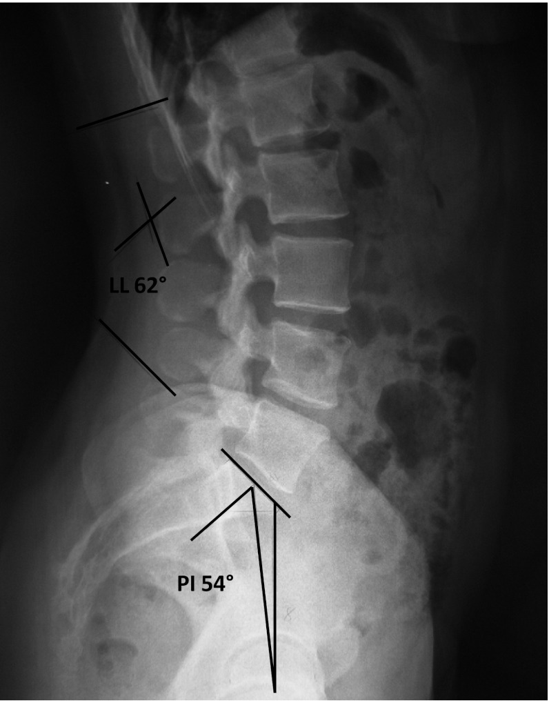
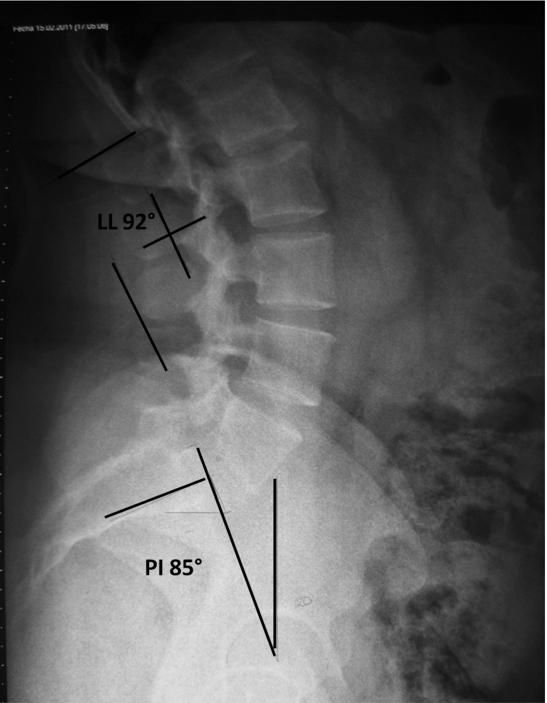
Each subject had a 30 × 90 cm lumbosacral lateral X-ray in a standardized upright position, arms lying forward horizontally on a support, and knees fully extended. Special care was taken to visualize both femoral heads on this X-ray. If the femoral heads did not completely overlap on the standing lateral film, then the midpoint of the line connecting both femoral head centers was alternatively used (Figs. 1, 2).
Fig. 1.
Lateral X-ray of the lumbar spine of a patient with a BMI of 22, PI 54°, SS 46°, PT 8°, and LL 62°
Fig. 2.
X-ray of the lumbar spine of a patient with a BMI of 37. Higher values of pelvic incidence and lumbar lordosis are found compared with patients with BMI <25, PI 85°, SS 65°, PT 20°, and LL 92°
Fixed and postural parameters were measured to explore the anatomic characteristics of the lumbopelvic junction. The following radiologic parameters were measured: LL (Cobb angle from the upper endplate of L1 to the upper endplate of S1), PT, SS, and PI, additionally, the patients were classified according to their type of LL. The angular parameters were expressed in degrees. The measurements were made manually, twice by two separated experienced individuals (S.R.V) (E.O.C).
Statistics
The mean values, range, and standard deviation (SD) were obtained for all the measurements. The comparison between categorical values was carried out with Chi-square test. The Kolmogorov–Smirnoff test was used to verify the normal data distribution. Comparison between different BMI means was carried out with one-way ANOVA test and the comparison between two means was made with Student t test. A Pearson correlation test was used to search for correlations between BMI and spino-pelvic values. A Spearman correlation test was used to analyze the correlation between BMI categories and LL types. Statistical significance levels were considered to be P < 0.05. All statistical analyses were carried out using SPSS, version 15.0 (SPSS Inc., Chicago, IL).
Results
The number of participants was 200 (n = 200), of these there were 51 (25.5 %) normal weight subjects, 93 (46.5 %) overweight, and 56 (28 %) obese individuals. The ages and weight by groups were: Normal weight group (29 years, 58.9 kg, range 45.2–78 kg), overweight group (31 years, 71.3 kg, range 47–101 kg), and obese group (28 years, 86.4 kg, range 65–113 kg). The analyzed groups were homogeneous in terms of age. The statistical analysis revealed no significant difference between the measurements for the intra and inter-observer comparisons.
The correlations between BMI and spino-pelvic values were poor: for PT 0.02 (P = 0.37), SS and PI 0.06 (P = 0.18).
The mean values and SDs for the measured parameters are shown in Table 1.
Table 1.
Spino-pelvic values, distributed by BMI groups
| Variable | Normal weight, mean (SD) | Overweight, mean (SD) | Obese, mean (SD) | P value |
|---|---|---|---|---|
| LL | 60.4 (13.9) | 59.4 (13.4) | 61.1 (14.9) | 0.38 |
| PT | 15.4 (7.4) | 15.1 (8.8) | 17.1 (8.3) | 0.349 |
| SS | 41 (10.9) | 40 (10) | 42.2 (11.2) | 0.473 |
| PI | 56.5 (12.5) | 55.1 (13.5) | 59.3 (13.7) | 0.176 |
The correlation among different BMI categories and LL types was poor 0.06 (P = 0.34) (Table 2).
Table 2.
Cross-tabs showing the frequencies of different BMI categories and LL types
| LL type | Normal weight | Overweight | Obese | Total |
|---|---|---|---|---|
| 1 | 13 | 18 | 8 | 39 |
| 2 | 3 | 7 | 7 | 17 |
| 3 | 17 | 42 | 19 | 78 |
| 4 | 18 | 26 | 22 | 66 |
| Total | 51 | 93 | 56 | 200 |
The Kolmogorov–Smirnoff normality test accepted the normality for all the spino-pelvic parameters. Overall, spino-pelvic parameters angles increased in the obese group, but without statistically significant difference.
A dichotomous comparison was made between obese (n = 56) and non-obese individuals (n = 144), the measurements are shown in Table 3.
Table 3.
Spino-pelvic values among obese and non-obese individuals
| Variable | Non-obese, mean (SD) | Obese, mean (SD) | P value |
|---|---|---|---|
| LL | 59.7 (13.5) | 61.1 (14.9) | 0.533 |
| PT | 15.2 (8.3) | 17.1 (8.3) | 0.153 |
| SS | 40.3 (10.3) | 42.2 (11.2) | 0.271 |
| PI | 55.6 (13.1) | 59.3 (13.7) | 0.078 |
The differences are more pronounced when comparing obese patients with to those without obesity, but the P values cannot reach the statistical significance.
Comparing the frequencies of LL types between obese and non-obese patients, we did not find significant differences (P = 0.26) (Table 4).
Table 4.
Cross tabs showing the frequencies between LL types according to the presence of obesity or not
| LL type | Non-obese | Obese | Total |
|---|---|---|---|
| 1 | 31 | 8 | 39 |
| 2 | 10 | 7 | 17 |
| 3 | 59 | 19 | 78 |
| 4 | 44 | 22 | 66 |
| Total | 144 | 56 | 200 |
BMI does not differentiate fat distribution, therefore, we made a comparative analysis making two groups based on the presence of elevated abdominal circumference or not. The comparative results of spino-pelvic values are shown in Table 5.
Table 5.
Spino-pelvic values based on the presence of central obesity
| Variable | Normal abdominal circumference, mean (SD) | Elevated abdominal circumference, mean (SD) | P value |
|---|---|---|---|
| LL | 59.9 (13.6) | 61.3 (15.6) | 0.635 |
| PT | 15.3 (7.2) | 16.1 (9.2) | 0.547 |
| SS | 41.1 (9.8) | 40.7 (11.2) | 0.788 |
| PI | 56.5 (11.4) | 56.8 (14.8) | 0.878 |
Comparing the frequencies of LL types between patients with abdominal obesity and without it, we did not find significant differences (P = 0.89) (Table 6).
Table 6.
Cross tabs showing the frequencies between LL types according to the presence of elevated abdominal circumference or not
| LL type | Normal abdominal circumference | Elevated abdominal circumference | Total |
|---|---|---|---|
| 1 | 33 | 6 | 39 |
| 2 | 14 | 3 | 17 |
| 3 | 64 | 14 | 78 |
| 4 | 52 | 14 | 66 |
| Total | 163 | 37 | 200 |
Discussion
Duval-Beaupère and co-workers [12] have demonstrated that PI is an important anatomic parameter that describes the positional configuration of the pelvis (PI = SS + PT) and of the sacrum. The spine reacts to this position by adapting through LL, the amount of lordosis increasing as the SS increases to balance the trunk in the upright position [16].
Relatively, little is known about the importance of spino-pelvic parameters in human musculoskeletal disorders. The spino-pelvic parameters have clinical implications; [2, 9, 10, 13] an association between PI and spondylolisthesis has been reported in many publications.
The risk of early distal discopathy increases in patients with low PI and flat back [15].
Lumbar lordosis types 3 and 4 have a bigger LL, mainly type 4. They are generally associated with a horizontal pelvis with a high-grade PI and SS, with a risk of isthmic spondylolisthesis through a ‘‘sliding’’ mechanism [15].
When differences in spine biomechanics of obese subjects are investigated, only a moderate link between low back pain and BMI appears [11].
Because biomechanics play an important role in the initiation and progression of several spine pathologies, it is imperative to have an understanding of spino-pelvic alignment among the obese patients. The present study provides an objective analysis of the correlation between BMI and spino-pelvic parameters.
Our findings revealed that the differences of the spino-pelvic parameters among different BMI categories did not reach statistical significance.
However, when comparing obese versus non-obese, we observed a slight increase in all the spino-pelvic parameters, including the LL. This finding implies that among obese people, the lumbosacral junction is subject to greater shear loads. Whereas the normal anatomy of the spine makes it well suited to resist axial load and anterior shear, the greater shear loads among obese subjects may lead to a less stable lumbosacral junction.
There have been several investigations that conclude that during stance, obese patients show a hyperextension of the lumbar spine, [8, 14] similar to the anterior translation of the center of mass described by Whitcome in pregnant women [19]. Therefore, we hypothesized that elevated abdominal circumference and gravity effect could influence the spino-pelvic parameters; interestingly, we only observed a slight elevation of LL among the patients with abdominal obesity that did not reach statistical significance. In our opinion, obese subjects, as women at early stages of pregnancy, seem to compensate the forward translation of the center of mass only with slight increases of LL and PI.
We sought to examine the correlation between the different obesity parameters and LL types described by Roussouly [15], hypothesizing that the abdominal obesity could have an influence over lumbar spine shape, but consistently we found poor correlations.
The main limitation of the study is that the impact of the BMI and central obesity over the whole spine sagittal alignment was not determined.
According to the differences observed, we fully recognize that the association between obesity and increased spino-pelvic parameters is weak. Possibly a larger study sample, including a larger sample of class II obesity (BMI 35–39.9) and class III obesity (BMI >40.0) should provide an interesting correlation. This association has yet to be fully investigated and discussed.
Conclusions
This is the first accurate analysis that compares spino-pelvic parameters on BMI subgroups and the presence of central obesity. The spino-pelvic parameters increased slightly among obese subjects, but the differences did not reach statistical difference. Apparently, the BMI and obesity do not have an important influence over the spino-pelvic parameters, looking for a definitive answer such relationship needs further research.
Conflict of interest
None.
References
- 1.Aro S, Leino P. Overweight and musculoskeletal morbidity: a ten-year follow-up. Int J Obes. 1985;9:267–275. [PubMed] [Google Scholar]
- 2.Barrey C, Jund J, Perrin G, Roussouly P. Spinopelvic alignment of patients with degenerative spondylolisthesis. Neurosurgery. 2007;61:981–986. doi: 10.1227/01.neu.0000303194.02921.30. [DOI] [PubMed] [Google Scholar]
- 3.Berthonnaud É, Roussouly P, Dimnet J. The parameters describing the shape and the equilibrium of the set back pelvis and femurs in sagittal view. Innov Techn Biol Med. 1998;19:411–426. [Google Scholar]
- 4.Berthonnaud É, Dimnet J, Roussouly P, Labelle H. Analysis of the sagittal balance of the spine and pelvis using shape and orientation parameters. J Spinal Disord. 2005;18:40–47. doi: 10.1097/01.bsd.0000117542.88865.77. [DOI] [PubMed] [Google Scholar]
- 5.Berthonnaud É, Labelle H, Roussouly P, Grimard G, Vaz G, Dimnet J. A variability study of computerized sagittal spinopelvic radiological measurements of trunk balance. J Spinal Disord. 2005;18:66–71. doi: 10.1097/01.bsd.0000128345.32521.43. [DOI] [PubMed] [Google Scholar]
- 6.Boulay C, Tardieu C, Hecquet J, Benaim C, Mouilleseaux B, Marty C, et al. Sagittal alignment of spine and pelvis regulated by pelvic incidence: standard values and prediction of lordosis. Eur Spine J. 2006;15:415–422. doi: 10.1007/s00586-005-0984-5. [DOI] [PMC free article] [PubMed] [Google Scholar]
- 7.Fanuele JC, Abdu WA, Hanscom B, Weinstein JN. Association between obesity and functional status in patients with spine disease. Spine. 2002;27:306–312. doi: 10.1097/00007632-200202010-00021. [DOI] [PubMed] [Google Scholar]
- 8.Gilleard W, Smith T. Effect of obesity on posture and hip joint moments during a standing task, and trunk forward flexion motion. Int J Obes. 2007;31:267–287. doi: 10.1038/sj.ijo.0803430. [DOI] [PubMed] [Google Scholar]
- 9.Hanson DS, Bridwel KH, Rhee J, et al. Correlation of pelvic incidence with low and high-grade isthmic spondylolisthesis. Spine. 2002;27(18):2026–2029. doi: 10.1097/00007632-200209150-00011. [DOI] [PubMed] [Google Scholar]
- 10.Labelle H, Roussouly P, Berthonnaud E, Transfeldt E, O Brien M, Hresko T, Chopin D, Dimmet J. Spondylolisthesis, pelvic incidence and sagittal spino-pelvic balance: a correlation study. Spine. 2004;29(18):2049–2054. doi: 10.1097/01.brs.0000138279.53439.cc. [DOI] [PubMed] [Google Scholar]
- 11.Leboeuf-Yde C, Kyvic KO, Bruun NH. Low back pain and lifestyle. Part II-obesity. Information from a population-based sample of 29,242 twin subjects. Spine. 1999;15:779–783. doi: 10.1097/00007632-199904150-00009. [DOI] [PubMed] [Google Scholar]
- 12.Legaye J, Duval-Beaupere G, Hecquet J, et al. Pelvic incidence: a fundamental pelvic parameter for three-dimensional regulation of spinal sagittal curves. Eur Spine J. 1998;7:99–103. doi: 10.1007/s005860050038. [DOI] [PMC free article] [PubMed] [Google Scholar]
- 13.Marty C, Boisaubert B, Descamps H, et al. The sagittal anatomy of the sacrum among young adults, infants and spondylolisthesis patients. Eur Spine J. 2002;11:119–125. doi: 10.1007/s00586-001-0349-7. [DOI] [PMC free article] [PubMed] [Google Scholar]
- 14.O’Sullivan PB, Dankaerts W, Burnett AF, Farrell GT, Jefford E, Naylor CS, O’Sullivan KJ. Effect of different upright sitting postures on spinal-pelvic curvature and trunk muscle activation in a pain-free population. Spine. 2006;31(19):E707–E712. doi: 10.1097/01.brs.0000234735.98075.50. [DOI] [PubMed] [Google Scholar]
- 15.Roussouly P, Pinheiro-Franco JL. Biomechanical analysis of the spino-pelvic organization and adaptation in pathology. Eur Spine J. 2011;20(5):609–618. doi: 10.1007/s00586-011-1928-x. [DOI] [PMC free article] [PubMed] [Google Scholar]
- 16.Vaz G, Roussouly P, Berthonnaud E, Dimnet J. Sagittal morphology and equilibrium of pelvis and spine. Eur Spine J. 2002;11:80–87. doi: 10.1007/s005860000224. [DOI] [PMC free article] [PubMed] [Google Scholar]
- 17.Vialle R, Levassor N, Rillardon L, Templier A, Skalli W, Guigui P. Radiographic analysis of the sagittal alignment and balance of the spine in asymptomatic subjects. J Bone Jt Surg Am. 2005;87:260–267. doi: 10.2106/JBJS.D.02043. [DOI] [PubMed] [Google Scholar]
- 18.Vismara L, Menegoni F, Zaina F, Galli M, Negrini S, Capodaglio P. Effect of obesity and low back pain on spinal mobility: a cross sectional study in women. J Neuroeng Rehabil. 2010;18(7):3. doi: 10.1186/1743-0003-7-3. [DOI] [PMC free article] [PubMed] [Google Scholar]
- 19.Whitcome KK, Shapiro JL, Lieberman DL. Fetal load and the evolution of lumbar lordosis in bipedal hominins. Nature. 2007;450(7172):1075–1078. doi: 10.1038/nature06342. [DOI] [PubMed] [Google Scholar]


