Abstract
Tonalli A (TnaA) is a Drosophila melanogaster protein with an XSPRING domain. The XSPRING domain harbors an SP-RING zinc-finger, which is characteristic of proteins with SUMO E3 ligase activity. TnaA is required for homeotic gene expression and is presumably involved in the SUMOylation pathway. Here we analyzed some aspects of the TnaA location in embryo and larval stages and its genetic and biochemical interaction with SUMOylation pathway proteins. We describe that there are at least two TnaA proteins (TnaA130 and TnaA123) differentially expressed throughout development. We show that TnaA is chromatin-associated at discrete sites on polytene salivary gland chromosomes of third instar larvae and that tna mutant individuals do not survive to adulthood, with most dying as third instar larvae or pupae. The tna mutants that ultimately die as third instar larvae have an extended life span of at least 4 to 15 days as other SUMOylation pathway mutants. We show that TnaA physically interacts with the SUMO E2 conjugating enzyme Ubc9, and with the BRM complex subunit Osa. Furthermore, we show that tna and osa interact genetically with SUMOylation pathway components and individuals carrying mutations for these genes show a phenotype that can be the consequence of misexpression of developmental-related genes.
Introduction
SUMOylation is a post-translational protein modification that can change the location, stability, activity or the interactions of the protein targets involved in many cellular processes, including cell death, cell cycle, signal transduction, and gene expression [1]. SUMOylation is the addition of SUMO (Small Ubiquitin-related MOdifier) to lysine residues of the target protein in the consensus amino acid sequence ΨKxE (Ψ represents a hydrophobic amino acid) [2]. Hundreds of proteins are SUMOylated in Drosophila [3]. The SUMOylation pathway starts with processing of an immature SUMO protein by the Ulp/SENP family of proteases. Next, the activating enzyme E1 (an Aos1/Uba2 heterodimer) generates a mature SUMO-adenylate intermediary which then forms a thioesther bond between the catalytic cysteine of Uba2 and SUMO. SUMO is next transferred to the E2 conjugating enzyme (Ubc9), which transfers SUMO to the target proteins. The SUMO E3 ligases function by stimulating the activity of Ubc9 or by facilitating the formation of an Ubc9-substrate complex. Finally, proteins of the Ulp/SENP family proteases make this whole process reversible [4].
The tna gene was identified in a genetic screen designed to find brahma (brm)-interacting genes [5]. brm encodes the SNF2 type-ATPase of the BRM chromatin remodeling complexes [6], [7]. The osa gene encodes an exclusive subunit of one type of BRM complexes [6], [8], [9]. Besides interacting with brm, tna interacts even stronger with osa. All three genes (brm, osa, and tna) are required for proper expressions of the homeotic genes [5]. Homeotic genes determine the identity of body segments in Drosophila [10], [11].
The role of various components of the SUMOylation pathway have been studied in Drosophila development [12], [13]. tna is involved in homeotic gene expression but little is known about the proteins encoded by this locus. tna expresses a at least one putative isoform called TnaA [5]. This isoform has an XSPRING (eXtended SP-RING) domain that harbors a zinc finger of the SP-RING type {Siz/PIAS (Protein Inhibitors of Activated STAT [Signal Transducers and Activator of Transcription])–RING (Really Interesting New Gene)}. This zinc finger is present in one of the four major groups of proteins that have SUMO E3 ligase activity [1]. The only SP-RING finger proteins with putative SUMO E3 ligase activity that have been identified in the Drosophila proteome are Su(var)2–10 [14] and TnaA [5].
Here we show that TnaA physically interacts with both Ubc9 (the SUMO E2 conjugating enzyme) and with Osa (a putative in vivo target). We determined the dynamics of different TnaA species throughout development and showed that TnaA is an embryonic nuclear protein and is also present at discrete bands on polytene salivary gland chromosomes of third instar larvae. We also found that defects in tna cause larval lethality, abnormalities in the whole protein profile and an extension of the lifespan at this stage. Finally, we found genetic interactions between tna and osa and genes encoding the SUMOylation pathway components.
Materials and Methods
Ethics Statement
All animal handling was approved by the Instituto de Biotecnología Bioethics Comittee, Permit Number 226 (2009/12/04), which follows NOM-062 animal welfare mexican law. All efforts were made to minimize animal suffering. Animals were sacrificed by CO2 euthanasia.
Protein Extraction and Analyses
Soluble protein extracts for the developmental Western were obtained from 1 g of Ore-R individuals from each developmental stage with Trizol (Invitrogen). For cellular localization of the TnaA proteins, soluble nuclear (SNF) and cytoplasmic fractions were obtained from Ore-R embryo collections of 3–21 hour postfertilization [15]. The SNF was also used for the TnaA coimmunoprecipitation (Co-IP) assays. For Osa Co-IP assays, a total soluble protein fraction was obtained from Ore-R embryo collections of 3–21 hour postfertilization [16]. Protein extracts from salivary glands of third instar larvae were obtained by collecting the glands in PBS buffer plus Complete protease inhibitors [EDTA-free protease inhibitor tablet (ROCHE)], and boiling them for 5 minutes in sample loading buffer. The proteins were separated by SDS-PAGE and electrotransfered to nitrocellulose membranes for Western blot analyses. Immunoblots were done according to standard procedures and proteins of interest were detected with specific antibodies using different chemoluminiscence kits (Supersignal West Pico Chemiluminescent Substrate from Thermo scientific, ECL Plus Western Blotting Detection System or ECL Advanced Western Blotting Detection kit from Amersham, GE Healthcare, USA), according to manufactureŕs instructions.
Affinity-purified primary TnaANH2 and TnaAXSPRING antibodies were used at a 1∶100 dilution. Anti-β-tubulin (E7, Developmental Studies Hybridoma Bank) and anti-Osa (Developmental Studies Hybridoma Bank) were used at 1∶3000 and 1∶1000 dilutions, respectively. The antibodies anti-Cdk7 (ds17, Santa Cruz), anti-RNA Pol II (8WG16, Covance) and anti-Hsp70 (ab2787, ABCAM) were used at 1∶1000, 1∶500 and 1∶600, respectively.
Production and Affinity Purification of TnaA Antibodies
To generate antibodies against different TnaA regions, we used the TnaA cDNA that contains the TnaA translated exons from the ZAP1 clone [5] that represent the TnaA RD transcript [17]. The TnaA cDNA clone was digested with BamHI and two fragments were independently subcloned into the pGEX2T vector to generate glutathione S-transferase (GST) fusion proteins harboring the TnaA amino-termini (amino acids 159–432, GST-TnaANH2–1) and the XSPRING domain (amino acids 433–856, GST-TnaAXSPRING1). GST-fusion proteins were expressed and purified [18] to inject Winstar rats to raise polyclonal antibodies [19]. The antibodies from total sera were affinity-purified [20].
Pull-down and Immunoprecipitation Assays
All the clones used in this work were nucleotide-sequenced. The Drosophila Ubc9 cDNA (BDGP Gold collection of Drosophila Genomics Resource Center) was amplified with the Forward: 5′-AGTTCGGAGAATTCTCCGGCATTGCTATTACACG-3′ and Reverse: 5′-CGGAATCCTCGAGGCG-CTTCTCGTACTCCAG-3′ primers, and cloned in the EcoRI and XhoI sites of the pGEX-4T vector. Pull-down assays were done as described previously [21]. Immunoprecipitations were done on SNF or total protein extracts from 3–21 hour postfertilization Ore-R embryos [22]. In these assays we made two preclearings steps and we used the Buffer PD (20 mM HEPES, pH 7.9, 100 mM NaCl, 1 mM EDTA, 4 mM MgCl2, 1 mM DTT, 0.1% NP-40, 10% glycerol and 0.2 mM PMSF).
Yeast Two-hybrid Assays
Drosophila TnaA and Ubc9 cDNAs were cloned in the EcoRI and SalI, and EcoRI and XhoI sites of the pGBKT7 and pGADT7 vectors, respectively. pGBKT7-TnaA was digested with BamHI and religated to obtain the TnaANH2–2 fragment (1–432 aa). TnaAXSPRING2 (379–927 aa) and TnaACOO (929–1073 aa) were obtained after the digestion of the full-length pGBKT7-TnaA with NcoI and each fragment was cloned separately in NcoI-digested pGBKT7. TnaAQLess (306–696 aa) and TnaASP-COO QLess (711–999 aa) were obtained by cloning in pGBKT7 digested with NdeI and EcoRI, PCR fragments obtained from the TnaA cDNA clone [5] using the primers: a) TnaAQLess Forward 5′-GGAATTCCATATGCGACGAATGGC. CCCATATC-3′ and Reverse 5′-CGAGAATTCATCTG-GCCCGGCATTC-3′, b) TnaASP-COO QLess Forward 5′-GGACAGGCTCATATGGCCAAGATCTCATTGAAGTGC-3′ and Reverse 5′-GCAGAATTCCGTTT. GGGGCGAGTTGTG-3′. OsaC2 cDNA harbouring aminoacids 1951–2600 from the Osa protein was synthesized from polyA+ RNA from Ore-R embryos 3–21 hour postfertilization, according to [23]. The OsaC2 PCR fragment was synthesized with the Forward: 5′-GGAATTCTCCATATGAACTACACGATGGT. CACG-3′ and Reverse: 5′-CGTGAATTCCGTACCGCAGCTGTTGCTGTTG-3′ primers and cloned in pGADT7 with NdeI and EcoRI. Yeast two-hybrid assay was performed using the BD Matchmaker library construction and screening kit (Biosciences-Clontech). The interaction between baits and preys were tested evaluating the reporter genes ADE2 and HIS3 using the media QDO (SD-Trp/−Leu/−Ade/−His) supplemented with 3 mM 3-AT (3-amino-1,2,4-triazole).
Fly Strains, Genetic Procedures, and Larval Staging
Unless otherwise noted, all mutations are described in Flybase [17]. Briefly, tna1, tna5, osa1 and osa2 are EMS-induced mutations. In tna1 Gln 566 changed to a stop codon [5]. tna5 was recovered after EMS mutagenesis in a genetic screen to identify brm-interacting mutations (J. A. K., unpublished results). The lesion in the lwr5 allele (Arg 104 to His) is located in a region that has been involved in the interaction between ubiquitin-conjugating enzymes with the HECT or RING ubiquitin E3 ligases [24]. The lwr4–3 and lwr13 were both derived from imprecise excision of P-elements inserted in the 5′ regulatory zone [25], [26]. smt304493 is a P-element insertion 10 bp upstream of the first exon of smt3 [27]. Fly cultures and crosses were performed according to standard procedures. Flies were raised on cornmeal-molasses media at 25°C unless otherwise noted. Media were supplemented with 0.05% of bromophenol blue to stage third instar larvae according to the gut dye clearance [28].
Immunostaining of Ring Glands, Salivary Glands, and Polytene Chromosomes of Third Instar Larvae
Immunostaining of ring and salivary glands were done as described by [29], and the immunostaining of polytene salivary gland chromosomes was done as reported by [30]. For immunostaining of polytene salivary gland chromosomes, the TnaAXSPRING antibodies were preabsorbed with fixed 0–3 hour embryos [31]. Polytene chromosomes and salivary and ring glands images were captured on a Leitz DMIRB inverted photoscope equipped with a Leica TCS Nt laser confocal imaging system, a Zeiss Inverted Axiovert fluorescent microscope, a Leica Aristaplan fluorescent microscope or an Olympus Inverted confocal FV1000 microscope. Images were processed using Image J.
Results
TnaA130 and TnaA123 in Space and Time throughout Development
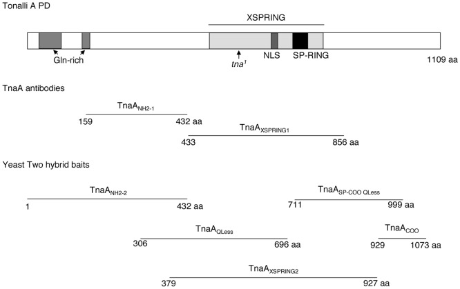
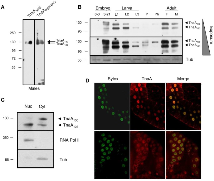
The tna gene produces several large transcripts that are differentially expressed from embryo through adult stages [5], [17]. The main large transcript is 6.1 kb and it peaks at the pupal stage [5]. Translation of this transcript predicts a protein product of 127 kDa that we named TnaA [5]. To study TnaA, we prepared two affinity-purified antibodies: TnaANH2 that was raised against the amino-terminal region and TnaAXSPRING that was raised against the XSPRING domain (Fig. 1 and Material and Methods). Both antibodies recognize the same proteins on adult male soluble extracts and they were used indistinctly along this work (Fig. 2A).
Figure 1. TnaA protein, domains and fragments.
The TnaA protein (upper section). TnaA domains are indicated. Nuclear localization signal is NLS. The stop codon in the tna1 allele [5] is indicated by an arrow. TnaA fragments used to produce TnaA antibodies from GST fusion proteins are shown (middle section). TnaA fragments fused to the yeast GAL4-binding domain to use as baits in two-hybrid assays (lower section).
Figure 2. Expression and location of TnaA proteins throughout Drosophila development.
(A) TnaANH2 and TnaAXSPRING antibodies detect the same proteins. TnaA proteins detected by full-range Western analysis in an adult male soluble protein extract with TnaANH2 and TnaAXSPRING antibodies (1∶100 dilution). (B) TnaA developmental Western. Detection of TnaA130 and Tna123 isoforms in soluble extracts isolated from embryos (0–3 and 3–21 hour), 1st, 2nd and 3rd instar larvae (L1, L2 and L3), pupae (P), pharate (Ph) and female and male adults (F and M). β-tubulin was used as a protein loading control (bottom). The TnaAXSPRING and β-tubulin antibodies were used 1∶100 and 1∶1000, respectively. (C) TnaA123 is mainly nuclear. Detection of TnaA proteins in nuclear (Nuc) and cytoplasmic (Cyt) soluble fractions isolated from embryos 3–21 hour. The largest RNA polymerase II subunit and β-tubulin were used as controls of nuclear and cytoplasmic fractions, respectively. TnaANH2, RNA polymerase II, and β-tubulin antibodies were used 1∶120, 1∶500, and 1∶1000, respectively. (D) Immunostaining of TnaA in salivary (upper panel) and ring glands (lower panel) of Ore-R third instar larvae with TnaAXSPRING (3∶5, red), DNA (Sytox, green) and merge (yellow). We detected no signal when immunostaining was done with secondary antibody only (not shown).
Two main TnaA protein products, one of 130 kDa (TnaA130) and another one of 123 kDa (TnaA123) are present in varying abundance throughout development (Fig. 2B). The abundance does not correspond to the tna mRNA expression pattern [5] suggesting postranscriptional regulation. We sometimes observe another product heavier than TnaA130 in embryos of 3–21 h (Fig. 2B). These three Tna species we found, are consistent with the three Tna polypeptides described in Flybase [17]. Nevertheless we cannot discard the possibility that TnaA could be postranslationally modified. For example, we determined using the SUMOsp 2.0 program [32] that TnaA has two putative SUMOylation sites and one putative SUMO Interacting Motif (SIM) [33] (data not shown). In extracts isolated from 0–3 hour embryos, we detected very low levels of TnaA130, while TnaA123 was not detected. In extracts isolated from 3–21 hour embryos, we detected a TnaA form larger than TnaA130, and the levels of both TnaA130 and TnaA123 increased, reaching maximums in the first larval instar. Decreases in the abundances of both proteins were observed in second and third instar larvae, with the levels of TnaA123 higher than those of TnaA130. Both forms abundance decreased substantially in pupae and TnaA130 was observed again at the pharate stage meanwhile TnaA123 is not detected. In adult flies of both sexes, TnaA130 and TnaA123 were both highly abundant at about equal levels. The appearance of TnaA123 was always preceded by the presence of TnaA130.
Next, we investigated the subcellular location of the TnaA proteins in nuclear and cytoplasmic fractions from 3–21 hour embryos (Fig. 2C, upper panel). The largest subunit of RNA polymerase II and β-tubulin were used to test the purity of the fractions (Fig. 2C, middle and lower panels). We found that TnaA123 was enriched in the nuclear fraction whereas TnaA130 was enriched in the cytoplasmic fraction (Fig. 2C). It has been shown that SUMO is present in prothoracic gland nuclei [29] in third instar larvae. tna mutant individuals arrest development at the larval-pupal transition which is where less TnaA protein is expressed (see ahead). This suggests that TnaA may be expressed in specific tissues relevant for metamorphosis. We immunostained salivary (Fig. 2D, upper panel) and ring glands (Fig. 2D, lower panel) from third instar larvae with the TnaAXSPRING antibody and we found that TnaA was present most highly within the nucleus of the secretory cells of salivary glands and in prothoracic gland cells.
TnaA is Critical for Larval Development
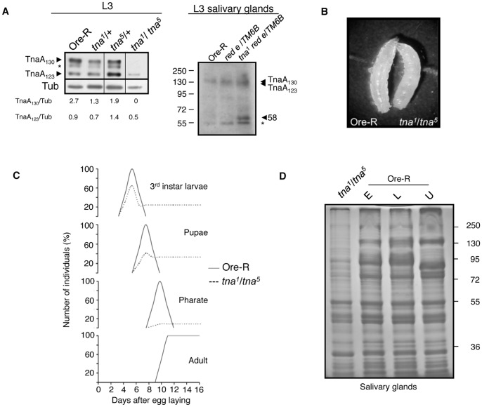
While we can detect TnaA130 and TnaA123 in Ore-R and in tna1/+ or tna5/+ individuals, TnaA130 is barely detectable and TnaA123 decreases dramatically in tna1/tna5 larvae (Fig. 3A, left panel). The tna1 mutation changes Gln 566 to a stop codon, is recessive lethal [5] and behaves as a dominant negative. tna1 is a much stronger dominant enchancer of osa1 than is a deficiency of the tna region (Table 1). tna1 would produce a truncated protein of 62 kDa that we have been able to observe in heterozygous tna1/+ salivary glands soluble extracts (Fig. 3A, right panel). The molecular lesion of tna5 has not been determined, but it behaves genetically as a hypomorphic allele and its product can be detected in tna1/tna5 third instar larvae extracts (Fig. 3A, left panel).
Figure 3. Characterization of tna1/tna5 individuals.
(A) TnaA proteins in tna1/tna5 third instar larvae (left panel). Note the absence of TnaA130 and the reduction of TnaA123 levels. Western blot probed with the TnaAXSPRING antibody (1∶100) was done with similar amount of proteins from total third instar larvae extracts of the indicated genotype. The band marked with an asterisk (*) appears sometimes depending on extracts and running gel conditions and may represent a processed protein product. Narrow vertical lines indicate edition of lanes from the same film with samples from flies with other tna genotypes not relevant to this work. β-tubulin was used as loading control (antibody used 1∶1000). The levels of TnaA130 and TnaA123 were compared with the level of β-tubulin using Image-J. Western blot of tna1 red e/TM6B salivary gland extracts probed with TnaANH2 antibody (right panel) detected TnaA130, TnaA123 (that comigrated in this gel) and a 58 kDa band close to the predicted size of a truncated Tna-1 protein (62 kDa). Salivary gland extracts from OreR individuals and from individuals harbouring the tna1 parental (red e) and balancer (TM6B) chromosomes were used as controls. The asterisk indicates a crossreacting band present in all the genotypes tested. (B) tna1/tna5 and Ore-R third instar larvae. (C) Survival percentage of tna1/tna5 individuals at different stages of development. Ore-R (−) and tna1/tna5 (–) survival percentages are indicated. More than 100 third instar larvae were counted for each genotype. Heterozygous tna individuals have survival rates similar to those for Ore-R. (D) Protein profile of tna1/tna5 salivary glands. Note the differences between the tna1/tna5 and the Ore-R total protein profiles. SDS-PAGE of soluble protein extracts from 10 pairs of larval salivary glands stained with Coomasie blue. Ore-R salivary glands extracts from early (E) and late (L) or unstaged (U) third instar larvae that were loaded as references.
Table 1. Genetic interactions of tna and osa with SUMOylation pathway genes.
| Genotype | Number of flies with HWOa | Penetrance (%)a |
| +/osa1 | 9/265 | 3 |
| +/osa2 | 0/303 | 0 |
| +/smt304493 | 0/389 | 0 |
| +/lwr4–3 | 1/133 | 1 |
| +/lwr5 | 0/297 | 0 |
| +/lwr13 | 0/341 | 0 |
| +/tna1 | 116/624 | 19 |
| tna1/osa1 | 327/334 | 98 |
| Df(3L)lxd6/osa1 | 16/115 | 14 |
| tna1/osa2 | 140/334 | 42 |
| bsmt304493/+; tna1/+ | 44/249 | 18 |
| csmt304493/+; tna1/+ | 66/118 | 56 |
| lwr4–3/+; tna1/+ | 69/150 | 46 |
| lwr5/+; tna1/+ | 71/282 | 25 |
| lwr13/+; tna1/+ | 177/343 | 52 |
| smt304493/+; osa1/+ | 57/264 | 22 |
| smt304493/+; osa2/+ | 0/216 | 0 |
| lwr5/+; osa1/+ | 1/147 | 1 |
| lwr13/+; osa1/+ | 0/124 | 0 |
| lwr5/+; osa2/+ | 0/212 | 0 |
| lwr13/+; osa2/+ | 0/256 | 0 |
For expressivity of held-out wing phenotype (HWO) see Fig. 6.
Reciprocal crosses were done were done in all cases with no observed differences except in the crosses with tna1 males and smt304493 femalesb, or with smt304493 males and tna1 femalesc. At least 100 flies were examined for each genotype. Flies that do not present the held-out wing phenotype include tna3, tna5, or tna− deficiencies Df(3L)vin2 and Df(3L)lxd6, osa2, lwr5, lwr4–3, lwr13, smt304493 heterozygous individuals, lwr/+;osa/+, smt304493/+;osa2/+ and all the transheterozygous combinations between tna3, tna5, or Df(3L)vin2 and Df(3L)lxd6 with smt3 and lwr alleles.
To better understand tna function we studied the lethality of tna1/tna5 animals. The tna1/tna5 larvae (Fig. 3B) did not have melanotic tumors as observed in lwr or aos1 mutant individuals [34], [35], [36], nor are they a larger size as observed for smt3 knockdowned larvae [29]. We found that 65% of tna1/tna5 individuals reach the third instar larval stage (Fig. 3C), but only 41% pupated and only 8% of the expected individuals reached the pharate stage. No tna1/tna5 individuals eclosed as adults (Fig. 3C). We also noticed that the tna1/tna5 third instar larvae that did not pupate often survived long after their heterozygous tna1/+ or tna5/+ siblings larvae pupated. Some of these tna1/tna5 larvae have an extended lifespan of at least two weeks (Fig. 3C). A similar extension of larval lifespan was previously observed in animals with reduced levels of SUMO [29], Aos1 (one of the E1 subunits) [34] or Ubc9 (E2) [35], [36].
Given the abnormal behavior of tna1/tna5 larvae and knowing that the TnaA profile is altered (Fig. 3A), we characterized the protein profile of their salivary glands (Fig. 3D). We staged the larvae by feeding them with bromophenol blue [28] and divided them in early (blue) and late (white) larvae. All tna1/tna5 larvae remained as early larvae (blue). They were collected 24 hours after they crawled from the food to obtain their salivary glands and we determined their protein profile (Fig. 3D). Although tna1/tna5 larvae remained blue, the protein profile differed from both the early and late wild-type Ore-R salivary glands obtained under the same conditions. Differences in the quantity and quality of proteins present in tna1/tna5 salivary glands fall mostly in the range over 72 kDa (Fig. 3D).
TnaA is Chromatin-associated at Discrete Sites on Polytene Salivary Gland Chromosomes
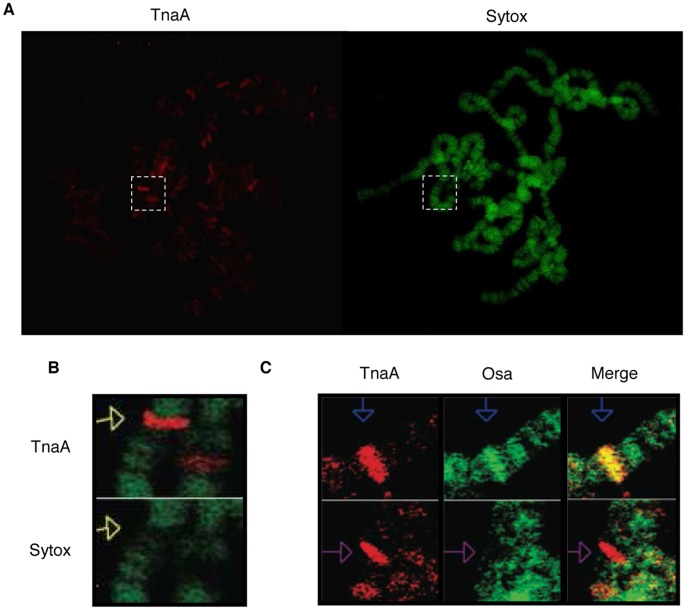
We have shown that TnaA123 is nuclear in Drosophila embryos (Fig. 2C) and that TnaA (probably TnaA123) is mainly nuclear in salivary and ring glands from third instar larvae (Fig. 2D). We immunostained polytene salivary gland chromosomes of third instar larvae and found that TnaA is associated with discrete sites (Fig. 4A). The number of TnaA sites suggests that TnaA might be required for the transcription of more than just the homeotic genes. Interestingly, most of the TnaA signals detected on polytene salivary gland chromosomes are located in interbands which are thought to have decondensed chromatin where transcription can occur (Fig. 4B). Because of the strong genetic interactions between tna and osa [5], we coimmunostained for TnaA and Osa on polytene salivary gland chromosomes. TnaA colocalizes with Osa at some sites, but not at others (Fig. 4C, upper and bottom panels). We do not know whether this is because TnaA is not required at all genes regulated by Osa, or whether it is due to an interaction between TnaA and Osa that is more transient than Osa localization.
Figure 4. TnaA is located on polytene salivary gland chromosomes of third instar larvae and sometimes colocalizes with Osa.
(A) Immunostaining of TnaA in Ore-R (wild type) polytene salivary gland chromosomes of third instar larvae. TnaAXSPRING antibody (1∶50, red) and DNA (Sytox, green). Amplification in B is indicated (pointed white rectangle). (B) TnaA is located in chromatin interbands. (C) TnaA and Osa colocalize in some sites on polytene salivary gland chromosomes of third instar larvae (blue arrows in the top panels) but in others do not (purple arrows in the bottom panels). TnaAXSPRING antibody (1∶50, red) and Osa (1∶50, green). No signal was detected when no primary antibody was added (data not shown).
TnaA Physically Interacts with Ubc9 and with Osa
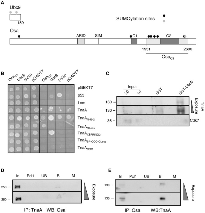
SUMO E3 ligases function for selection of SUMOylation targets and/or for enhancement of the SUMO conjugation process. TnaA has an SP-RING zinc finger that is also present in a subclass of SUMO E3 ligases that includes the PIAS proteins in mammals [37] and Su(var)2–10 in Drosophila [14]. Since the SP-RING in the PIAS proteins physically interacts with Ubc9 [38], [39], we explored whether TnaA physically interacts with Drosophila Ubc9, using yeast two-hybrid assays and pull-down assays.
For the yeast two-hybrid assays we first used the full-length TnaA protein (Fig. 1) fused to the yeast GAL4-DNA binding domain as “bait”, and the full-length Drosophila Ubc9 protein (Fig. 5A) fused to the GAL4-activation domain as “prey”. We found that the full-length TnaA protein was able to activate the transcription of at least two reporter genes in the absence of a “prey” (Fig. 5B), and as a consequence the full-length TnaA protein could not be used to test for the Ubc9 interaction in this assay. We then split the TnaA protein into five fragments that cover the whole TnaA protein (Fig. 1). Two out of the five fragments contain the SP-RING zinc finger (TnaAXSPRING2 and TnaASP-COO Qless). The other fragments have different TnaA regions that include the two glutamine-rich domains (TnaANH2–2), the bipartite nuclear location signal (TnaAQLess) and the carboxy-ending (TnaACOO). We found that the TnaAXSPRING2 fragment interacted with Ubc9 in the yeast two-hybrid assay while the other fragments, including TnaASP-COO QLess, did not interact (Fig. 5B). These results show that the TnaA SP-RING zinc finger is necessary but not sufficient for the TnaA interaction with Ubc9 in this assay.
Figure 5. TnaA interacts with Drosophila Ubc9 and with Osa.
(A) Schemes of Drosophila Ubc9 and OsaC2 used in biochemical assays. In the Osa protein, the ARID, the C1 and C2 domains (grey boxes), the SUMO interacting motif (SIM) and the OsaC2 fragment (dark line) are indicated. Forward (black circles) and inverted (gray circles) putative SUMOylation consensus sites in these proteins are indicated. For TnaA baits see Fig. 1. (B) TnaA interaction with Ubc9 and OsaC2 in yeast two-hybrid assays. Yeast colony complementation of growth controls in SD-Trp/−Leu media due to the presence of pGBKT7 (Trp+) and pGADT7, (Leu+) plasmids (left) in the same yeast cells. Interaction assay in QDO +3-AT (SD-Trp/−Leu/−Ade/−His +3-AT) media (right). Growth is observed when baits and preys interact, allowing GAL4 reconstitution with the consequent ADE2 and HIS3 reporter genes transcription. Baits were TnaA fragments (Fig. 1) fused to the DNA-binding domain of GAL4 in pGBKT7. Ubc9 and OsaC2 were preys fused to the GAL4 activation domain in pGADT7. Human p53 (p53) and Lamin C (Lam) interactions with SV40 are positive and negative controls, respectively. (C) TnaA interaction with Ubc9 by pull-down. The assays were done with 10 µg of each GST or GST-Ubc9 as baits and with 500 µg of soluble nuclear fraction from 3–21 hour embryos. 10 and 20% of the extract are shown as Input. TnaA was detected by Western analysis with TnaAXSPRING antibody (1∶100) when GST-Ubc9 was used as bait. The 130 kDa weight marker is indicated (left) and increasing exposures of the same membrane are shown. Cdk7 was detected only in the Input lanes (antibody dilution, 1∶1000). (D) Coimmunoprecipitation of Osa with TnaA antibodies from nuclear extracts obtained from 3–21 hour embryos. TnaAXSPRING antibodies (1 µg), and 3–21 hour embryos soluble nuclear fraction (500 µg) were used. The Western was revealed with the Osa antibody (1∶1000). Input, (In), preclearing 1 (Pcl1), unbound (Ub), bound (B). Immunoprecipitation with the equivalent amount of a preimmune serum instead of TnaAXSPRING antibody was used as Mock (M). Both panels show films with increasing exposure time of the same membrane. (E) Coimmunoprecipitation of TnaA with Osa antibodies from total extracts obtained from 3–21 hour embryos. Osa antibodies (1 µg), and 3–21 hour embryos soluble nuclear fraction (3.7 mg) were used. The Western was revealed with the TnaANH2 antibody (1∶120). Lanes are labeled as above. The equivalent amount of an irrelevant antibody was used as mock (M). Molecular weight markers are indicated (left).
Osa is a subunit of some BRM complexes, and the osa gene strongly interacts with tna [5]. Since it was found that Osa is modified by SUMO in Drosophila embryos [3], we thought that TnaA might be involved in Osa SUMOylation. We searched for SUMOylation consensus sites (ψKxE) in the Osa protein sequence (2713 aa) using the SUMOsp 2.0 program [32] and found eight putative SUMOylation sites (Fig. 5A), six of them located within a segment located from amino acids 1951 to 2600 surrounding the C2 domain [40]. We will refer to the fragment with the six putative SUMOylation sites as OsaC2 in this work. We synthesized the OsaC2 cDNA from polyA+ RNA of 3–21 hour embryos and fused it to the GAL4-activation domain to use as “prey” in the yeast two-hybrid assay. We tested the six TnaA baits already described (including full-length TnaA), and found that baits harbouring the SP-RING (TnaAXSPRING2 or TnaASP-COO QLess) did not interact with the OsaC2 prey. Although TnaANH2–2 (and to a lesser extent, full-length TnaA) interacted with OsaC2, these baits also interacted with pGADT7 or pGADT7-SV40 negative control samples, preventing us from concluding whether the interactions with OsaC2 are bona fide. In contrast, we found that the TnaAQless bait cleanly interacts physically with OsaC2 (Fig. 5B).
Although the TnaAXSPRING2 region interacted physically with Ubc9 in the yeast two-hybrid assays, we wanted to test for TnaA/Ubc9 physical interactions in Drosophila embryos. We performed pull-down assays using as bait a purified GST-Ubc9 fusion protein incubated with a nuclear protein extract from 3–21 hour embryos where we know TnaA is present (Fig. 2B). After extensive stringent washing, the presence of TnaA amongst the GST-Ubc9-interacting proteins was assessed by Western analyses with the TnaAXSPRING antibody (Fig. 5C). As expected, we found that full-length TnaA from nuclei of Drosophila embryos interacts with full length GST-Ubc9, confirming the results that we obtained with the yeast two-hybrid assays using TnaA fragments and further suggesting that these proteins interact in vivo.
In all reported cases it is known that only a fraction of the whole pool of a SUMOylatable protein in a cell is SUMOylated, either because of spatial restrictions (the target protein should be located where the SUMO and the SUMOylation enzymes are) or because fine regulation constricts the amount of the SUMOylated protein [4]. We showed that OsaC2 interacts with a fragment of TnaA (TnaAQLess) in a yeast two-hybrid assay (Fig. 5B). To test whether this interaction can be observed with the full-length proteins in Drosophila embryos, we performed TnaA or Osa coimmunoprecipitation assays from total or nuclear protein extracts from 3–21 hour embryos. For this purpose, we first showed that the TnaAXSPRING and Osa antibodies are able to immunoprecipitate TnaA and Osa, respectively (Fig. S1), and that the control proteins Hsp70 and Cdk7 do not coimmunoprecipitate with TnaA or with Osa, respectively (Fig. S2). Interestingly, we found that TnaA coimmunoprecipitates with a fraction of Osa found in nuclear protein extracts from 3–21 hour embryos (Fig. 5D), and that reciprocally, Osa coimmunoprecipitates with TnaA from a total protein extract of 3–21 hour embryos (Fig. 5E). Since we found that TnaA interacts physically with Osa and with Ubc9 (Fig. 5) we tried to test whether TnaA has SUMO E3 ligase activity on the OsaC2 fragment using a mammalian in vitro assay (Active Motif kit). Although OsaC2 is SUMOylated in this assay, we were not able to get convincing evidence that TnaA has SUMO E3 ligase activity under these conditions (data not shown).
tna and osa Genetically Interact with Components of the SUMOylation Pathway
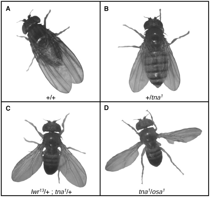
tna genetically interacts with brm and osa [5]. Transheterozygous adult flies carrying mutations in combinations of any of these three genes have a strong held-out wing phenotype [5], [8] (Fig. 6). This phenotype appears to result from reduced expression from the P2 promoter of the homeotic gene Antp [8]. The interactions with tna might be a consequence of reduced SUMOylation of Osa (and/or Brm) proteins. If so, mutations in other components of the SUMOylation pathway might also show genetic interactions. We generated transheterozygous flies carrying mutant alleles of either the SUMO E2 conjugating enzyme Ubc9 (lwr5, lwr4–3, and lwr13) [25], [26] or SUMO (smt304493) [27] in combination with mutant alleles of tna (tna1 and tna5) or osa (osa1 and osa2). All of these individuals have at least one wild type copy of each gene to allow survival to the adult stage.
Figure 6. SUMOylation pathway mutations enhance held-out wing phenotype of tna and osa flies.
Flies with different held-out wing phenotype expressivity. Fly genotype is indicated in each picture. Penetrance of the held-out wing phenotype in each genotype is in Table 1 (A) Wild type fly (B) Slight held-out wing phenotype of +/tna1 flies. The same phenotype is presented by +/osa1 individuals. (C) Stronger held-out wing phenotype of smt3/+;tna1/+ individuals. The same phenotype is presented by lwr/+;tna1/+ or smt304493/+;osa1/+ individuals. (D) Strongest held-out wing phenotype of tna1/osa1 individuals. This phenotype is also presented by tna1/osa2 individuals.
Individuals carrying tna alleles other than tna1 (tna3 or tna5), or deficiencies uncovering the tna region [Df(3L)vin2 or Df(3L)lxd6] do not show the held-out wing phenotype. This phenotype is also not shown by transheterozygous individuals carrying tna alleles other than tna1 or the tna deficiencies in combination with smt3 or lwr alleles. In contrast, we found that smt304493, lwr4–3 and lwr 13 enhance both the penetrance and the expressivity of the held-out wing phenotype of tna1 individuals (from 19% with weak expressivity to 56%, 46% and 52% with stronger expressivity, respectively, Fig. 6 and Table 1). Thus, we confirmed genetically that tna interacts with the SUMOylation pathway genes smt3 and lwr. Interestingly, we found a maternal effect in the enhancement of the tna1 held-out wings phenotype in double heterozygous smt304493/+; tna1/+ individuals. The enhancement is only observed when there is not maternal contribution of tna (Table 1). We did not observe this maternal effect with the other genes tested. Since we observed strong genetic interactions between tna and osa (Fig. 6) [5], therefore we tested whether the SUMOylation genes lwr and smt3 also interact with osa (osa1 and osa2 in Table 1 and Fig. 6). We found that smt304493 augmented the penetrance and the expressivity of the weak held-out wing phenotype of osa1 (Fig. 6, Table 1) but that it did not interact with the weaker osa2 allele. None of the lwr alleles tested interacted with osa1 or osa2, suggesting that Ubc9 activity in these heterozygous individuals is sufficient to reach appropriate SUMOylation levels.
Discussion
The presence of the SP-RING, the physical interaction of the SUMO E2 conjugating enzyme Ubc9 with TnaA, and the genetic interaction of tna with genes encoding SUMOylation pathway proteins suggest that TnaA may be involved in the SUMOylation pathway to activate transcription. TnaA may also have other functions not directly related to SUMOylation. These other functions may or may not act together with SUMOylation to positively regulate gene expression.
TnaA Function in Gene Expression Involving the SUMOylation Pathway
Gene expression involves the integration of many regulatory mechanisms. Recently, many examples of SUMOylation and/or ubiquitylation during transcriptional regulation have been described [4]. These examples include the clearance of activators to favor transcription cycles in inducible genes [41] and the assembly of different proteins into a complex [42], [43]. Most of the tna interacting genes (osa, brahma, moira, kohtalo, skuld, and kismet) [5] encode subunits of complexes involved in chromatin remodeling and transcription by RNA polymerase II, suggesting that SUMOylation may be important at multiple aspects of gene regulation in Drosophila. Typically, SUMO-tagged proteins are recognized by a binding partner that contains a SIM (SUMO Interacting Motif) [33]. All of the proteins encoded by the tna interacting genes listed above have more than one SIM and SUMOylation sites (data not shown) and could be either SUMOylation targets, readers of the SUMO mark, or proteins that help TnaA exert its function(s).
SUMO E3 ligases are required for the enhancement and/or for the specifity of the SUMOylation tagging on targets. In this work we utilized different approaches to show that TnaA is involved in the SUMOylation pathway possibly as a SUMO E3 ligase. We showed a TnaA physical interaction with Ubc9 and genetic interactions between tna and osa with SUMOylation pathway genes. SUMOylated Osa is found in early embryos (0–3 hour) [3] and embryonic TnaA and Osa coimmunoprecipitate reciprocally (this work). We also showed that a GST-Ubc9 fusion physically interacts with native nuclear TnaA from Drosophila embryos. Hence, we suggest that Osa is a good candidate to be a TnaA-SUMOylation target in vivo. Our data suggest that TnaA-dependent SUMOylation of Osa and/or of other target(s), particularly proteins associated with Osa (e.g. other BRM complex subunits, histones, or others, see ahead), may be required for correct gene expression including homeotic genes. Osa is a large protein of around 280 kDa with an ARID domain which binds AT-rich sequences, LXXLL domains [8] that could help it to interact with nuclear receptors and has eight putative SUMOylation target sequences, six of them in the OsaC2 fragment (Fig. 5A). In humans there are three proteins related to Osa, BAF250a, BAF250b and BAF200/ARID2 [44] and it was reported that BAF250b could be in a complex that has E3 ubiquitin ligase activity on histone H2B [45].
Originally tna was identified in a screen to find Brm-interacting proteins [5]. Although we did not study here whether Brm can be SUMOylated, it has been reported that mammalian SUMO-2 can be acetylated at K33 to inhibit some SUMO-SIM interactions [46]. Interestingly, these authors also show that the bromodomain of p300, besides recognizing acetylated histones [47], can bind the SUMO acetylated form, opening the question of whether other bromodomains, such as the one present in the Brahma protein, would be able to recognize a putative Drosophila acetylated SUMO when present in any of its interactor proteins.
TnaA may also be promoting homeotic gene expression by inactivation through SUMOylation of a PcG protein. Indeed, SUMOylation of the PcG protein Scm (encoded by the Sex comb on midleg gene) decreases its levels at the PRE (Polycomb Response Element) located upstream the Ubx homeotic gene. SUMO compromised animals show a reduction of Ubx expression and it has been suggested that TnaA may be involved in Scm SUMOylation to promote homeotic gene expression [48].
Other TnaA Interactors and SUMO-independent Functions of TnaA
We found that TnaA130 is mainly cytoplasmic and TnaA123 is mainly nuclear. Although most studied SUMO enzymes and targets are in the nucleus, there are some examples of SUMOylation of proteins in the cytoplasm [49]. As TnaA130 always precedes the appearance of TnaA123 through development (developmental Western, Fig. 2B), we think that TnaA may be processed to enter the nucleus to SUMOylate its targets. Notably, SUMOylation pathway proteins with well known nuclear activities also SUMOylate targets in the cytoplasm [50]. Thus, with what we know at present, we cannot discard the possibility that TnaA130 can also function in the cytoplasm. We also found that tna interacts with the γTub23C gene that encodes an isoform of γ-tubulin [51] and with taranis (tara) [5]. The significance of the interaction of tna with tara and γTub23C is currently unknown.
It is possible that TnaA could be necessary for BRM complex(es) function(s) regardless of SUMOylation, and that independently, SUMOylation could be required for function of other BRM complex(es) components. We cannot neither rule out the possibility that TnaA may have other functions independent of its possible role in the SUMOylation pathway, as has been reported for the PIAS proteins, known SP-RING SUMO E3 ligases [52], [53], [54]. The SP-RING plays a key role in this PIAS activity. The TnaA SP-RING is immersed in a 300-aminoacid region that we called the XSPRING domain that is shared with the vertebrate proteins Zimp7 and Zimp10 [KIAA1886 and KIAA1224 respectively, 5]. Although TnaA is related to the PIAS proteins because it has an SP-RING, it does not have the SAP (Scaffold attachment factor-A/B, Acinus and PIAS domain) nor the PINIT motifs that are PIAS signature domains.
The SAP and PINIT motifs in the PIAS proteins confer functions related to structural anchoring and transcriptional regulation. In mammals it has been shown that PIAS1 promotes the transcriptional repressive activity of Msx1 through regulating its location in a SUMO-independent way [53], it controls the stability of Msx1 by preventing its ubiquitination [55] and it regulates the transcriptional activity of GATA4 [56]. Similarly, in Xenopus, XPIASy down-regulate XSmad2 transcriptional activity independently from XPIASy SUMO E3 ligase activity [57].
Although human Zimp7, human Zimp10, and Drosophila TnaA do not have these other PIAS signature motifs they have transcriptional activation domains [58], [59] (Fig. 5B). The presence of a transcriptional activation domain could explain why we could not use the TnaA-Gal4 DNA-binding domain fusion in the yeast two-hybrid system (Fig. 5B). This suggests that TnaA, besides its possible role in the SUMOylation pathway, has other functions in Drosophila transcriptional activation.
TnaA in Drosophila Development
We described a genetic interaction between tna, osa, and SUMOylation pathway genes. TnaA interacts physically with Ubc9 through the SP-RING supporting the genetic interaction data. Animals derived from osa and tna mutant germline clones die at different stages of development. While the osa ones do not survive embryogenesis [8] the tna ones die mostly as third instar larvae [5]. A pool of Osa is found SUMOylated in embryos of 0–3 hour of development when zygotic expression has not started [3] and TnaA is barely detectable (overexposure of Fig. 2B, data not shown). Moreover, when we studied the tna and smt3 interaction, we found a tna maternal effect. The held-out wings phenotype in smt3/+; tna/+ adults is observed when the mother is tna defective, but we do not observe this when the mothers have low dosages of SUMO (Table 1). We think it is probable that SUMOylated Osa plays a role at early stages of development. SUMOylation of embryonic Osa can happen in the maternal germline or in the embryo with the help of the maternally-inherited SUMOylation pathway machinery. This machinery may include TnaA if TnaA is involved in SUMOylation or another protein with a SUMO-related function. It is also possible that smt3/+; tna/+ embryos derived from smt3 mothers do not present the held-out wings phenotype because the SUMOylation pathway can compensate even with low dosages of SUMO. On the other hand, if TnaA is related to SUMOylation, embryos derived from tna mothers would lack correct SUMOylation of specific targets (such as Osa) causing later the appearance of the held-out wings phenotype.
Why do tna mutant animals die at later stages of development? One possibility is that proteins other than TnaA can exert its function on particular targets, such as Osa, or that they could only impact the TnaA targets in earlier stages of development, but not in later stages. SUMO is required for metamorphosis [29]. As the majority of tna mutant animals die as larvae or pupae and cannot proceed to metamorphosis (Fig. 3C) [5], and as TnaA is in prothoracic gland nuclei of third instar larvae (Fig. 2D) obvious candidates for regulation by tna would be the ecdysone-pathway, ecdysone-regulated or patterning genes.
The relevance of SUMOylation (and of genes like tna) in different developmental processes is just starting to emerge. The requirement of SUMOylation and of tna to maintain gene expression makes that the next challenges will be to find the SUMOylation and tna targets in vivo and to understand the consequences of this modification in proteins involved in chromatin dynamics and in gene expression.
Supporting Information
The TnaAXSPRING and Osa antibodies immunoprecipitate TnaA and Osa proteins, respectively. (A) TnaA was immunoprecipitated from 3–21 hour embryo-soluble nuclear fraction (500 µg) using TnaAXSPRING antibody (1 µg). The Western was revealed with TnaAXSPRING (1∶100). The three panels correspond to films with increasing exposure times. Input (In), Preclearing 1 (Pcl1), Unbound (Ub), and Bound (B). Mock (M) where the immunoprecipitation was done with the equivalent amount of a preimmune serum instead of TnaAXSPRING. (B) Osa protein was immunoprecipitated from 3–21 hour embryos soluble extract (3.7 mg) with the Osa antibody (1 µg). For Osa detection, the Western was revealed with Osa antibody (1∶1000). Lanes are labeled as above. The equivalent amount of an irrelevant antibody was used as mock. Molecular weight markers are indicated (left).
(TIF)
Negative controls of TnaA and Osa immunoprecipitations. (A) TnaA antibodies do not coimmunoprecipitate Hsp70 (Bound, lane B) from a 3–21 hour embryos soluble nuclear fraction (immunoprecipitation shown in Fig. S1A), meanwhile Hsp70 is present in the input (In) and unbound (Ub) samples. The other lanes are preclearing 1 (Pcl1), and mock (M) samples. (B) The Osa antibody do not coimmunoprecipitate Cdk7 (immunoprecipitation shown in Fig. S1B). The assays were done as in (A). Lanes are labeled as above.
(TIF)
Acknowledgments
We thank the Bloomington Drosophila Stock Center for providing stocks. We also thank Luis Gutiérrez for TnaA cDNA clones in pGEX vectors, J. Riesgo-Escovar for providing the Gold collection Ubc9 clone, V. Valadez-Graham for assistance in this work, Carmen Muñoz for technical assistance and R. Vera, and J. A. Kassis for critical reading of the manuscript. We thank the two anonymous reviewers for valuable suggestions.
Funding Statement
This work was supported by funds from Dirección General Asuntos del Personal Académico (DGAPA) grant IN208808 (http://dgapa.unam.mx/html/papiit/papit.html), and Consejo Nacional Ciencia y Tecnología (CONACyT) grant 99654 (http://www.conacyt.gob.mx) to M. V. and in part by the Intramural Research Program of the United States National Institutes of Health, National Institute of Child Health and Human Development (NICHD) (http://dir.nichd.nih.gov/dirweb/home.html). The funders had no role in study design, data collection and analysis, decision to publish, or preparation of the manuscript.
References
- 1. Geiss-Friedlander R, Melchior F (2007) Concepts in SUMOylation: a decade on. Nat Rev Mol Cell Biol 10: 947–956. [DOI] [PubMed] [Google Scholar]
- 2. Rodríguez MS, Dargemont C, Hay RT (2001) SUMO-1 conjugation in vivo requires both a consensus modification motif and nuclear targeting. J Biol Chem 276: 12654–12659. [DOI] [PubMed] [Google Scholar]
- 3. Nie M, Xie Y, Loo JA, Courey AJ (2009) Genetic and proteomic evidence for roles of Drosophila SUMO in cell cycle control, Ras signaling, and early pattern formation. PLoS One 4: e5905. [DOI] [PMC free article] [PubMed] [Google Scholar]
- 4. Gareau JR, Lima CD (2010) The SUMO pathway: emerging mechanisms that shape specificity, conjugation and recognition. Nat Rev Mol Cell Biol 11: 861–871. [DOI] [PMC free article] [PubMed] [Google Scholar]
- 5. Gutiérrez L, Zurita M, Kennison JA, Vázquez M (2003) The Drosophila trithorax group gene tonalli (tna) interacts genetically with the Brahma remodeling complex and encodes an SP-RING finger protein. Development 130: 343–354. [DOI] [PubMed] [Google Scholar]
- 6. Kennison JA, Tamkun JW (1988) Dosage-dependent modifiers of Polycomb and Antennapedia mutations in Drosophila . Proc Natl Acad Sci USA 85: 8136–8140. [DOI] [PMC free article] [PubMed] [Google Scholar]
- 7. Tamkun JW, Deuring R, Scott MP, Kissinger M, Pattatucci AM, et al. (1992) brahma: a regulator of Drosophila homeotic genes structurally related to the yeast transcriptional activator SNF2/SWI2. Cell 68: 561–572. [DOI] [PubMed] [Google Scholar]
- 8. Vázquez M, Moore L, Kennison JA (1999) The trithorax group gene osa encodes an ARID-domain protein that genetically interacts with the Brahma chromatin-remodeling factor to regulate transcription. Development 126: 733–742. [DOI] [PubMed] [Google Scholar]
- 9. Mohrmann L, Langenberg K, Krijgsveld J, Kal AJ, Heck AJ, et al. (2004) Differential targeting of two distinct SWI/SNF-related Drosophila chromatin-remodeling complexes. Mol Cell Biol 24: 3077–3088. [DOI] [PMC free article] [PubMed] [Google Scholar]
- 10. Lawrence PA, Morata G (1994) Homeobox genes: their function in Drosophila segmentation and pattern formation. Cell 78: 181–189. [DOI] [PubMed] [Google Scholar]
- 11. Maeda R, Karch F (2006) The ABC of the BX-C: the bithorax complex explained. Development 133: 1413–1444. [DOI] [PubMed] [Google Scholar]
- 12. Talamillo A, Sanchez J, Barrio R (2008) Functional analysis of the SUMOylation pathway in Drosophila . Biochem Soc Trans 36: 868–873. [DOI] [PubMed] [Google Scholar]
- 13. Lomelí H, Vázquez M (2011) Emerging roles of the SUMO pathway in development. Cell Mol Life Sci 68: 4045–4064. [DOI] [PMC free article] [PubMed] [Google Scholar]
- 14. Hari K L, Cook KR, Karpen GH (2001) The Drosophila Su(var)2–10 locus regulates chromosome structure and function and encodes a member of the PIAS protein family. Genes Dev 15: 1334–1348. [DOI] [PMC free article] [PubMed] [Google Scholar]
- 15. Kamakaka RT, Tyree CM, Kadonaga JT (1991) Accurate and efficient RNA polymerase II transcription with a soluble nuclear fraction derived from Drosophila embryos. Proc Natl Acad Sci USA 88: 1024–1028. [DOI] [PMC free article] [PubMed] [Google Scholar]
- 16. Elfring LK, Daniel C, Papoulas O, Deuring R, Sarte M, et al. (1998) Genetic analysis of brahma: the Drosophila homolog of the yeast chromatin remodeling factor SWI2/SNF2. Genetics 148: 251–265. [DOI] [PMC free article] [PubMed] [Google Scholar]
- 17. McQuilton P, St Pierre SE, Thurmond J, Consortium F (2012) FlyBase 101-the basics of navigating FlyBase. Nucleic Acids Res 40: D706–D714. [DOI] [PMC free article] [PubMed] [Google Scholar]
- 18.Smith D, Corcoran L (2001) Expression and purification of glutathione-S-transferase fusion proteins. In: Ausubel F, Brent R, Kingston R, Moore D, Seidman J, Smith J, Struhl K, Editors. Current Protocols in Molecular Biology. New York: John Wiley and Sons. 16.7.1–16.7. [DOI] [PubMed]
- 19.Cooper H, Paterson Y (2004) Preparation of polyclonal antisera. In: Bonifacio J, Dasso M, Harford J, Lippincott-Schwartz J, Yamada K, Editors. Current Protocols in Cell Biology. New York: John Wiley and Sons. 16.2.1–16.2.8.
- 20. Koelle MR, Horvitz HR (1996) EGL-10 regulates G protein signaling in the C. elegans nervous system and shares a conserved domain with many mammalian proteins. Cell 84: 115–125. [DOI] [PubMed] [Google Scholar]
- 21. Vikis HG, Guan KL (2004) Glutathione-S- based assays for studying protein-protein interactions. Methods Mol Biol 261: 175–186. [DOI] [PubMed] [Google Scholar]
- 22. Leclerc V, Tassan JP, O'Farrell PH, Nigg EA, Leopold P (1996) Drosophila Cdk8, a kinase partner of Cyclin C that interacts with the large subunit of RNA polymerase II. Mol Biol Cell 7: 505–513. [DOI] [PMC free article] [PubMed] [Google Scholar]
- 23.Sambrook J, Fritsch E, Maniatis T (1989) Molecular Cloning: A Laboratory Manual. New York: Cold Spring Harbor Laboratory Press. 7.26 p.
- 24. Tang Z, Hecker CM, Scheschonka A, Betz H (2008) Protein interactions in the SUMOylation cascade: lessons from X-ray structures. FEBS J 275: 3003–3015. [DOI] [PubMed] [Google Scholar]
- 25. Apionishev S, Malhotra D, Raghavachari S, Tanda S, Rasooly RS (2001) The Drosophila Ubc9 homologue lesswright mediates the disjunction of homologues in meiosis I. Genes Cells. 6: 215–224. [DOI] [PubMed] [Google Scholar]
- 26. Sun X, Huang L, Van Doren MR, Tanda S (2003) Isolation of amorphic alleles of the lesswright gene by P element-mediated male recombination in Drosophila melanogaster . Drosophila Information Service 86: 79–83. [Google Scholar]
- 27. Schnorr JD, Holdcraft R, Chevalier B, Berg CA (2001) Ras1 interacts with multiple new signaling and cytoskeletal loci in Drosophila eggshell patterning and morphogenesis. Genetics 159: 609–622. [DOI] [PMC free article] [PubMed] [Google Scholar]
- 28. Andres AJ, Thummel CS (1994) Methods for quantitative analysis of transcription in larvae and prepupae. Methods Cell Biol 44: 565–573. [DOI] [PubMed] [Google Scholar]
- 29. Talamillo A, Sánchez J, Cantera R, Pérez C, Martín D, et al. (2008) Smt3 is required for Drosophila melanogaster metamorphosis. Development 135: 1659–1668. [DOI] [PubMed] [Google Scholar]
- 30. Corona DF, Armstrong JA, Tamkun JW (2003) Genetic and cytological analysis of Drosophila chromatin-remodeling factors. Methods Enzymol 377: 70–85. [DOI] [PubMed] [Google Scholar]
- 31.Rebay I, Fehon R (2000) Generating antibodies against Drosophila proteins. In: Sullivan W, Ashburner M, Hawley R, editors. Drosophila Protocols. New York: Cold Spring Harbor Laboratory Press. 389–411.
- 32. Ren J, Gao X, Jin C, Zhu M, Wang X, et al. (2009) Systematic study of protein SUMOylation: development of a site-specific predictor of SUMOsp 2.0. Proteomics 9: 3409–3412. [DOI] [PubMed] [Google Scholar]
- 33. Kerscher O (2007) SUMO junction-what's your function? New insights through SUMO-interacting motifs. EMBO Rep 8: 550–555. [DOI] [PMC free article] [PubMed] [Google Scholar]
- 34. Kanakousaki K, Gibson MC (2012) A differential requirement for SUMOylation in proliferating and non-proliferating cells during Drosophila development. Development 139: 2751–2762. [DOI] [PubMed] [Google Scholar]
- 35. Huang L, Ohsako S, Tanda S (2005) The lesswright mutation activates Rel-related proteins, leading to overproduction of larval hemocytes in Drosophila melanogaster . Dev Biol 280: 407–420. [DOI] [PubMed] [Google Scholar]
- 36. Chiu H, Ring BC, Sorrentino RP, Kalamarz M, Garza D, et al. (2005) dubc9 negatively regulates the Toll-NF-κB pathways in larval hematopoiesis and drosomycin activation in Drosophila . Dev Biol 288: 60–72. [DOI] [PubMed] [Google Scholar]
- 37. Kahyo T, Nishida T, Yasuda H (2001) Involvement of PIAS1 in the SUMOylation of tumor suppressor p53. Mol Cell 8: 713–718. [DOI] [PubMed] [Google Scholar]
- 38. Nishida T, Yasuda H (2002) PIAS1 and PIASxα function as SUMO E3 ligases toward androgen receptor and repress androgen receptor-dependent transcription. J Biol Chem 277: 41311–41317. [DOI] [PubMed] [Google Scholar]
- 39. Takahashi Y, Kikuchi Y (2005) Yeast PIAS-type Ull1/Siz1 is composed of SUMO ligase and regulatory domains. J Biol Chem 280: 35822–35828. [DOI] [PubMed] [Google Scholar]
- 40. Wu JI, Lessard J, Crabtree GR (2009) Understanding the words of chromatin regulation. Cell 136: 200–206. [DOI] [PMC free article] [PubMed] [Google Scholar]
- 41. Rosonina E, Duncan SM, Manley JL (2010) SUMO functions in constitutive transcription and during activation of inducible genes in yeast. Genes Dev 24: 1242–1252. [DOI] [PMC free article] [PubMed] [Google Scholar]
- 42. van Wijk SJ, Muller S, Dikic I (2011) Shared and unique properties of ubiquitin and SUMO interaction networks in DNA repair. Genes Dev 25: 1763–1769. [DOI] [PMC free article] [PubMed] [Google Scholar]
- 43. Iyengar S, Farnham PJ (2011) KAP1 protein: an enigmatic master regulator of the genome. J Biol Chem 286: 26267–26276. [DOI] [PMC free article] [PubMed] [Google Scholar]
- 44. Hargreaves DC, Crabtree GR (2011) ATP-dependent chromatin remodeling: genetics, genomics and mechanisms. Cell Res 21: 396–420. [DOI] [PMC free article] [PubMed] [Google Scholar]
- 45. Li XS, Trojer P, Matsumura T, Treisman JE, Tanese N (2010) Mammalian SWI/SNF-A subunit BAF250/ARID1 is an E3 ubiquitin ligase that targets histone H2B. Mol Cell Biol 30: 1673–1688. [DOI] [PMC free article] [PubMed] [Google Scholar]
- 46. Ullmann R, Chien CD, Avantaggiati ML, Muller S (2012) An acetylation switch regulates SUMO-dependent protein interaction networks. Mol Cell 46: 759–770. [DOI] [PMC free article] [PubMed] [Google Scholar]
- 47. Dhalluin C, Carlson JE, Zeng L, He C, Aggarwal AK, et al. (1999) Structure and ligand of a histone acetyltransferase bromodomain. Nature 399: 491–496. [DOI] [PubMed] [Google Scholar]
- 48. Smith M, Mallin DR, Simon JA, Courey AJ (2011) Small ubiquitin-like modifier (SUMO) conjugation impedes transcriptional silencing by the Polycomb group repressor Sex Comb on Midleg. J Biol Chem 286: 11391–11400. [DOI] [PMC free article] [PubMed] [Google Scholar]
- 49. Takahashi Y, Iwase M, Strunnikov AV, Kikuchi Y (2008) Cytoplasmic SUMOylation by PIAS-type Siz1-SUMO ligase. Cell Cycle 7: 1738–1744. [DOI] [PubMed] [Google Scholar]
- 50. Castillo-Lluva S, Tatham MH, Jones RC, Jaffray EG, Edmondson RD, et al. (2010) SUMOylation of the GTPase Rac1 is required for optimal cell migration. Nat Cell Biol 12: 1078–1085. [DOI] [PMC free article] [PubMed] [Google Scholar]
- 51. Vázquez M, Cooper MT, Zurita M, Kennison JA (2008) γTub23C interacts genetically with Brahma chromatin-remodeling complexes in Drosophila melanogaster . Genetics 180: 835–843. [DOI] [PMC free article] [PubMed] [Google Scholar]
- 52. Sharrocks AD (2006) PIAS proteins and transcriptional regulation-more than just SUMO E3 ligases? Genes Dev 20: 754–758. [DOI] [PubMed] [Google Scholar]
- 53. Lee H, Quinn JC, Prasanth KV, Swiss VA, Economides KD, et al. (2006) PIAS1 confers DNA-binding specificity on the Msx1 homeoprotein. Genes Dev 20: 784–794. [DOI] [PMC free article] [PubMed] [Google Scholar]
- 54. Rytinki MM, Kaikkonen S, Pehkonen P, Jaaskelainen T, Palvimo JJ (2009) PIAS proteins: pleiotropic interactors associated with SUMO. Cell Mol Life Sci 66: 3029–3041. [DOI] [PMC free article] [PubMed] [Google Scholar]
- 55. Song YJ, Lee H (2011) PIAS1 negatively regulates ubiquitination of Msx1 homeoprotein independent of its SUMO ligase activity. Mol Cells 32: 221–226. [DOI] [PMC free article] [PubMed] [Google Scholar]
- 56. Belaguli NS, Zhang M, García AH, Berger DH (2012) PIAS1 is a GATA4 SUMO ligase that regulates GATA4-dependent intestinal promoters independent of SUMO ligase activity and GATA4 SUMOylation. PLoS One 7: e35717. [DOI] [PMC free article] [PubMed] [Google Scholar]
- 57. Daniels M, Shimizu K, Zorn AM, Ohnuma S (2004) Negative regulation of Smad2 by PIASy is required for proper Xenopus mesoderm formation. Development 131: 5613–5626. [DOI] [PubMed] [Google Scholar]
- 58. Huang CY, Beliakoff J, Li X, Lee J, Sharma M, et al. (2005) hZimp7, a novel PIAS-like protein, enhances androgen receptor-mediated transcription and interacts with SWI/SNF-like BAF complexes. Mol Endocrinol 19: 2915–2929. [DOI] [PubMed] [Google Scholar]
- 59. Lee J, Beliakoff J, Sun Z (2007) The novel PIAS-like protein hZimp10 is a transcriptional co-activator of the p53 tumor suppressor. Nucleic Acids Res 35: 4523–4534. [DOI] [PMC free article] [PubMed] [Google Scholar]
Associated Data
This section collects any data citations, data availability statements, or supplementary materials included in this article.
Supplementary Materials
The TnaAXSPRING and Osa antibodies immunoprecipitate TnaA and Osa proteins, respectively. (A) TnaA was immunoprecipitated from 3–21 hour embryo-soluble nuclear fraction (500 µg) using TnaAXSPRING antibody (1 µg). The Western was revealed with TnaAXSPRING (1∶100). The three panels correspond to films with increasing exposure times. Input (In), Preclearing 1 (Pcl1), Unbound (Ub), and Bound (B). Mock (M) where the immunoprecipitation was done with the equivalent amount of a preimmune serum instead of TnaAXSPRING. (B) Osa protein was immunoprecipitated from 3–21 hour embryos soluble extract (3.7 mg) with the Osa antibody (1 µg). For Osa detection, the Western was revealed with Osa antibody (1∶1000). Lanes are labeled as above. The equivalent amount of an irrelevant antibody was used as mock. Molecular weight markers are indicated (left).
(TIF)
Negative controls of TnaA and Osa immunoprecipitations. (A) TnaA antibodies do not coimmunoprecipitate Hsp70 (Bound, lane B) from a 3–21 hour embryos soluble nuclear fraction (immunoprecipitation shown in Fig. S1A), meanwhile Hsp70 is present in the input (In) and unbound (Ub) samples. The other lanes are preclearing 1 (Pcl1), and mock (M) samples. (B) The Osa antibody do not coimmunoprecipitate Cdk7 (immunoprecipitation shown in Fig. S1B). The assays were done as in (A). Lanes are labeled as above.
(TIF)


