Abstract
Purpose
In this retrospective study we evaluated the short- to medium-term results after 20 Coonrad-Morrey revision total elbow arthroplasties (TEAs).
Methods
We included a consecutive series of revision TEAs performed at our institution from 2004 to 2010. At a mean follow-up of 4.4 years, patients were evaluated using the Mayo Elbow Performance Score (MEPS), the Oxford Elbow Score (OES) and standard radiographs.
Results
The mean age at revision TEA was 65.8 years. The median time of implant survival for primary prosthesis was 9.5 years. The mean post-operative MEPS was 79. The mean OES was 58, 66 and 53 for function, pain and social-psychological dimensions, respectively. At follow-up the range of motion had improved significantly. There were two cases of radiolucent lines and two cases of minor bushing wear; however, none of the implants were clinically loose. In one case deep infection led to a further revision. Two patients had post-operative ulnar nerve paraesthesia.
Conclusions
Results after revision TEA using the Coonrad-Morrey prosthesis are acceptable with a low short- to midterm failure rate. Revision improves range of motion and provides pain relief. One case of deep infection with recurrent revision is of concern. The treatment can be used as an option for failed TEA.
Introduction
Revision total elbow arthroplasty (TEA) is a challenging procedure. Loss of soft tissue support and bone stock leads to a more unstable elbow joint. The linked TEA provides better stability than the unlinked TEA that depends on soft tissue support for stability [1]. Previous studies have shown acceptable results with the linked Coonrad-Morrey TEA for both primary and revision surgery [2–6].
Primary TEA used to be a salvage procedure only for low-demand patients but is increasingly being used for other indications including fractures, osteoarthritis and instability [7, 8]. TEA is technically demanding [9] and regardless of design, reports on primary TEA have stated an overall five-year failure rate between eight and ten percent and approximately 15 % after ten years [10, 11]. Though the complication rate is decreasing, revision TEA will remain a challenge in the future [12–14]. Increasing implantation rates in younger patients may lead to an increase in revision procedures. Therefore, it is essential to evaluate the results after revision TEA in order to improve decision-making in relation to revision TEA surgery.
The purpose of this study is to evaluate the clinical and functional results after revision TEA using the Coonrad-Morrey linked TEA.
Materials and methods
In this retrospective study we reviewed a consecutive series of 20 revision TEAs in 19 patients performed with the linked Coonrad-Morrey TEA (Table 1). Revisions were performed at the Shoulder and Elbow Clinic, University Hospital Herlev, Denmark in the period from 2004 to 2010.
Table 1.
Patient characteristics
| Case | Age at revision (years) | Sex | Indication for primary TEA | Primary TEA | Survival of primary TEA (years) | Indication for revision TEA |
|---|---|---|---|---|---|---|
| 1 | 69 | M | RA | Souter-Strathclyde | 15.75 | Loosening |
| 2 | 65 | F | RA | Souter-Strathclyde | 11.75 | Loosening |
| 3a | 41 | F | RA | Capitellocondylar | 14.17 | Loosening |
| 3b | 39 | F | RA | Coonrad-Morrey | 14.92 | Loosening of spline |
| 4 | 66 | F | Fracture | Coonrad-Morrey | 1.17 | Infection |
| 5 | 57 | F | Osteoarthritis | Coonrad-Morrey | 1.42 | Loosening |
| 6 | 88 | F | RA | Capitellocondylar | 15 | Fracture |
| 7 | 70 | F | RA | Capitellocondylar | 10.08 | Maltracking |
| 8 | 64 | F | Osteoarthritis | Coonrad-Morrey | 4 | Loosening |
| 9 | 59 | F | RA | Capitellocondylar | 5.42 | Ulnar stem fracture |
| 10 | 74 | F | RA | Souter-Strathclyde | 9.25 | Loosening |
| 11 | 76 | M | Fracture | Pritchard | 17.5 | Loosening |
| 12 | 69 | F | RA | Souter-Strathclyde | 9.33 | Loosening |
| 13 | 78 | F | RA | Souter-Strathclyde | 9.58 | Loosening |
| 14 | 77 | F | Non-union | Coonrad-Morrey | 0.5 | Loosening |
| 15 | 74 | M | RA | Coonrad-Morrey | 0.67 | Loosening |
| 16 | 78 | F | Non-union | Coonrad-Morrey | 5.58 | Fracture |
| 17 | 47 | F | RA | Coonrad-Morrey | 8.58 | Ulnar stem fracture |
| 18 | 47 | F | RA | Souter-Strathclyde | 13 | Loosening |
| 19 | 77 | F | RA | Souter-Strathclyde | 12.33 | Loosening |
RA rheumatoid arthritis
The mean age at revision TEA was 65.8 years. The median survival of the primary TEA was 9.5 years. Four different prostheses were revised, including Souter-Strathclyde (Stryker UK, Newbury, UK) (n = 7), Capitellocondylar (Johnson & Johnson, Warsaw, IN, USA) (n = 4), Pritchard II (DePuy, Warsaw, IN, USA) (n = 1) and Coonrad-Morrey (Zimmer, Warsaw, IN, USA) (n = 8). Indications for primary TEA were: rheumatoid arthritis (RA, n = 14), fracture (n = 2), osteoarthritis (n = 2) and non-union following fracture (n = 2). Indications for revision TEAs were loosening of stem (n = 14), loosening of spline (n = 1), fracture (n = 2), deep infection (n = 1), ulnar stem fracture (n = 1) and maltracking (n = 1). Two of the procedures were second revision TEAs, primarily revised due to loosening; both index revisions were performed at another hospital.
Nine linked and 11 unlinked TEAs were revised. The average preoperative arc of motion was 80° (SD = 24°), with extension averaging 31° (SD = 19°) and flexion averaging 111° (SD = 19°). Average supination and pronation were 47° (SD = 16°) and 52° (SD = 14°), respectively.
Clinical and radiographic assessments
At a mean follow-up of 4.4 years (range 2.6–8 years) the revision TEAs were evaluated radiographically with conventional anteroposterior and lateral X-rays. Radiographs were evaluated for signs of fracture of bone or implant, bushing wear and loosening. Loosening was defined as described by Morrey and Adams and is specified in Table 2 [15].
Table 2.
Definition of loosening
| Type | Definition |
|---|---|
| 0 | No radiolucent line or one less than 1 mm wide and involving less than 50 % of the bone-cement interface |
| 1 | A radiolucent line 1 mm wide and involving less than 50 % of the bone-cement interface |
| 2 | A radiolucent line more than 1 mm wide and involving more than 50 % of the bone-cement interface |
| 3 | A radiolucent line more than 2 mm wide and traversing the entire bone-cement interface |
| 4 | Gross loosening of the implant |
Patients were clinically assessed with the Mayo Elbow Performance Score (MEPS). Results of the MEPS are divided into four categories: excellent (>90 points), good (75–89 points), fair (60–74 points) and poor (<60 points). Clinical examination included goniometric assessment of range of motion (ROM), extension, flexion, supination and pronation. Furthermore, we assessed longitudinal, rotational and varus-valgus stability and determined motor or sensory loss. Patient-related outcome measures were evaluated using the Oxford Elbow Score (OES) [16, 17]. The OES was translated into Danish prior to this study following the instructions by Beaton et al. [18]. The native authors have accepted the back-translation and the Danish version has been validated but not yet published. Finally, all patients were asked whether they were very satisfied, satisfied, less satisfied or unsatisfied with the TEA at follow-up.
Statistical analysis
Statistical software R version 2.12.2 was used for statistical analysis. Preoperative and post-operative ROMs were normally distributed on a histogram, and change was tested with the use of Student’s paired t test (p < 0.05 being significant). Kaplan-Meier analysis was used to assess the implant survival rate. Failure was defined as partial or complete removal or exchange of the components.
Operative technique
The linked Coonrad-Morrey prosthesis with cement fixation of both stems was used in all revisions. The implant allows 7–10° of varus-valgus movement and 7–10° of axial rotation. All patients were positioned on the side with a tourniquet applied to the upper arm. A posterior skin incision was made followed by identification of the ulnar nerve. Decompression of the ulnar nerve was performed. The nerve was not transposed in any of the cases. The joint was exposed as described by Souter [19, 20]. In all cases five biopsies as per Kamme-Lindberg were sent for histological examination and culture [21]. Infection of the elbow joint was defined as more than one biopsy containing infected tissue. Second-generation cephalosporin was given intravenously preoperatively, and eight and 16 hours post-operatively. The cement-within-cement technique as described by Athwal and Morrey was used for the revisions [7]. A drain was always used and was removed again after 24 hours. The joint was left in a cast at 30° extension for 12 days. In the subsequent three months the patients followed a training programme guided by a physiotherapist.
Results
Clinical results
At follow-up the average MEPS was 79 (SD = 17, range 50–100 points) and was excellent for seven, good for four, fair for six and poor for three patients. Eight patients had no pain, seven had mild pain and five had moderate pain. The average score for the pain component of the MEPS was 30 (SD = 13) (range 15–45) and the average score for the functional component of the MEPS 20 (SD = 20) (range 0–25).
The average score for the pain component of the OES was 66 (SD = 27) (range 12.5–100). The average score for the functional component of the OES was 58 (SD = 29) (range 0–100). The average score for the social-psychological component of the OES was 53 (SD = 30) (range 0–100) (Table 3).
Table 3.
MEPS and OES scores
| Case | MEPS | OES-function | OES-pain | OESSP |
|---|---|---|---|---|
| 1 | 50 | 25 | 37.5 | 31.25 |
| 2 | 80 | 75 | 81.25 | 75 |
| 3a | 55 | 37.5 | 43.75 | 25 |
| 3b | 100 | 93.75 | 100 | 100 |
| 4 | 100 | 93.75 | 93.75 | 100 |
| 5 | 65 | 37.5 | 37.5 | 56.25 |
| 6 | 95 | 93.75 | 100 | 81.25 |
| 7 | 100 | 100 | 87.5 | 87.5 |
| 8 | 55 | 37.5 | 12.5 | 12.5 |
| 9 | 60 | 31.25 | 31.25 | 25 |
| 10 | 65 | 62.5 | 50 | 62.5 |
| 11 | 60 | 0 | 100 | 0 |
| 12 | 75 | 75 | 68.75 | 43.75 |
| 13 | 95 | 50 | 81.25 | 75 |
| 14 | 70 | 12.5 | 37.5 | 25 |
| 15 | 100 | 56.25 | 62.5 | 37.5 |
| 16 | 85 | 81.25 | 75 | 68.75 |
| 17 | 80 | 56.25 | 50 | 31.25 |
| 18 | 100 | 87.5 | 100 | 87.5 |
| 19 | 70 | 43.375 | 50 | 18.75 |
| Mean | 79 (SD = 17) | 58 (SD = 29) | 66 (SD = 27) | 53 (SD = 30) |
SP social-psychological
At follow-up ROM had increased significantly (Table 4). The average arc of motion at follow-up was 109° (SD = 22°) with flexion averaging 132° (SD = 13°) and extension averaging −23° (SD = 13°). The average arc of rotation at follow-up was 124° with supination averaging 57° (SD = 18°) and pronation averaging 66° (SD = 17°).
Table 4.
Clinical results
| Preoperative | Follow-up | Change in movement (p value) | |
|---|---|---|---|
| Extension deficit (mean) | 31° (SD = 19) | 23° (SD = 14) | 8° (p = 0.174) |
| Flexion (mean) | 111° (SD = 19) | 132° (SD = 12) | 21° (p < 0.001) |
| Arc of extension-flexion (mean) | 80° (SD = 25) | 109° (SD = 22) | 29° (p < 0.001) |
| Supination (mean) | 47° (SD = 16) | 57° (SD = 14) | 10° (p < 0.001) |
| Pronation (mean) | 52° (SD = 14) | 66° (SD = 10) | 14° (p = 0.002) |
| Arc of rotation, supination-pronation (mean) | 99° (SD = 24) | 124° (SD = 22) | 25° (p < 0.001) |
Eleven patients were very satisfied, seven patients were satisfied and one patient was unsatisfied with the TEA.
Radiographic results
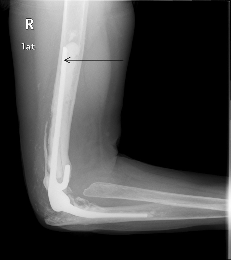
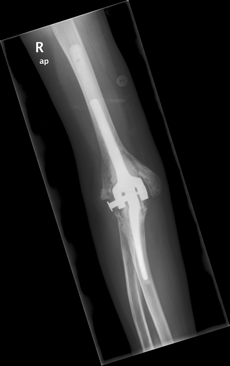
Of the 20 patients 18 were evaluated with standard elbow radiographs. Two patients were not radiographically assessed due to their medical conditions, but they were clinically assessed by a visit in their home. There were two cases of radiolucent lines type 1 (Fig. 1). Neither of these cases was clinically loose. Two cases of minor bushing wear were identified. One patient had loosening of the spline (Fig. 2), but scored 100 on the MEPS, had a ROM of 145° and did not have any symptoms. The patient was informed and offered surgery, but is currently not willing to attend further surgery. She is followed up once a year.
Fig. 1.
Radiolucent line around humeral stem of Coonrad-Morrey TEA
Fig. 2.
Loose spline between humeral and ulnar stems of Coonrad-Morrey TEA
Complications
One patient was revised 12 months after revision due to clinical deep infection despite negative culture. Though treated with antibiotics after soft tissue revision, the infection was not eradicated and removal of the prosthesis was performed after 12 months due to persistent infection. The implant was revised in a two-step procedure but the infection recurred. A new two-step procedure was performed successfully.
Three patients had ulnar nerve paraesthesia prior to revision TEA, and neither one resolved after revision. Two patients developed ulnar nerve paraesthesia post-operatively. One of these had resolved at follow-up. In one case the spline connecting the ulnar and humeral parts of the TEA was loose, but the patient did not want revision due to lack of symptoms.
Discussion
In this retrospective study we found a significant improvement in ROM with an increase in arc of extension-flexion from 80° preoperatively to 109° post-operatively and an increase in the arc of rotation from 99 to 124°. The five-year survival in this study was 95 %. The average MEPS of 79 points is defined as a good result. We have not found any studies on primary or revision TEA that uses the OES and therefore we can only compare our results to other studies in terms of MEPS and ROM.
In 1997, King et al. [2] described the results with 41 Coonrad-Morrey revision TEAs at a mean follow-up of six years. Indications for primary TEA were RA (n = 20), osteoarthritis (n = 20) and tumour (n = 1). The study reported an improvement in MEPS from 44 to 87 and an average post-operative arc of extension-flexion of 101°. King et al. found nine cases of radiolucent lines around the humeral component and six cases of radiolucent lines around the ulnar component. In three cases a second revision was performed due to bushing wear (n = 2) and periprosthetic fracture (n = 1). Furthermore, one patient had permanent removal of the TEA due to recurrent aseptic loosening.
In 2006, Sneftrup et al. [6] reported the results with a series of 24 Coonrad-Morrey revision TEAs. Indications for primary TEA were RA (n = 16), fracture (n = 4) and osteoarthritis (n = 4). At a mean follow-up of 4.1 years the mean MEPS was 85. The arc of flexion-extension was 101° and the arc of rotation was 109° and was comparable to the preoperative measurements. In this study ROM at follow-up was compared to ROM prior to primary TEA. Four patients were re-revised within 18 months and the five-year survival rate was 83.1 %. There were five cases of bushing wear and nine cases of radiolucent lines.
In 2007 Shi et al. reported a five-year survival rate of 64 % on 30 Coonrad-Morrey revision TEAs with an average follow-up of 5.7 years. The arc of extension-flexion improved from 88 to 109°, and the arc of rotation improved from 130 to 151°. The post-operative MEPS was 85 [5].
Finally, Athwal and Morrey published a study in 2006 on 26 Coonrad-Morrey revision TEAs after prosthetic fracture using either the cement-within-cement technique (14 cases) or the traditional method with removal of all cement (12 cases). The post-operative MEPS was 82 in the cement-within-cement group and 78 in the traditional group. At a mean follow-up of 5.1 years the arc of extension-flexion was 108° [7].
Only a few studies on the Coonrad-Morrey revision TEA have been published [2, 5–7]. The average follow-up is approximately five years and the material consists of between 20 and 41 patients. The five-year survival rate in this study is higher than previous studies as described by Sneftrup et al. and Shi et al., but long-term follow-up is needed for specific data on long-term survival rate. All studies except the study by Sneftrup et al. report improvement in ROM. All studies report comparable improvement in pain score. Deep infection is a major concern due to poor results and recurrent revisions. The incidence of deep infection after TEA is estimated to be between two and four percent [22, 23]. A negative culture in biopsies taken at revision does not seem to exclude infection and one has to rely on the clinical signs of deep infection [6, 24].
It has been concluded in some studies that revision with linked implants has a better outcome and survival rate than unlinked implants [3, 25]. The five-year survival rate after revision with the unlinked Souter-Strathclyde TEA has been reported by van der Lugt and Rozing to be 73.8 % and by Redfern et al. to be 86.7 % [26, 27]. Both studies conclude that the results are satisfactory. The results after linked and unlinked revision TEAs are comparable, but the linked Coonrad-Moorey TEA is a safe choice for revision in patients with or without sufficient soft tissue support. Currently available studies on revision TEAs mainly consist of small case series with short- to midterm follow-up. Long-term follow-up and/or multicentre studies comparing different implants would be of interest.
Conclusion
In this study revision with the linked Coonrad-Morrey TEA provides significant improvement in ROM, good pain relief for both index linked and unlinked implants and low failure rate. The results in this study are comparable to previous studies. Future reports with the OES could be of interest to compare patient-related outcome measures. The linked Coonrad-Morrey TEA provides a good option for revision in patients with a failed primary TEA.
References
- 1.Schmidt K, Hilker A, Miehlke RK. Differences in elbow replacement in rheumatoid arthritis. Orthopade. 2007;36(8):714–722. doi: 10.1007/s00132-007-1119-y. [DOI] [PubMed] [Google Scholar]
- 2.King GJ, Adams RA, Morrey BF. Total elbow arthroplasty: revision with use of a non-custom semiconstrained prosthesis. J Bone Joint Surg Am. 1997;79(3):394–400. doi: 10.1302/0301-620X.79B3.6642. [DOI] [PubMed] [Google Scholar]
- 3.Levy JC, Loeb M, Chuinard C, Adams RA, Morrey BF. Effectiveness of revision following linked versus unlinked total elbow arthroplasty. J Shoulder Elbow Surg. 2009;18(3):457–462. doi: 10.1016/j.jse.2008.11.016. [DOI] [PubMed] [Google Scholar]
- 4.Schneeberger AG, Meyer DC, Yian EH. Coonrad-Morrey total elbow replacement for primary and revision surgery: a 2- to 7.5-year follow-up study. J Shoulder Elbow Surg. 2007;16(3 Suppl):S47–S54. doi: 10.1016/j.jse.2006.01.013. [DOI] [PubMed] [Google Scholar]
- 5.Shi LL, Zurakowski D, Jones DG, Koris MJ, Thornhill TS. Semiconstrained primary and revision total elbow arthroplasty with use of the Coonrad-Morrey prosthesis. J Bone Joint Surg Am. 2007;89(7):1467–1475. doi: 10.2106/JBJS.F.00715. [DOI] [PubMed] [Google Scholar]
- 6.Sneftrup SB, Jensen SL, Johannsen HV, Søjbjerg JO. Revision of failed total elbow arthroplasty with use of a linked implant. J Bone Joint Surg Br. 2006;88(1):78–83. doi: 10.1302/0301-620X.88B1.16446. [DOI] [PubMed] [Google Scholar]
- 7.Athwal GS, Morrey BF (2006) Revision total elbow arthroplasty for prosthetic fractures. J Bone Joint Surg Am 88:2017–2026 [DOI] [PubMed]
- 8.Sanchez-Sotelo J. Total elbow arthroplasty. Open Orthop J. 2011;5:115–123. doi: 10.2174/1874325001105010115. [DOI] [PMC free article] [PubMed] [Google Scholar]
- 9.Futai K, Tomita T, Yamazaki T, et al. In vivo three-dimensional kinematics of total elbow arthroplasty using fluoroscopic imaging. Int Orthop. 2010;34(6):847–854. doi: 10.1007/s00264-010-0972-1. [DOI] [PMC free article] [PubMed] [Google Scholar]
- 10.Fevang BT, Lie SA, Havelin LI, Skredderstuen A, Furnes O. Results after 562 total elbow replacements: a report from the Norwegian Arthroplasty Register. J Shoulder Elbow Surg. 2009;18(3):449–456. doi: 10.1016/j.jse.2009.02.020. [DOI] [PubMed] [Google Scholar]
- 11.Skyttä ET, Eskelinen A, Paavolainen P, Ikävalko M, Remes V. Total elbow arthroplasty in rheumatoid arthritis: a population-based study from the Finnish Arthroplasty register. Acta Orthop. 2009;80(4):472–477. doi: 10.3109/17453670903110642. [DOI] [PMC free article] [PubMed] [Google Scholar]
- 12.Bernardino S. Total elbow arthroplasty: history, current concepts, and future. Clin Rheumatol. 2010;29(11):1217–1221. doi: 10.1007/s10067-010-1539-7. [DOI] [PubMed] [Google Scholar]
- 13.Celli A, Morrey BF. Total elbow arthroplasty in patients forty years of age or less. J Bone Joint Surg Am. 2009;91(6):1414–1418. doi: 10.2106/JBJS.G.00329. [DOI] [PubMed] [Google Scholar]
- 14.Voloshin I, Schippert DW, Kakar S, Kaye EK, Morrey BF. Complications of total elbow replacement: a systematic review. J Shoulder Elbow Surg. 2011;20(1):158–168. doi: 10.1016/j.jse.2010.08.026. [DOI] [PubMed] [Google Scholar]
- 15.Morrey BF, Adams RA. Semiconstrained arthroplasty for the treatment of rheumatoid arthritis of the elbow. J Bone Joint Surg Am. 1992;74(4):479–490. [PubMed] [Google Scholar]
- 16.Dawson J, Doll H, Boller I, Fitzpatrick R, Little C, Rees J, et al. Comparative responsiveness and minimal change for the Oxford Elbow Score following surgery. Qual Life Res. 2008;17(10):1257–1267. doi: 10.1007/s11136-008-9409-3. [DOI] [PubMed] [Google Scholar]
- 17.Dawson J, Doll H, Boller I, Fitzpatrick R, Little C, Rees J, et al. The development and validation of a patient-reported questionnaire to assess outcomes of elbow surgery. J Bone Joint Surg Br. 2008;90(4):466–473. doi: 10.1302/0301-620X.90B4.20290. [DOI] [PubMed] [Google Scholar]
- 18.Beaton DE, Bombardier C, Guillemin F, Ferraz MB. Guidelines for the process of cross-cultural adaptation of self-report measures. Spine (Phila Pa 1976) 2000;25(24):3186–3191. doi: 10.1097/00007632-200012150-00014. [DOI] [PubMed] [Google Scholar]
- 19.Souter WA. Arthroplasty of the elbow with particular reference to metallic hinge arthroplasty in rheumatoid patients. Orthop Clin North Am. 1973;4(2):395–413. [PubMed] [Google Scholar]
- 20.Souter WA. Surgery of the rheumatoid elbow. Ann Rheum Dis. 1990;49(Suppl 2):871–882. doi: 10.1136/ard.49.Suppl_2.871. [DOI] [PMC free article] [PubMed] [Google Scholar]
- 21.Kamme C, Lindberg L. Aerobic and anaerobic bacteria in deep infections after total hip arthroplasty: differential diagnosis between infectious and non-infectious loosening. Clin Orthop Relat Res. 1981;154:201–207. [PubMed] [Google Scholar]
- 22.Little CP, Graham AJ, Carr AJ. Total elbow arthroplasty: a systematic review of the literature in the English language until the end of 2003. J Bone Joint Surg Br. 2005;87(4):437–444. doi: 10.1302/0301-620X.87B4.15692. [DOI] [PubMed] [Google Scholar]
- 23.Yamaguchi K, Adams RA, Morrey BF. Infection after total elbow arthroplasty. J Bone Joint Surg Am. 1998;80(4):481–491. doi: 10.2106/00004623-199804000-00004. [DOI] [PubMed] [Google Scholar]
- 24.Vergidis P, Greenwood-Quaintance KE, Sanchez-Sotelo J, Morrey BF, Steinmann SP, Karau MJ, et al. Implant sonication for the diagnosis of prosthetic elbow infection. J Shoulder Elbow Surg. 2011;20(8):1275–1281. doi: 10.1016/j.jse.2011.06.016. [DOI] [PMC free article] [PubMed] [Google Scholar]
- 25.Ring D, Kocher M, Koris M, Thornhill TS. Revision of unstable capitellocondylar (unlinked) total elbow replacement. J Bone Joint Surg Am. 2005;87(5):1075–1079. doi: 10.2106/JBJS.D.02449. [DOI] [PubMed] [Google Scholar]
- 26.van der Lugt JC, Rozing PM. Outcome of revision surgery for failed primary Souter-Strathclyde total elbow prosthesis. J Shoulder Elbow Surg. 2006;15:208–214. doi: 10.1016/j.jse.2005.07.009. [DOI] [PubMed] [Google Scholar]
- 27.Redfern DR, Dunkley AB, Trail IA, Stanley JK. Revision total elbow replacement using the Souter-Strathclyde prosthesis. J Bone Joint Surg Br. 2001;83(5):635–639. doi: 10.1302/0301-620X.83B5.11268. [DOI] [PubMed] [Google Scholar]


