Abstract
Purpose
The most common fixation techniques for tibial avulsion fractures of the anterior cruciate ligament (ACL) described in the literature are screw and suture fixation. The fixation of these fractures with the TightRope® device might be an alternative. Up to now it has been commonly used in other injuries, such as acromioclavicular joint or syndesmosis ruptures. The purpose of this study was to evaluate the biomechanical properties of different fixation techniques for the reconstruction of tibial avulsion fractures.
Methods
Type III tibial avulsion fractures were simulated in 40 porcine knees. Each specimen was randomly assigned to one of four groups: (1) anterograde screw fixation, (2) suture fixation, (3) TightRope® fixation or (4) control group. The initial displacement, strength to failure and the failure mode were documented.
Results
The maximum load to failure was 1,345 ± 155.5 N for the control group, 402.5 ± 117.6 N for the TightRope® group, 367 ± 115.8 N for the suture group and 311.7 ± 120.3 N for the screw group. The maximum load to failure of the control group was significantly larger compared to all other groups. The initial dislocation was 0.28 ± 0.09 mm for the control group, 0.55 ± 0.26 mm for the TightRope® group, 0.84 ± 0.15 mm for the screw group and 1.14 ± 0.9 mm for the suture group. The initial dislocation was significantly larger for the suture group compared to the TightRope® and control groups.
Conclusions
The TightRope® fixation shows significantly lower initial displacement compared to the suture group. The TightRope® fixation might be an alternative for the repair of ACL tibial avulsion fractures that can be used arthroscopically.
Introduction
Tibial avulsion fractures of the anterior cruciate ligament (ACL) can lead to instability and inadequate refixation can cause extension and flexion limitation of the range of motion. Therefore it is mandatory to restore the biomechanics by anatomical reduction and internal fixation [1–3]. Arthroscopic treatment is the gold standard nowadays and has replaced open techniques [4, 5]. In the literature a number of different fixation methods are described; however, suture pull-out and screw fixation are the most commonly evaluated [4, 6–9].
Some recent studies have investigated the biomechanical stability of different arthroscopic fixation techniques for ACL tibial avulsion fractures [10–14]. However, there is no study that compares the initial fixation strength of ACL tibial avulsion fractures using the TightRope® device. Fixation with the TightRope® system is described frequently in the treatment of other ligamentous injuries, such as acromioclavicular joint ruptures [15] or syndesmosis ruptures [16]. A recent systematic review of the literature revealed similar results for the TightRope® system compared to syndesmotic screw or bolt fixation in the treatment of syndesmosis ruptures. Furthermore, the rate of implant removal was lower for the TightRope® fixation [17].
These findings led to the idea that the TightRope® fixation might be an alternative for the fixation of tibial avulsion fractures of the ACL. The hypothesis was that the TightRope® fixation of ACL tibial avulsion fractures would not yield inferior results in terms of biomechanical properties compared with commonly used fixations employing anterograde screw or pull-out suture.
Methods
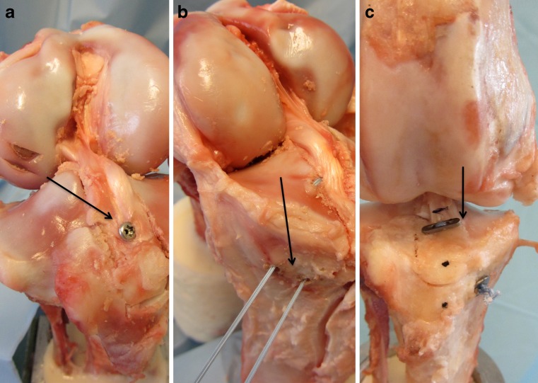
For the biomechanical testing, we used the knees of 40 German Landrace pigs. The pigs were one year old, fully grown and weighed between 100 and 120 lbs. The tibial neck and the femoral head were cut off and the shafts of the bones cemented into an aluminum holder using cold-curing methyl methacrylate resin (Technovit 4071, Heraeus Kulzer GmbH, Wehrheim, Germany). Each specimen was randomly assigned to one of four groups: (1) antegrade screw fixation (Fig. 1a), (2) suture fixation (Fig. 1b), (3) TightRope® fixation (Fig. 1c) or (4) control group.
Fig. 1.
a–c Anterograde screw fixation (a), suture fixation (b) and TightRope® fixation (c)
Type III tibial avulsion fractures (according to Meyers and McKeever [18]) were simulated in 30 knees using a custom-made template. The fragments were 30 mm (length) × 20 mm (width) × 8.5 mm (height). This set-up had previously been used by another study group [11]. Ten intact knees were used as a control group. All muscles and soft tissue were removed leaving only the femur-ACL-tibia complex intact. In group 1, a drill guide was placed in the middle of the bone fragment to replace and secure it. A guide pin (1 mm in diameter) was drilled from inside out. A 4 × 20 mm cannulated, partially-threaded screw was inserted to fix the fragment. In the second group, two K-wires were placed medial and lateral to the insertion point of the ACL in order to put the fracture in place using FiberWire 2 (Arthrex, Naples, FL, USA). The two ends of the strand were sutured over a bony bridge. The bone bridge had a width of ten millimetres. In group 3, the Arthrex ACL aiming device was placed in the middle of the fracture. A guide pin was drilled from outside in order to place the flip button of the TightRope® device on the fragment. The ends of the strand were fixed on the anterior tibial cortex. In group 4, no avulsion fracture was created.
The constructions were thawed at 4 °C for 24 hours prior to mechanical testing and kept moist using saline spray during the entire procedure. A material testing machine (Mini Bionix 858, MTS Systems Co., Minneapolis, MN, USA) was used for the mechanical evaluation of the constructions. The potted knees were rigidly fixed in a base platform setting the force direction angle to 0° to simulate a “worst-case scenario”.
The constructions were pre-tensioned with 5 N for 30 seconds prior to testing. Then, 20 cycles of mechanical loading between 0 and 40 N were applied at a repetition rate of 1 Hz. The increase in construction length was recorded. Length changes are reported between the minimum of the 1st and the maximum of the 20th cycle. After decreasing the preload from 40 to 10 N, and pausing for 30 seconds, a failure test with a ramp speed of one millimetre per second was performed. The maximum failure load, failure mode and dislocation of the constructions were analysed.
Statistical analysis
All mean values are reported with standard deviations. The four groups were compared using a one-way analysis of variance (ANOVA). Normality and equality of variance tests were conducted. If the normality test failed, a Kruskal-Wallis ANOVA by ranks was executed. All operations were performed using SigmaStat 15.0 (SPSS Inc., Chicago, IL, USA). A significance level of p < 0.05 was assumed.
Results
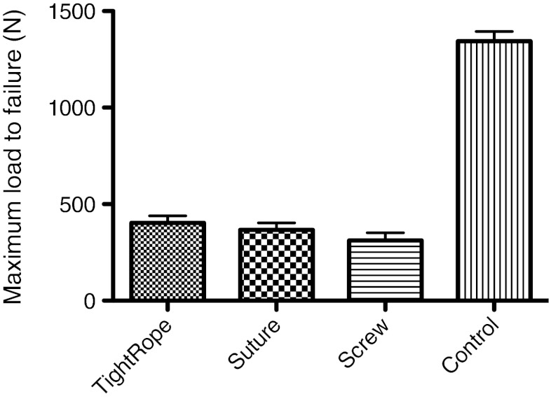
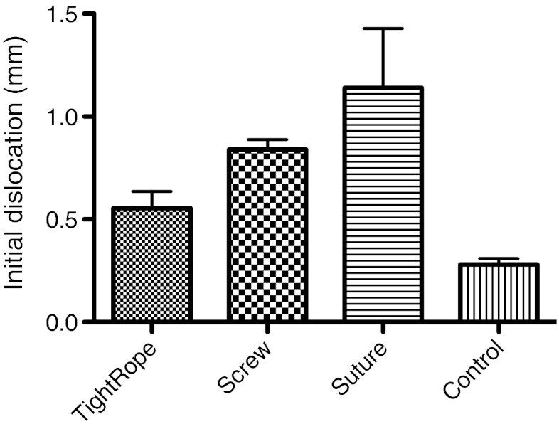
The maximum load to failure was 1,345 ± 155.5 N for the control group, 402.5 ± 117.6 N for the TightRope® group, 367 ± 115.8 N for the suture group and 311.7 ± 120.3 N for the screw group. The maximum load to failure of the control group was significantly larger than all other groups (p < 0.05; Fig. 2). The initial displacement was 0.28 ± 0.09 mm for the control group, 0.55 ± 0.26 mm for the TightRope® group, 0.84 ±0.15 mm for the screw group and 1.14 ± 0.9 mm for the suture group. The initial dislocation was significantly larger for the suture group compared to the TightRope® and control groups (p < 0.05; Fig. 3).
Fig. 2.
The maximum load to failure of the control group was significantly larger compared to all other groups (p < 0.05)
Fig. 3.
The initial dislocation was significantly larger for the suture group compared to the TightRope® and control groups (p < 0.05)
At the moment of failure the following failure modes were observed: In group 1 an avulsion of the screw occurred in 6/10 (60 %) of the specimens and a fracture of the fragment occurred in 4/10 (40 %) of the specimens. In group 2 a fracture of the fragment was observed in 6/10 (60 %) of the specimens and a fracture of the bone bridge was observed in 4/10 (40 %) of the specimens. In group 3 a fracture of the fragment occurred in 10/10 (100 %). In group 4, a rupture of the ACL occurred in 7/10 (70 %), a fracture of the condyle occurred in 1/10 (10 %) and a tibia eminence avulsion fracture occurred in 1/10 (10 %).
Discussion
The most important finding of this study is that the TightRope® fixation showed equal fixation strength and lower initial displacement compared to the suture group. The results of this biomechanical analysis support the authors’ working hypothesis. To the best of our knowledge, this is the first study presenting the biomechanical properties of the TightRope® fixation system in ACL avulsion fractures. Other authors have focused on the biomechanical properties of different suture anchors [12, 13], screw fixations [11], suture techniques and materials [11–14] or fixation devices such as the EndoButton® [12].
There are several study limitations, which have to be mentioned. In this biomechanical set-up only one force direction was applied. Nothing can be reported about the influence on flexion extension motion or any biological healing responses. This study only reports the “time zero” biomechanical properties of the tested fixation techniques. Nurmi et al. [19] criticised the use of porcine tibia due to the fact that graft slippage is underestimated and maximum load to failure is overestimated when the biomechanical properties of interference screws are evaluated. The differences between young human tibias and porcine tibias regarding the property of the cancellous bone might have influenced the screw fixation. This should not have influenced either the TightRope® fixation or the suture fixation, since they were fixed over a cortical bone bridge. Porcine bones were also used in recent studies [11, 12, 14] concerning the biomechanical properties of tibial avulsion fracture fixation techniques. This is due to their easy availability and the homogeneity of the bone quality. The results of our control group verify this in terms of a low standard deviation of the biomechanical properties.
A variety of testing protocols for simulated tibial avulsion fractures have been published recently. In et al. [13] applied a series of ten cycles between 0 and 30 N and a strain rate of 200 mm/min in human cadaver knees with an average age around 60 years. Hapa et al. [12] applied a series of 500 cycles between 0 and 100 N with a strain rate of 100 mm/min in ovine knees and Mahar et al. [14] applied 200 cycles between 0 and 150 N with a strain rate of 0.5 mm/s in porcine knees. We choose to limit the loading cycles to 20 since pilot testing with more cycles showed that most of the total displacement appears within the first 15 cycles. A higher cyclical loading force showed untimely failures, so that the maximum force during cyclical loading was limited to 40 N. The strain rate used in this study is comparable to the above-mentioned protocols.
A high initial fixation strength is essential to minimise the risk of residual laxity, which leads to a longer post-operative immobilisation that may cause arthrofibrosis and a limitation of range of motion [20, 21]. Hence, a sturdy fixation is the basis for a promising outcome after ACL avulsion fracture fixation. The knowledge of failure loads for fixation devices/techniques is important in general. It gives useful information about the resistance during unexpected events such as tripping or slipping. In addition, it sets limits to the early rehabilitation protocol. The maximum failure loads evaluated for the different groups in this study are comparable to the results presented by others. Eggers et al. [11] reported 457.1 N for a screw fixation and 599.6 N for a suture fixation. Hapa et al. [12] reported maximum failure loads for different suture fixations between 213 and 299 N as well as 314 N for an EndoButton fixation. In et al. [13] reported a maximum load to failure of 126.6 N for a screw fixation and 101.8 N for a suture anchor fixation. The variance of the results of the screw fixations might be caused by the different protocols and tissue used.
The initial dislocation of the fragment is also crucial. Montgomery et al. [21] reported in a retrospective case study (17 patients) that only 29 % progressed through physical therapy uneventfully. Complications correlate with initial displacement. The initial dislocation of the TightRope® fixation was 0.55 ± 0.26 mm. This is comparable or superior to the results throughout the literature [10–14].
The observed failure modes of the screw and the suture show results similar to other studies. A pull-out of the screw, a failure of the suture or a fragment fracture occurred in a comparable prevalence [11, 13, 14]. The TightRope® fixation failed in 100 % of the tests due to a fragment fracture. This might be due to a superior quality of the strands compared to the tested suture materials.
This study shows that a TightRope® fixation of tibial avulsion fractures is possible. This fixation technique showed biomechanical properties comparable to a screw and a suture fixation as well as other fixation techniques described throughout the literature.
Conclusion
The TightRope® fixation show significantly lower initial dislocation compared to the suture group and higher but not significantly different maximum load to failure compared to the commonly used suture and screw. Due to these facts the TightRope® fixation can be considered as a reliable alternative for the repair of ACL tibial avulsion fractures that can be used arthroscopically.
Acknowledgment
Arthrex supported the work. No financial biases exist for any author. The authors, their immediate family, and any research foundation with which they are affiliated did not receive other benefits from any commercial entity related to the subject of this article.
References
- 1.Ahn JH, Yoo JC. Clinical outcome of arthroscopic reduction and suture for displaced acute and chronic tibial spine fractures. Knee Surg Sports Traumatol Arthrosc. 2005;13(2):116–121. doi: 10.1007/s00167-004-0540-6. [DOI] [PubMed] [Google Scholar]
- 2.Griffith JF, Antonio GE, Tong CW, Ming CK. Cruciate ligament avulsion fractures. Arthroscopy. 2004;20(8):803–812. doi: 10.1016/j.arthro.2004.06.007. [DOI] [PubMed] [Google Scholar]
- 3.Hunter RE, Willis JA. Arthroscopic fixation of avulsion fractures of the tibial eminence: technique and outcome. Arthroscopy. 2004;20(2):113–121. doi: 10.1016/j.arthro.2003.11.028. [DOI] [PubMed] [Google Scholar]
- 4.Huang TW, Hsu KY, Cheng CY, Chen LH, Wang CJ, Chan YS, Chen WJ. Arthroscopic suture fixation of tibial eminence avulsion fractures. Arthroscopy. 2008;24(11):1232–1238. doi: 10.1016/j.arthro.2008.07.008. [DOI] [PubMed] [Google Scholar]
- 5.Kogan MG, Marks P, Amendola A. Technique for arthroscopic suture fixation of displaced tibial intercondylar eminence fractures. Arthroscopy. 1997;13(3):301–306. doi: 10.1016/S0749-8063(97)90025-6. [DOI] [PubMed] [Google Scholar]
- 6.Ahn JH, Lee YS, Lee DH, Ha HC. Arthroscopic physeal sparing all inside repair of the tibial avulsion fracture in the anterior cruciate ligament: technical note. Arch Orthop Trauma Surg. 2008;128(11):1309–1312. doi: 10.1007/s00402-007-0506-5. [DOI] [PubMed] [Google Scholar]
- 7.Delcogliano A, Chiossi S, Caporaso A, Menghi A, Rinonapoli G. Tibial intercondylar eminence fractures in adults: arthroscopic treatment. Knee Surg Sports Traumatol Arthrosc. 2003;11(4):255–259. doi: 10.1007/s00167-003-0373-8. [DOI] [PubMed] [Google Scholar]
- 8.Kim YM, Kim SJ, Yang JY, Kim KC. Pullout reattachment of tibial avulsion fractures of the anterior cruciate ligament: a firm, effective suture-tying method using a tensioner. Knee Surg Sports Traumatol Arthrosc. 2007;15(7):847–850. doi: 10.1007/s00167-007-0315-y. [DOI] [PubMed] [Google Scholar]
- 9.Lafrance RM, Giordano B, Goldblatt J, Voloshin I, Maloney M. Pediatric tibial eminence fractures: evaluation and management. J Am Acad Orthop Surg. 2010;18(7):395–405. doi: 10.5435/00124635-201007000-00002. [DOI] [PubMed] [Google Scholar]
- 10.Bong MR, Romero A, Kubiak E, Iesaka K, Heywood CS, Kummer F, Rosen J, Jazrawi L. Suture versus screw fixation of displaced tibial eminence fractures: a biomechanical comparison. Arthroscopy. 2005;21(10):1172–1176. doi: 10.1016/j.arthro.2005.06.019. [DOI] [PubMed] [Google Scholar]
- 11.Eggers AK, Becker C, Weimann A, Herbort M, Zantop T, Raschke MJ, Petersen W. Biomechanical evaluation of different fixation methods for tibial eminence fractures. Am J Sports Med. 2007;35(3):404–410. doi: 10.1177/0363546506294677. [DOI] [PubMed] [Google Scholar]
- 12.Hapa O, Barber FA, Süner G, Özden R, Davul S, Bozdağ E, Sünbüloğlu E. Biomechanical comparison of tibial eminence fracture fixation with high-strength suture, EndoButton, and suture anchor. Arthroscopy. 2012;28(5):681–687. doi: 10.1016/j.arthro.2011.10.026. [DOI] [PubMed] [Google Scholar]
- 13.In Y, Kwak DS, Moon CW, Han SH, Choi NY. Biomechanical comparison of three techniques for fixation of tibial avulsion fractures of the anterior cruciate ligament. Knee Surg Sports Traumatol Arthrosc. 2012;20(8):1470–1478. doi: 10.1007/s00167-011-1694-7. [DOI] [PubMed] [Google Scholar]
- 14.Mahar AT, Duncan D, Oka R, Lowry A, Gillingham B, Chambers H. Biomechanical comparison of four different fixation techniques for pediatric tibial eminence avulsion fractures. J Pediatr Orthop. 2008;28(2):159–162. doi: 10.1097/BPO.0b013e318164ee43. [DOI] [PubMed] [Google Scholar]
- 15.Scheibel M, Dröschel S, Gerhardt C, Kraus N. Arthroscopically assisted stabilization of acute high-grade acromioclavicular joint separations. Am J Sports Med. 2011;39(7):1507–1516. doi: 10.1177/0363546511399379. [DOI] [PubMed] [Google Scholar]
- 16.Naqvi GA, Shafqat A, Awan N. Tightrope fixation of ankle syndesmosis injuries: clinical outcome, complications and technique modification. Injury. 2012;43(6):838–842. doi: 10.1016/j.injury.2011.10.002. [DOI] [PubMed] [Google Scholar]
- 17.Schepers T. Acute distal tibiofibular syndesmosis injury: a systematic review of suture-button versus syndesmotic screw repair. Int Orthop. 2012;36(6):1199–1206. doi: 10.1007/s00264-012-1500-2. [DOI] [PMC free article] [PubMed] [Google Scholar]
- 18.Meyers MH, McKeever FM. Fracture of the intercondylar eminence of the tibia. J Bone Joint Surg Am. 1970;52(8):1677–1684. [PubMed] [Google Scholar]
- 19.Nurmi JT, Sievänen H, Kannus P, Järvinen M, Järvinen TLN. Porcine tibia is a poor substitute for human cadaver tibia for evaluating interference screw fixation. Am J Sports Med. 2004;32(3):765–771. doi: 10.1177/0363546503261732. [DOI] [PubMed] [Google Scholar]
- 20.May JH, Levy BA, Guse D, Shah J, Stuart MJ, Dahm DL. ACL tibial spine avulsion: mid-term outcomes and rehabilitation. Orthopedics. 2011;34(2):89. doi: 10.3928/01477447-20101221-10. [DOI] [PubMed] [Google Scholar]
- 21.Montgomery KD, Cavanaugh J, Cohen S, Wickiewicz TL, Warren RF, Blevens F. Motion complications after arthroscopic repair of anterior cruciate ligament avulsion fractures in the adult. Arthroscopy. 2002;18(2):171–176. doi: 10.1053/jars.2002.30433. [DOI] [PubMed] [Google Scholar]


