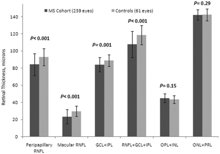
Figure 2.
Spectral-domain optical coherence tomography thicknesses (in μm) for eyes of patients with multiple sclerosis (MS) versus disease-free controls. Mean values for peripapillary retinal nerve fiber layer (RNFL), macular RNFL, ganglion cell layer, and inner plexiform layer (GCL+IPL), and the combination of macular RNFL+GCL+IPL were significantly lower for MS eyes versus controls (P values are from generalized estimating equation models, accounting for age and within-patient, intereye correlations). INL = inner nuclear layer; ONL = outer nuclear layer; OPL = outer plexiform layer; PRL = photoreceptor layer.

