Abstract
A case of furuncular myiasis was reported for the first time in a 29-year-old young Taiwanese traveler returning from an ecotourism in Peru. Furuncle-like lesions were observed on the top of his head and he complained of crawling sensations within his scalp. The invasive larva of botfly, Dermatobia hominis, was extruded from the furuncular lesion of the patient. Awareness of cutaneous myiasis for clinicians should be considered for a patient who has a furuncular lesion and has recently returned from a botfly-endemic area.
Keywords: Furuncular myiasis, Botfly, Traveler, Taiwan
1. Introduction
Furuncular myiasis can be caused by the human botfly Dermatobia hominis (D. hominis), the African tumbu fly Cordylobia anthropophaga, or the rodent botfly (Cuterebra spp.)[1]–[5]. Among these flies, D. hominis is the most common agent of both cutaneous myiasis and furuncular myiasis diagnosed in returning travelers[6]–[9]. This species is distributed through Mexico to Argentina of Central and South America, in areas of relatively high temperature and humidity[10]. Because D. hominis is not endemic in Taiwan and the caused lesion resembles a furuncle in the skin. Thus, the diagnosis of furuncular myiasis is never reported in Taiwan. Here, we reported the first human case of furuncular myiasis in a Taiwanese traveler returning from botfly-endemic areas.
2. Case report
A 29-year-old Taiwanese man living in Ilan City of northern Taiwan presented to our Surgical Emergency Division of Tri-Service General Hospital on 4 February 2012, with a chief complaint of painful nodule combined with perceived movement within the scalp of his head. Four weeks earlier, he had returned from an ecological trip to Peru. He started the trip from the Amazonas region (Iquitos) on 28 November 2011 and ended at the coast region (Trujillo) of Peru. He had spent two weeks in the Amazonas region and recalled a mosquito bite during his trip. The patient felt a regional swelling of lymph node on right side of his neck on 24 December 2011. After returning home on 5 January 2012, he observed a furuncle-like lesion on the top of his head and continued hearing of movement within his scalp. He had scratched the skin lesion and had been misdiagnosed as common arthropod bite by the local hospital.
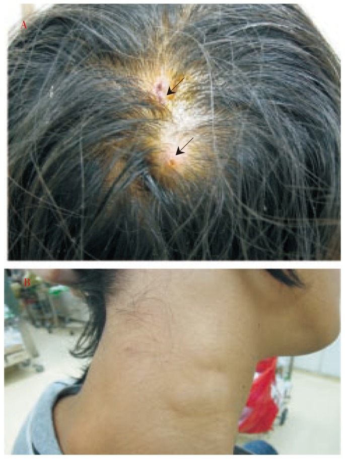
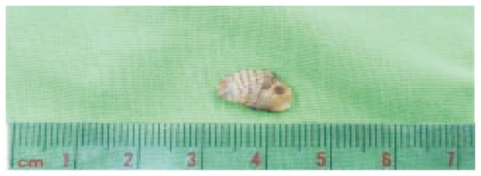
Physical examinations of this patient revealed that he had a 1.2 cm erythematous nodule with a central pore on the top of his head and a regional swelling of lymph node on his neck (Figure 1). After a general sterilization, the larva was gently extruded and grasped with forcep from the furuncle-like lesion[11]. A third instar larva of D. hominis was further identified by the entomological specialist based on its morphological characteristics (Figure 2). Because of the presumed bacterial infection, the patient was treated and prescribed with a 7-day course of antibiotic. The patient recovered completely with no further complication.
Figure 1. Physical examinations.

A: Furuncle-like nodules (arrows) located on the scalp of a 29-year-old man; B: A regional swelling of lymph node observed on his neck.
Figure 2. Morphological characteristics of a third instar larva of D. hominis extruded from the furuncular lesion of this patient, characterized with a size of 7 mm×12 mm and the rows of black spines that resist extraction of larva.

3. Discussion
Myiasis is defined as the invasion of a vertebrate host by immature stages of flies that feed on living tissues, body fluids, or ingested foods. Human myiasis is characterized with the invasion of human tissues by the larvae of the order Diptera (true flies). It occurs commonly in the areas with hot and humid climates. Species causing myiasis can be classified into three parasitological categories: obligatory, facultative, and accidental. An obligatory parasite requires living tissue for the development of invaded larvae[10]. Clinically, species classification is based on the infested area of the host body that includes cutaneous, enteric, ophthalmic, nasopharyngeal, auricular, and urogenital[1]. In general, cutaneous myiasis is the most common type and can be presented as furuncular, migratory, and wound myiasis, depending on the infesting larvae[3].
Furuncular myiasis caused by D. hominis is attributed to the deposition of its eggs by a carrier insect like mosquito, to the host tissues. Once in contact with the host skin, the host's body temperature stimulates the eggs to hatch and the first instar larva then painlessly burrow into host skin. After larval penetration, a small erythematous papule develops that later becomes a furuncular-like nodule and a central pore within the lesion allows exposure to air for larval respiration[12]. During a period of 5 to 10 weeks, the larva develops into the second and then third instars, and burrow deeper into the host's skin, forming a dome-shaped cavity[2]. Symptoms during the larval stage may include itching, a sensation of movement, lancinating pain, and a serosanguinous discharge[1],[3].
In our patient, he was infested by the larva of D. hominis recalled as a mosquito bite during his trip in the Amazonas region (Iquitos) of Peru. The third instar larva was extruded from the furuncular lesion after a period of 8 weeks later. This is fully accordant with the time period required for the larval development of D. hominis. Although this patient had a regional swelling of lymph node, no further complication was observed after antibiotic therapy. It is assumed that scratching of skin lesion by himself may contribute to the cause of secondary infection in this patient. Indeed, secondary infection in myiasis is unusual because bacteriostatic substances are produced in the gut of invaded larvae[13]. However, the most severe human complication caused by D. hominis myiasis had been reported as fatal cerebral myiasis, resulting from the larval infestation in the skin covering the fontanelles of infants and young children[14].
Furuncular myiasis caused by D. hominis is often mistaken as a common arthropod bite. Indeed, differential diagnoses including the cutaneous larva migrans and cutaneous leishmaniasis caused by other arthropods should be excluded[15]. In addition, a Tunga flea invades the host's skin may cause a furuncular nodule which can also resemble as furuncular myiasis. However, tungiasis is often observed on the toes and soles of the foot[16],[17], where furuncular myiasis caused by D. hominis is rarely found. Although medical device of ultrasonography and dermoscopy have been described as an aid in the diagnosis of furuncular myiasis caused by D. hominis[18],[19], diagnosis is made primarily on clinical observation of furuncle-like skin lesion with particular attention to the patient's travel history.
The medical management of cutaneous myiasis is straightforward. Effective treatment of myiasis consists of the removal of invasive larvae, cleaning of the wound, and treated with systemic antibiotics to control any possible associated infections. Surgery is usually unnecessary while the invasive larvae remain alive, and the protruding larva was grasped immediately with forcep and completely extracted with continuous tension together with manual pressure exerted on the side of the nodule. However, surgical removal will be used to remove the dead or decayed larvae from an affected site to prevent any possible secondary infections.
Although human myiasis is rarely found among humans in Taiwan[20]–[22], the increasing activities for ecotourism to South America of Taiwanese travelers enhance the risk of acquiring infection by the invasive larvae. This report provides the first case of furuncular myiasis in Taiwan. Because personal ecotourism to South America has become increasingly common for Taiwanese travelers. Awareness for cutaneous myiasis should be considered for a patient who has a furuncular lesion and has recently returned from a botfly-endemic area. This practical procedure may help to further diagnosis and prevention of this ectoparasitic infection in humans.
Acknowledgments
This work was supported in part by a grant from the National Science Council (NSC99-2314-B-760-001-MY2), Taipei, Taiwan.
Comments
Background
Cutaneous myiasis is defined as the invasion of live human and vertebrate animals by larvae of the order Diptera that feed on living tissues, body fluids, or ingested foods. Human myiasis is characterized with the invasion of human tissues, and it occurs commonly in areas with hot and humid climates. Furuncular myiasis can be caused by Dermatobia hominis, Cordylobia anthropophaga, or Cuterebra spp. This species is endemic through much of Central and South America. Its larvae are transmitted to vertebrate hosts by hematophagous insects, most commonly mosquitoes, on whose abdomens the female botfly has deposited her eggs. Among these flies, D. hominis is the most common agent of both cutaneous myiasis and furuncular myiasis diagnosed in returning travelers.
Research frontiers
This study describes a human case of furuncular myiasis for the first time in a Taiwanese traveler returning from an ecotourism in Peru. Furuncle-like skin lesions were observed on his scalp and an invasive larva of botfly was extruded from the furuncular lesion. Based on the entomological characteristics of botfly, diagnosis is accurate.
Related reports
Physical examination of this patient revealed that he had a 1.2 cm erythematous nodule with a central pore on the top of his head. This location of furuncle nodule is different from the previous report by Bhandari et al.(2007) which was described on the lower leg of patient. Extrution method for removing invasive live larva is reported as Li Loong et al. (1992).
Innovations and breakthroughs
Data regarding the first case of furuncular myiasis in Taiwan is significant value for the clinical diagnosis and medical entomology.
Applications
This report is a very important reference for the clinical diagnosis and treatment of furuncular myiasis in Taiwan. It may remind the clinicians to consider for a patient who has a furuncular lesion and has recently retured from a botfly endemic area.
Peer review
This is an interesting report regarding the clinical diagnosis and treatment of furuncular myiasis. Results of this study are highly interesting and may enhance the awareness of cutaneous myiasis for clinicians.
Footnotes
Foundation Project: Supported in part by a grant from the National Science Council Taipei, Taiwan (Grant No. NSC99-2314-B-760-001-MY2).
Conflict of interest statement: We declare that we have no conflict of interest.
References
- 1.Robbins K, Khachemoune A. Cutaneous myiasis: a review of the common types of myiasis. Int J Dermatol. 2010;49:1092–1098. doi: 10.1111/j.1365-4632.2010.04577.x. [DOI] [PubMed] [Google Scholar]
- 2.Delshad E, Rubin AI, Almeida L, Niedt GW. Cuterebra cutaneous myiasis: case report and world literature review. Int J Dermatol. 2008;47:363–366. doi: 10.1111/j.1365-4632.2008.03532.x. [DOI] [PubMed] [Google Scholar]
- 3.McGraw TA, Turiansky GW. Cutaneous myiasis. J Am Acad Dermatol. 2008;58:907–926. doi: 10.1016/j.jaad.2008.03.014. [DOI] [PubMed] [Google Scholar]
- 4.Marty FM, Whiteside KR. Myiasis due to Dermatobia hominis. N Engl J Med. 2005;352:23. doi: 10.1056/NEJMicm041049. [DOI] [PubMed] [Google Scholar]
- 5.Plotinsky RN, Talbot EA, Davis H. Cuterebra cutaneous myiasis, new Hampshire, 2004. Am J Trop Med Hyg. 2007;76:596–597. [PubMed] [Google Scholar]
- 6.House HR, Ehlers JP. Travel-related infections. Emerg Med Clin North Am. 2008;26:499–516. doi: 10.1016/j.emc.2008.01.008. [DOI] [PubMed] [Google Scholar]
- 7.Bhandari R, Janos DP, Sinnis P. Furuncular myiasis caused by Dermatobia hominis in a returning traveler. Am J Trop Med Hyg. 2007;76:598–599. [PMC free article] [PubMed] [Google Scholar]
- 8.Clyti E, Deligny C, Nacher M, del Giudice P, Sainte-Marie D, Pradinaud R, et al. et al. An urban epidemic of human myiasis caused by Dermatobia hominis in French Guiana. Am J Trop Med Hyg. 2008;79:797–798. [PubMed] [Google Scholar]
- 9.Solomon M, Benenson S, Baum S, Schwartz E. Tropical skin infections among Israeli travelers. Am J Trop Med Hyg. 2011;85:868–872. doi: 10.4269/ajtmh.2011.10-0471. [DOI] [PMC free article] [PubMed] [Google Scholar]
- 10.Fernandes LF, Pimenta FC, Fernandes FF. First report of human myiasis in goias state, Brazil: frequency of different types of myiasis, their various etiological agents, and associated factors. J Parasitol. 2009;95:32–38. doi: 10.1645/GE-1103.1. [DOI] [PubMed] [Google Scholar]
- 11.Li Loong PT, Lui H, Buck HW. Cutaneous myiasis: a simple and effective technique for extraction of Dermatobia hominis larvae. Int J Dermatol. 1992;31:657–659. doi: 10.1111/j.1365-4362.1992.tb03990.x. [DOI] [PubMed] [Google Scholar]
- 12.Boruk M, Rosenfeld RM, Alexis R. Human botfly infestation presenting as peri-auricular mass. Int J Pediatr Otorhinolaryngol. 2006;70:335–338. doi: 10.1016/j.ijporl.2005.06.025. [DOI] [PubMed] [Google Scholar]
- 13.Sampson CE, MaGuire J, Eriksson E. Botfly myiasis: case report and brief review. Ann Plast Surg. 2001;46:150–152. doi: 10.1097/00000637-200102000-00011. [DOI] [PubMed] [Google Scholar]
- 14.Rossi MA, Zucoloto S. Fatal cerebral myiasis caused by the tropical warble fly, Dermatobia hominis. Am J Trop Med Hyg. 1973;22:267–269. doi: 10.4269/ajtmh.1973.22.267. [DOI] [PubMed] [Google Scholar]
- 15.Mitropoulos P, Konidas P, Durkin-Konidas M. New world cutaneous leishmaniasis: updated review of current and future diagnosis and treatment. J Am Acad Dermatol. 2010;63:309–322. doi: 10.1016/j.jaad.2009.06.088. [DOI] [PubMed] [Google Scholar]
- 16.Veraldi S, Valsecchi M. Imported tungiasis: a report of 19 cases and review of the literature. Int J Dermatol. 2007;46:1061–1066. doi: 10.1111/j.1365-4632.2007.03280.x. [DOI] [PubMed] [Google Scholar]
- 17.Tsai KH, Fang CT, Lien JC. Periungual tungiasis in the Democratic Republic of Sao Tome and Principe. Am J Trop Med Hyg. 2012;86:742. doi: 10.4269/ajtmh.2012.11-0724. [DOI] [PMC free article] [PubMed] [Google Scholar]
- 18.Bakos RM, Bakos L. Dermoscopic diagnosis of furuncular myiasis. Arch Dermatol. 2007;143:123–124. doi: 10.1001/archderm.143.1.123. [DOI] [PubMed] [Google Scholar]
- 19.Quintanilla-Cedillo MR, Leon-Urena H, Contreras-Ruiz J, Arenas R. The value of Doppler ultrasound in diagnosis in 25 cases of furuncular myiasis. Int J Dermatol. 2005;44:34–37. doi: 10.1111/j.1365-4632.2004.02471.x. [DOI] [PubMed] [Google Scholar]
- 20.Tu WC, Chen HC, Chen KM, Tang LC, Lau SC. Intestinal myiasis caused by larvae of Telmatoscopus albipunctatus in a Taiwanese man. J Clin Gastroenterol. 2007;41:400–402. doi: 10.1097/01.mcg.0000212615.66713.ba. [DOI] [PubMed] [Google Scholar]
- 21.Hsiao FC, Chen YL, Chang LW. Umbilical myiasis in a healthy adult. South Med J. 2008;101:1054–1055. doi: 10.1097/SMJ.0b013e3181831456. [DOI] [PubMed] [Google Scholar]
- 22.Lee YT, Chen TL, Lin YC, Fung CP, Cho WL. Nosocomial nasal myiasis in an intubated patient. J Chin Med Assoc. 2011;74:369–371. doi: 10.1016/j.jcma.2011.06.001. [DOI] [PubMed] [Google Scholar]


