Introduction
Ectopic pregnancy is the leading cause of mortality in early pregnancy. The incidence of ectopic pregnancy is estimated to be between 1 and 2 %. The majority of these pregnancies are located in the fallopian tube. However, pregnancies also occur implanted in the cervix, ovary, previous cesarean scar, and abdomen [1]. The diagnosis and treatment of these unusual implantation sites present both diagnostic and therapeutic dilemmas. Risk factors include previous pelvic inflammatory disease, IUCD usage, endometriosis, and assisted reproductive technologies [2]. Diagnosis of ovarian pregnancy is made by ultrasound appearance of a wide echogenic ring on the ovary. Laparoscopic surgery is presently considered to be the treatment of choice. Ovarian pregnancy is a rare event. Reports vary from one in 2,100 pregnancies to one in 60,000, making ovarian pregnancy 1–3 % of all ectopic pregnancies [2].
Case Reports
No. 1
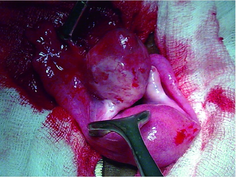
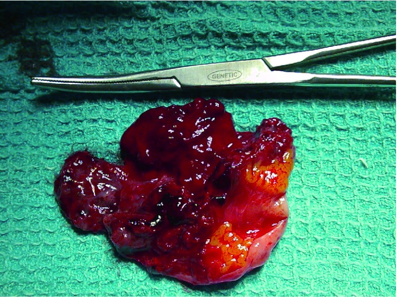
A 19-year-old primigravida, married for the past 8 months, presented with complaints of abdominal pain at 2 months of gestation. On examination, she was hemodynamically stable. Per-abdominal examination showed only mild supra-pubic tenderness. On per-vaginal examination, the uterus was bulky, and a tender cystic mass was palpable in the right fornix. Transvaginal ultrasound showed empty uterine cavity, with a gestation sac and live fetus (CRL 10.2 ➔ 7 + 1 week) in the right adnexa and minimal free fluid in the abdomen. She was taken up for a laparoscopy with a diagnosis of ectopic pregnancy, and at surgery, there was hemoperitoneum of 500 ml. The uterus was found to be of normal size, bilateral tubes were normal, right ovary was enlarged to 5 × 5 cm size and was bleeding markedly, and hence proceeded with laparotomy. The tubal serosa was intact on both sides with no tubo-peritoneal fistula. The left ovary was normal. The right ovary was connected to the utero-ovarian ligament and also to the pelvic wall by the suspensory ligament of ovary. It was enlarged and bleeding (Picture 1). On cut section, products of conception and minimal ovarian tissue were demonstrated (Picture 2). No portion of the ovary could be salvaged, and the ovariectomy specimen was sent for histopathology which was reported as ectopic gestation, right ovary. Post-operative period was uneventful.
Picture 1.
Bilateral ovary with bleeding right ovary
Picture 2.
Cut-section of the right ovary showing normal ovarian tissue and products of conception
No. 2
A 28-year-old unmarried girl, presented to casualty with complaints of abdominal pain for 2 days and vomiting for 3 days. She was also a known case of idiopathic thrombocytopenic purpura. On examination, she was hemodynamically stable. Per-abdominal examination showed supra-pubic tenderness. On per-vaginal examination, a tender cystic mass was palpable in the right fornix. Transvaginal ultrasound showed empty uterine cavity, with a gestation sac in the right adnexa and minimal free fluid in the abdomen. She was taken up for a laparoscopy with a diagnosis of ectopic pregnancy, and at surgery there was hemoperitoneum of 750 ml. The uterus was found to be of normal size, bilateral tubes were normal, right ovary was enlarged, and as it was bleeding markedly, and hence proceeded with laparotomy. She underwent wedge resection of the ovary, and the specimen was sent for histopathology which was reported as ectopic gestation, right ovary. Post-operative period was uneventful.
Discussion
Primary ovarian pregnancy is a rare form of ectopic pregnancy that must be demonstrated with use of the four criteria of Spiegelberg [3], which are (a) the fallopian tube, including the fimbria ovarica, is intact and clearly separate from the ovary; (b) the gestational sac definitely occupies the normal position of the ovary; (c) the sac is connected to the uterus by the utero-ovarian ligament; and (d) the ovarian tissue is unquestionably demonstrated in the wall of the sac.
In contrast to patients with tubal pregnancies, traditional risk factors such as pelvic inflammatory disease and prior pelvic surgery may not play a significant role in the etiology. Ovarian pregnancy is more frequent in ectopic pregnancies associated with the use of contraceptive intrauterine devices. In a study of six cases of ovarian pregnancy, Comstock [2] found abdominal pain and light vaginal bleeding to be common presenting symptoms.
Diagnosis is routinely made by ultrasound appearance of a wide echogenic ring on the ovary, frequently with a yolk sac or fatal parts [2]. Ovarian pregnancies are often confused with corpus luteum cysts [4]. Three-dimensional ultrasound imaging has been used to distinguish ovarian pregnancy from corpus luteum cysts. Doppler ultrasonography offers little additional diagnostic value because of the high vascularity of the ovary [2]. Diagnostic laparoscopy is frequently required to make the diagnosis of ovarian pregnancy, which is only later confirmed by histological examination of removed tissue [5]. At the time of surgery, ovarian pregnancies frequently resemble hemorrhagic cysts.
Treatment of ovarian pregnancy usually in the past was ipsilateral oophorectomy, but the trend has since shifted toward conservative surgery such as cystectomy or wedge resection performed at either laparotomy or laparoscopy. Medical management with methotrexate has been reported. Methotrexate may also be an option if there is persistent trophoblastic tissue after laparoscopy. If future fertility is desired, wedge resection may be considered. In many case reports, subsequent pregnancy has been uncomplicated [5].
We reviewed our institution data and saw that there were 21 cases of ectopic gestation over the preceding 4 months of which the following two (10 %) were ovarian.
Contributor Information
Priyankur Roy, Phone: +91-94433-30050, Email: priyankurroy@hotmail.com.
Bivas Biswas, Phone: +91-98431-19098, Email: mitali@cmcvellore.ac.in.
Ruby Jose, Phone: +91-416-2283397, Email: og2@cmcvellore.ac.in.
References
- 1.Bouyer J, Coste J, Fernandez H, et al. Sites of ectopic pregnancy: a 10 year population-based study of 1800 cases. Hum Reprod. 2002;17:3224–3230. doi: 10.1093/humrep/17.12.3224. [DOI] [PubMed] [Google Scholar]
- 2.Comstock C, Huston K, Lee W. The ultrasonographic appearance of ovarian ectopic pregnancies. Obstet Gynecol. 2005;105:42–46. doi: 10.1097/01.AOG.0000148271.27446.30. [DOI] [PubMed] [Google Scholar]
- 3.Spiegelberg O. Zur Casuistik den Ovarial-Schwangenschaft. Arch Gynaekol. 1878;13:73. doi: 10.1007/BF01991416. [DOI] [Google Scholar]
- 4.Bontis J, Grimbizis G, Tarlatzis BC, et al. Intrafollicular ovarian pregnancy after ovulation induction/intrauterine insemination: pathophysiological aspects and diagnostic problems. Hum Reprod. 1997;12:376–378. doi: 10.1093/humrep/12.2.376. [DOI] [PubMed] [Google Scholar]
- 5.Seinera P, DiGregorio A, Arisio R, et al. Ovarian pregnancy and operative laparoscopy: report of eight cases. Hum Reprod. 1997;12:608–610. doi: 10.1093/humrep/12.3.608. [DOI] [PubMed] [Google Scholar]


