Over the past 20 years the availability of aortic stent-grafts has allowed a major step change in the management of abdominal aortic aneurysms (AAA). Prior to this open surgical resection, first described by Charles Dubost in 1950, was the mainstay of AAA management(1). For elective cases open surgical repair has now largely been superseded by the deployment of a covered stent (stent-graft) through minimal surgical access in the common femoral arteries. Over the past few years there have been significant developments in stent-graft technology and an improved understanding of how best to utilise stent-grafts when treating aortic disease. Despite this, their role in the management of a patient with an AAA is still, at times, debatable. This article aims to discuss the modern day utilisation of aortic stent-grafts in patients with AAA.
Introduction
Based on data from the Health and Social Care Information Centre there were 63.9 million people registered with a general practitioner (GP) in the United Kingdom in 2007(2). For those aged between 65 and 80 years the incidence of an AAA is 7.6% in men(4) and lower (4.2%) for women(5). For an average General Practice of 6,300 patients(3) this would suggest that around 44 patients will have an AAA at any one time. If left untreated, Wilmink and Quick(6), estimate that a third of these aneurysms would eventually rupture. Rupture is usually lethal with overall mortality rates of up to 90% common(7). Currently around 7,000 people in England and Wales die each year as a result of ruptured AAA(8). Endovascular aortic aneurysm repair (EVAR) is a treatment option directed at removing the risk of rupture in patients with known AAA. Over recent years EVAR has developed into the most common method for the elective management of AAA(9) and large randomised controlled trials (RCTs) are evaluating its efficacy in those with rupture(10, 11). Potential advantages of EVAR over traditional open repair include reduced time under general anaesthesia, elimination of the pain and trauma associated with major abdominal surgery, reduced length of stay in the hospital and intensive care unit (ICU), and reduced blood loss(12).
Like open surgical repair, decisions regarding whether to use EVAR for the management of an AAA are still partly based on the maximum aneurysm diameter. The natural course of an AAA is continued expansion of between 2-3mm per year and a rupture risk which is exponentially related to AAA diameter(4, 13). Treatment recommendations are based on data from the UK Small Aneurysm Trial(14) and advise the referral of patients with large AAA (>5.5 cm) to a vascular specialist for consideration of surgery (15). Patients with smaller aneurysms, either found incidentally or in screening programmes, are now followed up with surveillance ultrasound (US) examinations generally run through Vascular Surgery units.
Technique
EVAR involves the internal lining of the aorta using a stent-graft. A stent-graft comprises a metallic (stainless steel or nitinol) skeleton covered with an impermeable (polytetrafluoroethylene or polyster) fabric and is implanted using fluoroscopic guidance through the femoral arteries. Sealing of the device against the aortic wall is achieved above (proximal) and below (distal) the aneurysm and this thereby excludes the aneurysm from the systemic circulation and aims to prevent subsequent rupture (Fig 1). Sealing of the stent-graft unlike a surgically sutured anastomosis is achieved by the radial force of the stent-graft on the aortic wall. Three configurations of stent-graft are currently available: tube, bifurcated and aorto-uni-iliac (AUI) (Fig 2). Bifurcated systems are used in the majority (90%) of cases(16-18) having the added advantage of being more stable within the aorta and avoiding the risks of supplying blood to the lower limbs via a single common iliac artery (CIA).
Fig 1.
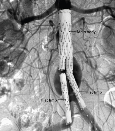
Diagram illustrating the components of a bifurcated modular aortic stent-graft used to treat infrarenal abdominal aortic aneurysms
Fig 2.
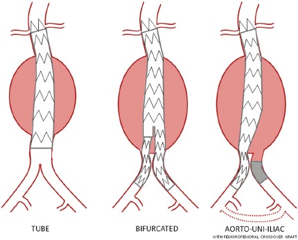
Configurations of aortic stent-graft
EVAR was first undertaken by a Ukranian surgeon Nicholas Volodos in 1987(19); however, it was a later publication by Juan Carlos Parodi in 1991(20) that was responsible for the widespread introduction of EVAR across the globe. In the UK, EVAR is typically performed by an endovascular team that includes a vascular surgeon and an interventional radiologist. Procedures may be performed under general, regional or local anaesthesia. The stent-graft is delivered into the aorta within a long flexible sheath of between 16F to 24F (8mm) in diameter which allows the stent-graft to be remotely positioned within the abdominal aorta. Surgical exposure and control of the common femoral arteries is the most common means of accessing the aorta. Totally percutaneous EVAR is now available with haemostasis achieved using a percutaneous suturing device. This technique requires careful patient selection with excessive vessel calcification and obesity being associated with technical failures and increased access site complications(21).
For modular devices, composed of two or more parts, the main component (main body) is inserted first. The undeployed device is then positioned so that the fabric at the proximal margin of the device is immediately below the most caudal renal artery. Once deployment has started, a check angiogram is undertaken to confirm the deployment position and the patency of the renal arteries. The main body is then released and the device extended so that both distal ends are within the CIAs (Fig 1).
CIA aneurysms are present in around 43% of patients with intact AAA(22). For these patients a decision is made on whether to extend one or both stent-graft limbs into the external iliac artery (EIA). If this is the case then it may be necessary to embolise the internal iliac artery (IIA) in order to prevent backfilling of the aneurysm. Occlusion of an IIA may cause significant buttock claudication and iliac branched devices (Fig 3) are now available which provide an option for preserving blood flow to the IIA(23). Whichever strategy is employed, every effort is made to preserve at least one IIA since bilateral IIA occlusion is associated with a risk of neurological and ischaemic complications.
Fig 3.
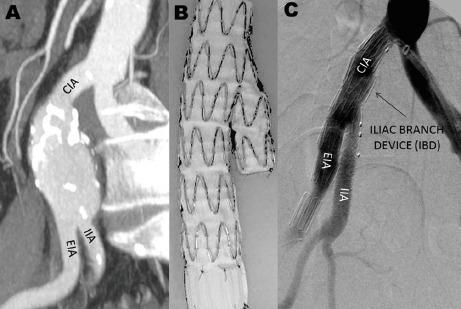
Iliac branched devices (IBD) are an option for treating an isolated CIA aneurysm or an aorto-iliac aneurysm avoiding the need for deliberate occlusion of the IIA. Image A, a CT maximum intensity projection (MIP) demonstrating an isolated CIA aneurysm. Image B shows an in vitro deployed IBD. Image C, post-procedural angiogram of an isolated CIA aneurysm (same case as image A) successfully treated with an IBD
Patient Selection
The majority of AAA are first diagnosed on ultrasound (US). If EVAR is considered then a further assessment of aneurysm morphology is necessary. For the majority of patients this will involve a thin-slice contrast-enhanced CT scan of the abdomen and pelvis. The CT data are then displayed as a series of virtual 3D aortic models. From this the diameter, length and quality of the aorta immediately below the renal arteries (aortic neck) can be assessed together with the suitability of the distal landing zones (CIA) and femoral artery access vessels (Fig 4). One of the fundamental aims of the EVAR procedure is to cover the entire aorto-iliac segment from below the renal arteries to the CIA bifurcation. Vessel length measurements are therefore very important and stent-grafts are planned to be of adequate length whilst allowing sufficient overlap of the modular components. Suitability for EVAR is also determined by the relevant stent-graft manufacturer's eligibility criteria set down in their ‘ Instructions for Use'. One of the strictest criteria surrounds the aortic neck which, for the majority of devices, must be parallel for 15 mm below the lowest renal artery, free from thrombus and excessive angulation. Off-label use of stent-grafts, by treating patients who do not fulfil such anatomical inclusion criteria, is associated with poorer outcomes but is known to be common practice(24).
Fig 4.
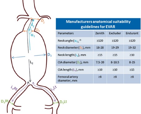
Criteria used to assess anatomical suitability for EVAR
It is also important that any preoperative imaging data are scrutinised for the presence of non-aortic pathology. Incidental pathology has been reported in over 75% of cases, with around 20% considered as clinically significant e.g. malignancy(25). In these situations unexpected findings may affect the decision to treat the aneurysm or alter the timing of repair.
Complications
Complications following EVAR can occur early or late and are a distinct limitation. Large RCTs have documented a 20-30% higher complication rate for EVAR when compared to open surgery(26, 27). This has led to some opposition to the widespread use of EVAR. Renal impairment, graft-related endoleak, device occlusion, migration, distal embolisation and femoral access site complications all may be encountered(28).
A decline in renal function is often seen after EVAR and usually reverts back to preoperative levels. Permanent renal damage may result from deliberate or unintentional coverage of a renal artery by the stent-graft fabric, toxic effects of the iodinated contrast media and cholesterol emboli. Graft occlusion may occur and is generally the result of poor blood flow due to either graft kinking or poor outflow(29). Infection of an aortic stent-graft and embolisation to the lower limb from arterial debris dislodged during implantation are both rare. Vascular access complications can occur and include haematoma, infection and seroma.
Persistent blood flow outside the stent-graft and within the aneurysm is defined by WHITE et al. (30) as an endoleak (Fig 5). The most serious endoleaks are types I and III which are associated with aneurysm enlargement and rupture. Secondary intervention to correct these endoleaks is almost always necessary. Whilst rupture has been reported with type II endoleaks these are considered to have a more benign course and conservative management is recommended unless there is evidence of continuing aneurysm enlargement.
Fig 5.
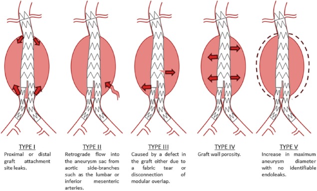
Endoleak classification system
Stent-grafts are subjected to distraction forces from the relentless force of pulsatile blood flow. These distraction forces act longitudinally and challenge the fixation of the graft and the modular overlap zones. Failure of fixation will lead to migration or disconnection of overlapping components with late type I or type III endoleaks and the risk of aortic rupture. Graft limb distortion with subsequent thrombosis can also arise secondary to device migration. Metallic component fractures and fabric tears have also generated additional durability concerns. Component fractures may lead to diminished stent strength and loss of radial force which can result in graft migration. The jagged ends of metal fractures can also result in fabric tears and subsequent endoleaks.
Secondary Intervention And Surveillance
The complication rate associated with EVAR has been described by some as its Achilles heel. The EUROSTAR registry reported EVAR outcomes for 2846 patients and highlighted reintervention rates at 1, 2, 3 and 4 years of 6, 9, 12 and 14 per cent(31). These rates are similar to those reported in the main UK and European RCTs (32). There is pathological evidence that the aneurysm wall atrophies with the depressurisation that results from an initially successful EVAR. If there is late repressurisation of the aneurysm due to endoleak then rupture may occur(Fig 6). Rupture should be an urgent consideration in any post-EVAR patient presenting with acute abdominal pain with or without collapse. In the UK EVAR trials a total of 27 post-EVAR ruptures have been reported(33). Eighty-two per cent of these ruptures occurred greater than 30-days following implantation and the majority of these (63%) were in patients with previously reported complications or signs of failed EVAR. In a review of the literature Schlösser et al.,(34) also reported on post-EVAR ruptures (n=270). In this series 35 patients had no prior abnormalities found during follow-up, 39/101 ruptures showed no change in aneurysm diameter during follow-up and 26/101 showed evidence of aneurysm regression. Research has identified types I and II (with sac enlargement) endoleaks, migration and graft kinking as risk factors for aortic rupture(33). With the possibility of rupture all EVAR patients are recommended to undergo regular follow-up imaging surveillance, especially for patients where secondary intervention would be considered.
Fig 6.
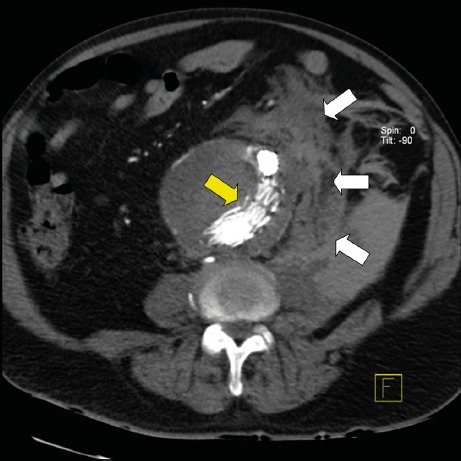
Post-EVAR CT image of a patient with a ruptured AAA. The aortic stent-graft (yellow arrow) can be seen within the aneurysm and there is extensive haemorrhage surrounding the aorta (white arrows)
Current EVAR surveillance protocols are mostly based on costly and time-consuming imaging procedures and aim to detect adverse events such as graft migration, endoleaks or aneurysm enlargement. These imaging procedures are either associated with serial radiation exposure or may be potentially harmful due to the use of iodine- or gadolinium-based contrast agents. CT is considered by many as the gold standard for imaging surveillance and is typically performed at 1, 6 and 12 months and then annually thereafter. With the economic and logistical drawbacks of repeat CT a demand has arisen for an alternative examination. Combined ultrasound and plain abdominal radiography (AXR) has emerged as an alternative with studies confirming their efficacy(35, 36).
Device Modifications
The main anatomical feature that limits the role of current devices is the quality and length of normal aorta below the renal arteries (aortic neck). For standard stent-grafts the fabric cannot be placed above the renal arteries without occluding them. If the aortic neck is small or non-existent then renal blood flow may be preserved by manufacturing holes or fenestrations in the fabric and sealing the aneurysm above the renal arteries. Fenestrated stent-grafts (Fig 7) have been available for several years and are now in widespread use. The locations of the fenestrations must be carefully planned using preoperative CT data and are manufactured to be at the correct height and correct position on the circumference of the graft.
Fig 7.
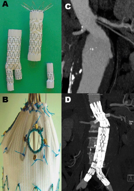
An in vitro deployed fenestrated stent-graft (Image A). Image B – a magnified view demonstrating a pre-planned fenestration within the proximal component. Image C – a preoperative CT scan of patient with a short and angulated aortic neck that is unsuitable for standard EVAR. Image D - postoperative CT scan (same patient as Image C) showing a deployed fenestrated stent-graft with exclusion of the AAA and patent visceral arteries
Fenestrated stent-grafts are not well suited if the fabric of the stent-graft cannot abut the aortic wall at the level of the visceral arteries. This is common in thoraco-abdominal aortic aneurysms (TAA) and for these situations custom made stent-grafts with side branch protrusions can be used(37). The idea is that the stent-graft side branches will provide a bridge to reduce the distance from the main body to the visceral artery orifice (Fig 8). Covered stents are inserted through the branches and help achieve a better seal due to a longer overlap zone.
Fig 8.
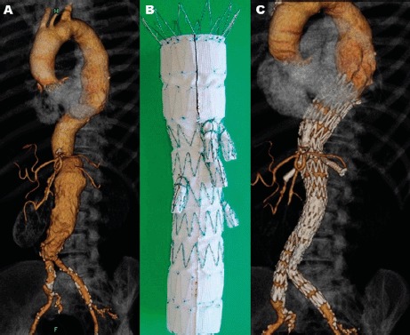
Image A demonstrates an AAA where apposition of the stent-graft against the aortic wall across the visceral arteries would be unlikely. For this situation a branched aortic stent-graft (Image B) was successfully utilised (Image C)
The utility of aortic stent-grafts has also extended to aneurysm disease above the diaphragm (TEVAR). Even with a lack of RCT evidence stent-grafts are now the preferred treatment option for aneurysms involving the thoracic aorta. The less invasive nature of TEVAR offers the potential for lower mortality and post-operative morbidity. Anatomical suitability is still a concern; many thoracic aneurysms lie close to or involve the origins of the great vessels arising from the aortic arch. Advances in fenestrated and branched stent-grafts have expanded the application of EVAR for these more complex aortic pathologies and those involving the thoracoabdominal aorta and aortic arch(38). Combined surgical debranching and stent-graft implantation procedures exist(39) and the implantation site can also be lengthened, whilst preserving side-branch blood flow, using a branched stent-graft(40).
In the early days of EVAR treatment decisions were relatively straightforward. With the introduction of these newer technologies there is an increased range of endovascular options for patients. Appropriate consideration of all of these newer techniques must be given when considering a patient for any intervention.
The combination of metal and currently available fabrics has resulted in cases of fabric degeneration with resulting endoleaks. Alternative devices which do not rely on a fabric mounted on a metallic frame are being considered. Such devices may allow the treatment of an AAA by modulating haemodynamic flow(41). In other areas research has focused on treating the aneurysm by the injection of polymers and elastomers. The Endologix Nellix system (Endologix, Irvine, CA) uses two polymer PTFE endobags that are inserted and seek to freeze the aneurysm and prevent any further morphological changes. Other researchers have attempted to fill the aneurysm with polymers whilst temporarily occluding the aortic lumen(42, 43). All of these technologies are currently in the early phases of development and are not part of routine care.
Current Evidence
The most important sources of evidence concerning EVAR are the UK EVAR trials (EVAR 1 and EVAR 2). In the EVAR 1 trial(44) 30-day mortality data showed a significant advantage in favour of EVAR (1.7%) when compared with open surgery (4.7%). All-cause mortality was similar for both procedures by 4-years (28%), however a 3% difference in aneurysm-related mortality, in favour of EVAR remained (4% versus 7%)(17). Post-operative complications were more frequent for EVAR (41%) but had a negligible difference on health-related quality of life (HRQL), being similar for both procedures. Over the long-term the benefit of a lower aneurysm-related mortality for EVAR was eventually lost due to a higher incidence of fatal ruptures in the EVAR group(26). New complications were still occurring up to 8 years following treatment.
EVAR 2 was designed to investigate the role of EVAR in patients deemed unfit for open surgery(45). The 30-day operative mortality for the EVAR group was 7.3%, higher than in the EVAR 1 trial. This trial did not demonstrate a survival benefit for EVAR in patients unfit for open repair, EVAR was costly and did not improve HRQL. With long-term follow-up there is a benefit from EVAR in terms of a lower aneurysm-related mortality (3.6 vs 7.3 deaths per 100 person-years)(46). No differences in total mortality rates have been demonstrated and the differences in aneurysm-related mortality are limited since this cohort has a limited life expectancy with few surviving >8 years.
Similar results to EVAR 1 have been published by the Dutch Randomised Endovascular Aneurysm Management (DREAM) trial group(47). 30-day mortality rates of 1.2% (EVAR) and 4.6% (open repair) are consistent with the UK trial as is the persistent reduction in aneurysm-related deaths in the EVAR group. Again, no overall survival difference was observed during the mid and long-terms(27). By six years, higher incidences of complications and reinterventions were still being reported in the EVAR group.
The more recent US OVER (Veteran Affairs Open vs. Endovascular Repair) trial demonstrated a lower 30-day mortality for EVAR (0.5%) when compared with open repair (3.0%)(48). This survival advantage, unlike in other studies, was maintained by 2-years. The more recent French ACE trial(49) reported no significant differences in 30-day mortality between open surgery and EVAR (0.6% vs 1.3%). This continued at one and three years but there were higher reintervention rates for EVAR (24% versus 14%). The French study opposes the findings of the three previous RCTs which all favoured EVAR in terms of short-term mortality. Even the large case-matched Medicare analysis (45,660 patients) showed a reduction of post-operative mortality favouring EVAR (1.2% vs. 4.8%)(50). Differences between the ACE trial results and the remaining EVAR mortality data can be attributed to numerous factors which may include study design, experience of the centres and the overall national standards of care.
Three multi-centre trials are currently recruiting or have recently completed evaluating open repair and EVAR in patients with ruptured aneurysms. The Amsterdam Acute Aneurysm (AJAX) trial started in 2004(51) and initially failed to show any differences between techniques for ruptured AAA. Recruitment was extended twice and the study finally completed in 2010 and showed no difference in mortality between the two techniques(52) . The Paris-based Endovasculaire vs Chirurgie dans les Anevrysmes Rompus (ECAR) trial started in 2008 and aims to recruit 190 patients(53). The larger and more recent UK Immediate Management of the Patient with Rupture: Open versus Endovascular Repair (IMPROVE) trial started in 2009 and aims to recruit 600 patients(10).
One of the main criteria for AAA treatment is maximum aneurysm diameter. Data from the UK Small Aneurysm Trial(14) reported a 5.8% 30-day operative mortality rate for AAA between 4.0-5.5 cm and concluded that there is no long-term survival advantage from early surgery. With the lower 30-day mortality for EVAR questions have arisen whether this treatment criterion should now be revised. The European CAESAR (Comparison of Surveillance versus Aortic Endografting for Small Aneurysm Repair) trial failed to demonstrate any advantage of early EVAR over traditional surveillance(54). 30-day mortality rates for EVAR were low (0.6%) with overall aneurysm-related mortality and rupture rates low and comparable between the groups. The study did, however, raise concern that up to 60% of surveillance patients will require treatment within 3 years and that 16% of these patients may lose anatomical suitability for EVAR during this time. Similarly, the US PIVOTAL (Positive Impact of Endovascular Options for Treating Aneurysms Early) trial showed that early all-cause mortality for both EVAR and surveillance groups were equal (4.1%)(55).
Conclusion
Over the past three decades the number of patients treatable by EVAR has grown. What started as a series of devices constructed in the operating theatre has evolved into mass produced ‘off-the-shelf’ systems which can treat a range of patients. Not only has anatomical eligibility increased but other vascular diseases are now being treated using a stent-graft. The endovascular treatment of complex type B dissections, traumatic aortic transections and aorto-enteric fistulae is possible. Pushing the boundaries of both patient and disease selection does, however, bring additional uncertainties. Studies published over recent years have shown that ‘off-label’ device use brings poorer outcomes and post-EVAR rupture will occur in a limited number of patients. Nevertheless, we have seen huge developments in EVAR technologies and their applicability. Devices are now deployable on smaller delivery systems, are repositionable within the aorta and can conform to more challenging anatomy. Durability issues still exist and appropriate diligence is needed during follow-up. With this in mind, manufacturers are now starting to explore alternatives to a metallic skeleton covered by an impermeable fabric. Open surgery dominated for over forty years but we are currently living in the endovascular era and we anticipate further improvements in the safety and applicability of endovascular therapy for aortic aneurysms.
The authors have no conflicts of interest.
References
- 1.Naef AP. The mid-century revolution in thoracic and cardiovascular surgery: Part 5. Interact Cardiovasc Thorac Surg. 2004;3(3):415–22. doi: 10.1016/j.icvts.2004.05.001. [DOI] [PubMed] [Google Scholar]
- 2.Health and Social Care Information Centre. Average Practice ListSizes in the UK. London: HMSO; 2008. [Google Scholar]
- 3.Office for National Statistics. Population Ageing in the United Kingdom, its Constituent Countries and the European Union. London: HMSO; 2012. [Google Scholar]
- 4.Ashton HA, Buxton MJ, Day NE, Kim LG, Marteau TM, Scott RA, et al. The Multicentre Aneurysm Screening Study (MASS) into the effect of abdominal aortic aneurysm screening on mortality in men: a randomised controlled trial. Lancet. 2002;360(9345):1531–9. doi: 10.1016/s0140-6736(02)11522-4. [DOI] [PubMed] [Google Scholar]
- 5.Singh K, Bønaa KH, Jacobsen BK, Bjørk L, Solberg S. Prevalence of and risk Factors for abdominal aortic aneurysms in a population-based Study: The Tromsø Study. Am J Epidemiol. 2001;154(3):236–244. doi: 10.1093/aje/154.3.236. [DOI] [PubMed] [Google Scholar]
- 6.Wilmink AB, Quick CR. Epidemiology and potential for prevention of abdominal aortic aneurysm. Br J Surg. 1998;85(2):155–62. doi: 10.1046/j.1365-2168.1998.00714.x. [DOI] [PubMed] [Google Scholar]
- 7.Bengtsson H, Bergqvist D. Ruptured abdominal aortic aneurysm: a population-based study. J Vasc Surg. 1993;18(1):74–80. doi: 10.1067/mva.1993.42107. [DOI] [PubMed] [Google Scholar]
- 8.Office for National Statistics . Leading causes of Deaths in England and Wales, 2011. London: HMSO; 2012. [Google Scholar]
- 9.Jackson RS, Chang DC, Freischlag JA. Comparison of long-term survival after open vs endovascular repair of intact abdominal aortic aneurysm among Medicare beneficiaries. JAMA. 2012;307(15):1621–8. doi: 10.1001/jama.2012.453. [DOI] [PubMed] [Google Scholar]
- 10.Improve Trial. Powell JT, Thompson SG, Thompson MM, Grieve R, Nicholson AA, et al. The Immediate Management of the Patient with Rupture: Open Versus Endovascularrepair (IMPROVE) aneurysm trial— ISRCTN 48334791 IMPROVE trialists. Acta Chir Belg. 2009;109(6):678–80. doi: 10.1080/00015458.2009.11680516. [DOI] [PubMed] [Google Scholar]
- 11.Hoornweg LL, Wisselink W, Vahl A, Balm R. Amsterdam Acute Aneurysm Trial Collaborators. The Amsterdam Acute Aneurysm Trial: suitability and application rate for endovascular repair of ruptured abdominal aortic aneurysms. Eur J Vasc Endovasc Surg. 2007;33(6):679–83. doi: 10.1016/j.ejvs.2006.12.011. [DOI] [PubMed] [Google Scholar]
- 12.National Institute for Health and Clinical Excellence. Endovascular stent-graftsfor the treatment of abdominal aortic aneurysms [TAG167] London: National Institute for Health and Clinical Excellence; 2009. [Google Scholar]
- 13.Brady AR, Thompson SG, Fowkes FG, Greenhalgh RM, Powell JT. Participants UK Small Aneurysm Trial Participants. Abdominal aortic aneurysm expansion: risk factors and time intervals for surveillance. Circulation. 2004;110(1):16–21. doi: 10.1161/01.CIR.0000133279.07468.9F. [DOI] [PubMed] [Google Scholar]
- 14.The UK Small Aneurysm Trial Participants Mortality results for randomised controlled trial of early elective surgery orultrasonographic surveillance for small abdominal aortic aneurysms. Lancet. 1998;352(9141):1649–55. [PubMed] [Google Scholar]
- 15.Metcalfe D, Holt PJ, Thompson MM. The management of abdominal aortic aneurysms. BMJ. 2011;342:d1384. doi: 10.1136/bmj.d1384. [DOI] [PubMed] [Google Scholar]
- 16.Vallabhaneni SR, Harris PL. Lessons learnt from the EUROSTAR registry on endovascular repair of abdominal aortic aneurysm repair. Eur J Radiol. 2001;39(1):34–41. doi: 10.1016/s0720-048x(01)00340-0. [DOI] [PubMed] [Google Scholar]
- 17.EVAR Trial Participants Endovascular aneurysm repair versus open repair in patients with abdominal aortic aneurysm (EVAR trial 1): randomised controlled trial. Lancet. 2005;365(9478):2179–86. doi: 10.1016/S0140-6736(05)66627-5. [DOI] [PubMed] [Google Scholar]
- 18.Prinssen M, Verhoeven EL, Buth J, Cuypers PW, van Sambeek MR, Balm R, et al. A randomized trial comparing conventional and endovascular repair of abdominal aortic aneurysms. N Engl J Med. 2004;351(16):1607–18. doi: 10.1056/NEJMoa042002. [DOI] [PubMed] [Google Scholar]
- 19.Volodos NL, Karpovich IP, Shekhanin VE, Troian VI, Iakovenko LF. [A case of distant transfemoral endoprosthesis of the thoracic artery using a self-fixing synthetic prosthesis in traumatic aneurysm] Grudn Khir. 1988;(6):84–6. [PubMed] [Google Scholar]
- 20.Parodi JC, Palmaz JC, Barone HD. Transfemoral intraluminal graft implantation for abdominal aortic aneurysms. Ann Vasc Surg. 1991;5(6):491–9. doi: 10.1007/BF02015271. [DOI] [PubMed] [Google Scholar]
- 21.Smith ST, Timaran CH, Valentine RJ, Rosero EB, Clagett GP, Arko FR. Percutaneous access for endovascular abdominal aortic aneurysmrepair: can selection criteria be expanded? Ann Vasc Surg. 2009;23(5):621–6. doi: 10.1016/j.avsg.2008.09.002. [DOI] [PubMed] [Google Scholar]
- 22.Hinchliffe RJ, Alric P, Rose D, Owen V, Davidson IR, Armon MP, et al. Comparison of morphologic features of intact and ruptured aneurysms of infrarenal abdominal aorta. J Vasc Surg. 2003;38(1):88–92. doi: 10.1016/s0741-5214(03)00079-x. [DOI] [PubMed] [Google Scholar]
- 23.Karthikesalingam A, Hinchliffe RJ, Holt PJ, Boyle JR, Loftus IM, Thompson MM. Endovascular aneurysm repair with preservation of the internal iliac artery using the iliac branch graft device. Eur J Vasc Endovasc Surg. 2010;39(3):285–94. doi: 10.1016/j.ejvs.2009.11.018. [DOI] [PubMed] [Google Scholar]
- 24.Schanzer A, Greenberg RK, Hevelone N, Robinson WP, Eslami MH, Goldberg RJ, et al. Predictors of abdominal aortic aneurysm sac enlargement after endovascular repair. Circulation. 2011;123(24):2848–2855. doi: 10.1161/CIRCULATIONAHA.110.014902. [DOI] [PubMed] [Google Scholar]
- 25.Indes JE, Lipsitz EC, Veith FJ, Gargiulo NJ, 3rd, Privrat AI, Eisdorfer J, et al. Incidence and significance of nonaneurysmal-related computed tomography scan findings in patients undergoing endovascular aortic aneurysm repair. J Vasc Surg. 2008 Aug;48(2):286–90. doi: 10.1016/j.jvs.2008.03.057. [DOI] [PubMed] [Google Scholar]
- 26.United Kingdom EVAR Trial Investigators. Greenhalgh RM, Brown LC, Powell JT, Thompson SG, Epstein D, et al. Endovascular versus open repair of abdominal aortic aneurysm. N Engl J Med. 2010;362(20):1863–71. doi: 10.1056/NEJMoa0909305. [DOI] [PubMed] [Google Scholar]
- 27.De Bruin JL, Baas AF, Buth J, Prinssen M, Verhoeven EL, Cuypers PW, et al. Long-term outcome of open or endovascular repair of abdominal aortic aneurysm. N Engl J Med. 2010;362(20):1881–9. doi: 10.1056/NEJMoa0909499. [DOI] [PubMed] [Google Scholar]
- 28.Maleux G, Koolen M, Heye S. Complications after endovascular aneurysm repair. Semin Intervent Radiol. 2009;26(1):3–9. doi: 10.1055/s-0029-1208377. [DOI] [PMC free article] [PubMed] [Google Scholar]
- 29.Cochennec F, Becquemin JP, Desgranges P, Allaire E, Kobeiter H, Roudot-Thoraval F. Limb graft occlusion following EVAR: clinical pattern, outcomes and predictive factors of occurrence. Eur J Vasc Endovasc Surg. 2007;34(1):59–65. doi: 10.1016/j.ejvs.2007.01.009. [DOI] [PubMed] [Google Scholar]
- 30.White GH, Yu W, May J. Endoleak—-a proposed new terminology to describe incomplete aneurysm exclusion by an endoluminal graft. J Endovasc Surg. 1996;3(1):124–5. doi: 10.1583/1074-6218(1996)003<0124b:>2.0.CO;2. [DOI] [PubMed] [Google Scholar]
- 31.Hobo R, Buth J, EUROSTAR collaborators Secondary interventions following endovascular abdominal aortic aneurysm repair using current endografts. A EUROSTAR report. J Vasc Surg. 2006;43(5):896–902. doi: 10.1016/j.jvs.2006.01.010. [DOI] [PubMed] [Google Scholar]
- 32.Brown LC, Greenhalgh RM, Powell JT, Thompson SG, EVAR Trial Participants Use of baseline factors to predict complications and reinterventions after endovascular repair of abdominal aortic aneurysm. Br J Surg. 2010;97(8):1207–17. doi: 10.1002/bjs.7104. [DOI] [PubMed] [Google Scholar]
- 33.Wyss TR, Brown LC, Powell JT, Greenhalgh RM. Rate and predictability of graft rupture after endovascular and open abdominal aortic aneurysm repair: data from the EVAR Trials. Ann Surg. 2010;252(5):805–12. doi: 10.1097/SLA.0b013e3181fcb44a. [DOI] [PubMed] [Google Scholar]
- 34.Schlösser FJ, Gusberg RJ, Dardik A, Lin PH, Verhagen HJ, Moll FL, et al. Aneurysm rupture after EVAR: can the ultimate failure be predicted? Eur J Vasc Endovasc Surg. 2009;37(1):15–22. doi: 10.1016/j.ejvs.2008.10.011. [DOI] [PubMed] [Google Scholar]
- 35.Harrison GJ, Oshin OA, Vallabhaneni SR, Brennan JA, Fisher RK, McWilliams RG. Surveillance after EVAR based on duplex ultrasound and abdominal radiography. Eur J Vasc Endovasc Surg. 2011;42(2):187–92. doi: 10.1016/j.ejvs.2011.03.027. [DOI] [PubMed] [Google Scholar]
- 36.Karthikesalingam A, Al-Jundi W, Jackson D, Boyle JR, Beard JD, Holt PJ, et al. Systematic review and meta-analysis of duplex ultrasonography, contrast-enhanced ultrasonography or computed tomography for surveillance after endovascular aneurysm repair. Br J Surg. 2012;99(11):1514–23. doi: 10.1002/bjs.8873. [DOI] [PubMed] [Google Scholar]
- 37.Chuter TA, Gordon RL, Reilly LM, Pak LK, Messina LM. Multi- branched stent-graft for type III thoracoabdominal aortic aneurysm. J Vasc Interv Radiol. 2001;12(3):391–2. doi: 10.1016/s1051-0443(07)61924-1. [DOI] [PubMed] [Google Scholar]
- 38.Brozzi NA, Roselli EE. Endovascular therapy for thoracic aortic aneurysms: state of the art in 2012. Curr Treat Options Cardiovasc Med. 2012;14(2):149–63. doi: 10.1007/s11936-012-0169-6. [DOI] [PubMed] [Google Scholar]
- 39.Cires G, Noll RE, Albuquerque FC, Tonnessen BH, Sternbergh WC., 3rd Endovascular debranching of the aortic arch during thoracic endograft repair. J Vasc Surg. 2011;53(6):1485–91. doi: 10.1016/j.jvs.2011.01.053. [DOI] [PubMed] [Google Scholar]
- 40.Chuter TA, Schneider DB. Endovascular repair of the aortic arch. Perspect Vasc Surg Endovasc Ther. 2007;19(2):188–92. doi: 10.1177/1531003507304165. [DOI] [PubMed] [Google Scholar]
- 41.Chocron S, Vaislic C, Kaili D, Bonneville JF. Multilayer stents in the treatment of thoraco-abdominal residual type B dissection. Interact Cardiovasc Thorac Surg. 2011;12(6):1057–9. doi: 10.1510/icvts.2010.257394. [DOI] [PubMed] [Google Scholar]
- 42.Uflacker R, Brothers T. Filling of the aneurysmal sac with DEAC- glucosamine in an animal model of abdominal aortic aneurysm following stent-graft repair. JCardiovasc Surg (Torino) 2006;47(4):425–36. [PubMed] [Google Scholar]
- 43.Bosman WM, van der Steenhoven TJ, Hinnen JW, Kaptein BL, de Vries AC, Brom HL, et al. Aortic customize: a new alternative endovascular approach to aortic aneurysm repair using injectable biocompatible elastomer. An in vitro study. J Vasc Surg. 2010;51(5):1230–7. doi: 10.1016/j.jvs.2009.08.093. [DOI] [PubMed] [Google Scholar]
- 44.Greenhalgh RM, Brown LC, Kwong GP, Powell JT, Thompson SG, EVAR Trial Participants Comparison of endovascular aneurysm repair with open repair in patients with abdominal aortic aneurysm (EVAR trial 1), 30-day operative mortality results: randomised controlled trial. Lancet. 2004;364(9437):843–8. doi: 10.1016/S0140-6736(04)16979-1. [DOI] [PubMed] [Google Scholar]
- 45.EVAR Trial Participants Endovascular aneurysm repair and outcome in patients unfit for open repair of abdominal aortic aneurysm (EVAR trial 2): randomised controlled trial. Lancet. 2005;365(9478):2187–92. doi: 10.1016/S0140-6736(05)66628-7. [DOI] [PubMed] [Google Scholar]
- 46.United Kingdom EVAR Trial Investigators. Greenhalgh RM, Brown LC, Powell JT, Thompson SG, Epstein D. Endovascular repair of aortic aneurysm in patients physically ineligible for open repair. N Engl J Med. 2010;362(20):1872–80. doi: 10.1056/NEJMoa0911056. [DOI] [PubMed] [Google Scholar]
- 47.Blankensteijn JD, de Jong SE, Prinssen M, van der Ham AC, Buth J, van Sterkenburg SM, et al. Two-year outcomes after conventional or endovascular repair of abdominal aortic aneurysms. N Engl J Med. 2005;352(23):2398–405. doi: 10.1056/NEJMoa051255. [DOI] [PubMed] [Google Scholar]
- 48.Lederle FA, Freischlag JA, Kyriakides TC, Padberg FT, Matsumura JS, Kohler TR, et al. Outcomes following endovascular vs open repair of abdominal aortic aneurysm: a randomized trial. JAMA. 2009;302(14):1535–42. doi: 10.1001/jama.2009.1426. [DOI] [PubMed] [Google Scholar]
- 49.Becquemin JP, Pillet JC, Lescalie F, Sapoval M, Goueffic Y, Lermusiaux P, et al. A randomized controlled trial of endovascular aneurysm repair versus open surgery for abdominal aortic aneurysms in low- to moderate- risk patients. J Vasc Surg. 2011;53(5):1167–73e1. doi: 10.1016/j.jvs.2010.10.124. [DOI] [PubMed] [Google Scholar]
- 50.Schermerhorn ML, O'Malley AJ, Jhaveri A, Cotterill P, Pomposelli F, Landon BE. Endovascularvs. openrepairof abdominal aortic aneurysms in the Medicare population. N Engl J Med. 2008;358(5):464–74. doi: 10.1056/NEJMoa0707348. [DOI] [PubMed] [Google Scholar]
- 51.Amsterdam Acute Aneurysm Trial Collaborators Amsterdam Acute Aneurysm trial: background, design, and methods. Vascular. 2006;14(3):130–5. doi: 10.2310/6670.2006.00027. [DOI] [PubMed] [Google Scholar]
- 52.CX Daily News. AJAX confirms no difference between EVAR and open repair for ruptured aneurysms. http ://www.cxvascular.com/cx-latest-news/cx-latest-news/ajax-confirms-no-difference-between-evar-and-open-repair-for-ruptured-aneurysms(accessed 7 December 2012) [Google Scholar]
- 53.Desgranges P, Kobeiter H, Castier Y, Sénéchal M, Majewski M, Krimi A. The Endovasculaire vs Chirurgie dans les Anevrysmes Rompus PROTOCOL trial update. J Vasc Surg. 2010;51(1):267–70. doi: 10.1016/j.jvs.2009.10.128. [DOI] [PubMed] [Google Scholar]
- 54.Cao P, De Rango P, Verzini F, Parlani G, Romano L, Cieri E, CAESAR Trial Group Comparison of surveillance versus aortic endografting for small aneurysm repair (CAESAR): results from arandomised trial. Eur J Vasc Endovasc Surg. 2011;41(1):13–25. doi: 10.1016/j.ejvs.2010.08.026. [DOI] [PubMed] [Google Scholar]
- 55.Ouriel K, Clair DG, Kent KC, Zarins CK. Positive Impact of Endovascular Options for treating Aneurysms Early (PIVOTAL) Investigators. Endovascular repair compared with surveillance for patients with small abdominal aortic aneurysms. J Vasc Surg. 2010;51(5):1081–7. doi: 10.1016/j.jvs.2009.10.113. [DOI] [PubMed] [Google Scholar]


