Abstract
This in vitro study aims to evaluate the crystal and surface microstructure of dental enamel after cold-light bleaching treatment. Twelve sound human premolars were cross-split into four specimens, namely, mesio-buccal (Group LP), disto-buccal (Group P), mesio-lingual (Group NP) and disto-lingual (Group L) specimens. These four groups were treated using the standard cold-light bleaching procedure, a bleaching agent, a peroxide-free bleaching agent and cold-light, respectively. Before and after treatment, all specimens were analyzed by high-resolution, micro-area X-ray diffraction and scanning electron microscopy. Using a spectrometer, tooth color of all specimens was measured before and after treatment. The phase of the enamel crystals was identified as hydroxyapatite and carbonated hydroxyapatite. After treatment, specimens in Groups LP and P showed significantly weaker X-ray diffraction peaks, significant reduction in crystal size and crystallinity, significant increase in L* but decrease in a* and b*, and obvious alterations in the surface morphology. However, specimens in Groups NP and L did not show any significant changes. The cold-light bleaching treatment leads to demineralization in the enamel surface. The acidic peroxide-containing bleaching agent was the major cause of demineralization, whereas cold-light did not exhibit significant increase or decrease effect on this demineralization.
Keywords: dental enamel, light, micro-area X-ray diffraction, tooth bleaching
Introduction
Tooth bleaching, sometimes described as ‘tooth whitening', is a popular treatment modality in dentistry. It offers a conservative and well-accepted treatment option for discolored teeth compared with the resin-bonded composites, porcelain veneers or crowns.1 Current bleaching mechanisms are based on the application of hydrogen peroxide-releasing agents. Hydrogen peroxide may be applied directly, or it may be produced from carbamide peroxide. Carbamide peroxide, also called urea peroxide, is an adduct of hydrogen peroxide and urea. When dissolved in water or saliva, carbamide peroxide produces hydrogen peroxide and urea, which is further decomposed to carbon dioxide and ammonia. As the active agent of bleaching procedure, hydrogen peroxide penetrates the tooth, produces free radicals and interacts with chromophore molecules, oxidizing them.1,2 Many laboratory and clinical studies have demonstrated that bleaching agents containing hydrogen peroxide or carbamide peroxide are effective for bleaching discolored teeth.3,4,5 Carbamide peroxide can continuously produce hydrogen peroxide with less irritation, so it is generally used as home-bleaching agent. The common concentration of carbamide peroxide is 10%.
Numerous previous studies have evaluated the adverse effects of peroxide-containing bleaching products on tooth enamel and/or dentin. However, these studies have conflicting results. Some studies have reported no significant deleterious effects on the surface micro-hardness, morphology, chemistry or ultrastructure of the enamel and/or dentine after bleaching treatment.6,7,8,9,10,11 On the contrary, other studies have indeed observed deleterious effects on the enamel and/or dentine, such as decrease in micro-hardness,12,13,14,15,16 alteration of surface morphology,15,17,18 changes in chemical composition and structure,19,20,21 loss of calcium and phosphorus,21,22 and decrease in the fracture resistance23,24 and elastic modulus.16
The cold-light bleaching technique is a novel bleaching technique widely applied in mainland China. It is a light-activated, in-office bleaching technique using blue light (wavelength between 480 nm and 520 nm). The cold-light lamp is equipped with filter to exclude the harmful infrared (wavelength λ>750 nm) and ultraviolet (λ<380 nm). The exclusion of infrared reduces its thermal pulp damage. The ultraviolet is excluded to reduce the risk of possible side effects of ultraviolet radiation on living cells. This technique has proven effective in bleaching discolored teeth.25,26 However, the effect of the cold-light bleaching treatment on enamel has not yet been studied.
Using a novel method, our study examined the effects of the cold-light bleaching technique on enamel. This method is able to precisely analyze the changes in enamel crystals, which are essential components of the enamel. The purpose of this in vitro study was to evaluate the adverse effects of cold-light bleaching treatment on enamel crystals. We expected that the novel method would provide research community with additional information to eliminate the conflict on the adverse effects of tooth bleaching.
Materials and methods
Tooth selection
Twelve sound premolars, extracted for orthodontic reasons, were selected for this study. All twelve premolars had no apparent evidence of extrinsic staining, caries lesions, dental fluorosis, enamel hypoplasia, cracks or other defects on the buccal and lingual surfaces. The tooth surfaces were cleaned under high-pressure water for 10 min to remove the materia alba. Then, the teeth were stored in 0.1% thymol solution at 4 °C until the preparation for testing.
Sample preparation
The 12 teeth were randomly marked from 1 to 12 and washed in running deionized water for 24 h to completely remove thymol residue. Using a low speed, water-cooled saw (Isomet; Buehler Ltd, Lake Bluff, IL, USA), the root of each tooth was removed and the crown was cross-split bucco-lingually and mesio-distally into four specimens, namely, mesio-buccal, disto-buccal, mesio-lingual and disto-lingual specimens. As a result, four specimens were obtained from each tooth. The specimens from the first 10 teeth were collected for micro-area X-ray diffraction (XRD) analysis and they were divided into the following four groups (n=10):
Group LP: mesio-buccal specimens, treated with cold-light combined with peroxide-containing bleaching agent; Group P: disto-buccal specimens, treated with peroxide-containing bleaching agent alone; Group NP: mesio-lingual specimens, treated with peroxide free bleaching agent; Group L: disto-lingual specimens, treated with cold-light alone.
The specimens from tooth number 11 and number 12 were collected for scanning electron microscopy (SEM) analysis. The mesio-buccal specimen of tooth number 11 was treated in the same manner as the Group LP specimens. The mesio-lingual specimen of tooth number 11 was treated in the same manner as the Group P specimens. The mesio-buccal specimen of tooth number 12 was treated in the same manner as the Group NP specimens. The mesio-lingual specimen of tooth number 12 was treated in the same manner as the Group L specimens. The disto-buccal and disto-lingual specimens of tooth number 11 and number 12 were stored in deionized water at 4 °C without any treatment.
The specimens were embedded in denture base resin with either the buccal or the lingual enamel surface exposed. The samples were then stored in deionized water at 4 °C until the next treatment.
Bleaching procedure
All specimens were washed under running deionized water for 30 s and dried with compressed air for 5 s before the bleaching treatment. Different bleaching agents were prepared for the different groups. Agent for Group LP and Group P was prepared by mixing the 35% hydrogen peroxide solution and silicon dioxide catalyst (BEYOND II; Beyond Technology Ltd, Centennial, CO, USA), while agent for Group NP was prepared by mixing deionized water with the same silicon dioxide catalyst. The pH of the 35% hydrogen peroxide solution and the bleaching agents was measured three times using a pH meter (PT-10; Sartorius Ltd, Göttingen, Germany). The averages of pH value of 35% hydrogen peroxide solution, agents for Groups LP, P and NP were 6.0, 5.0, 5.0 and 5.9, respectively. The specimens of Group LP were treated according to the standard bleaching procedures, as follows: (i) the specimens surfaces were painted with whitening agent to form a uniform 2–3 mm thick layer; (ii) the head of cold-light lamp (BY-0398; Beyond Technology Ltd) was placed vertically 1 cm above the tooth surfaces and the lamp was switched on for 8 min; (iii) the whitening agent was removed, and then steps (i)–(iii) were repeated twice; and finally (iv) the bleaching agent was removed and the tooth surfaces were cleaned with flowing deionized water for 1 min. The specimens of Group P and Group NP were treated with the same procedures as Group LP without the use of cold-light. The specimens of Group L were exposed under cold-light, as described for the specimens of Group LP in step (ii), for 24 min. After bleaching treatment, all specimens were stored in deionized water at 4 °C.
Color measurement
The colors of the specimens from the first 10 teeth were measured using a spectrometer (USB 2000; Ocean Optics, Dunedin, FL, USA). Before color measurement, all specimens were wetted with deionized water and then dried under compressed air for 5 s to ensure that no apparent water exists on the surface of specimens. Subsequently, the specimens were placed on dark paper. The illumination source in this study was a 3 600 mcd (IV) at 20 mA (IF) high-power white LED (NSPW300BS; Nichia, Tokushima, Japan). The light sensor was placed vertically above the specimens surfaces, and the illumination source was arranged at the position to ensure that the angle between incident ray and vertical axis or horizontal plane was 45°. The color of each specimen was measured three times to obtain a mean value. Prior to the bleaching treatment, the color of each specimen was measured to yield pre-treatment data. After the bleaching experiment, the color was measured again to yield post-treatment data. The color was recorded as L*a*b* Commission International de l'Eclairage (International Commission on Illumination) CIELAB coordinates. The values of L*, a* and b* were recorded, and the changes before and after the experiment, between these values were expressed as ΔL*, Δa* and Δb*, respectively.
High-resolution, micro-area XRD analysis
To evaluate the changes in the crystal phase and size of enamel after cold-light bleaching, specimens were analyzed before and after the bleaching procedure using a high-resolution, micro-area XRD (X'Pert PRO; PANalytical Ltd, Almelo, The Netherlands). The scanning range was from 20° to 60°, with a scanning speed of 0.212 2° per minute and a step size of 0.033 4° per minute. The radiation utilized was CuK–Alpha1 (λ=0.154 06 nm). The depth of X-ray penetration was less than 10 µm, and the area of light spot was 100 µm2. Micro-area XRD data were analyzed using MDI Jade 5.0 and Philips X'Pert HighScore 1.0 to evaluate the enamel crystal phase, crystal size and crystallinity. The enamel crystal size and crystallinity were calculated with MDI Jade 5.0. The ‘Peak Paint' function of MDI Jade 5.0 was used to calculate the peak area and the software would then automatically report the crystal size. The crystallinity was calculated according to the formula below: Crystallinity=(Diffraction Peaks Intensity/Total Intensity)×100%. The ‘Profile Fitting' function of MDI Jade 5.0 was used to fit the peaks one by one and the software would then automatically report the crystallinity. Crystal size and crystallinity data were analyzed using SPSS 10.0 for Windows, and the Student's t-test and analysis of variance (ANOVA) were used to test the statistical significance of the data (P<0.05).
SEM analysis
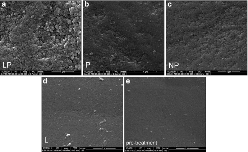
SEM (Inspect F; FEI Ltd, Eindhoven, The Netherlands), with a resolution power of 1.3 nm, was applied to observe the morphological changes in the enamel surface before and after the bleaching treatment. The micrographs are reproduced in Figure 1.
Figure 1.
SEM micrograph of specimen. (a) Post-treatment specimen of Group LP, ×80 000 original magnification; (b) post-treatment specimen of Group P, ×80 000 original magnification; (c) post-treatment specimen of Group NP, ×80 000 original magnification; (d) post-treatment specimen of Group L, ×80 000 original magnification; (e) pre-treatment specimen, ×80 000 original magnification. SEM, scanning electron microscopy.
Results
Color measurement
The mean values and standard deviation (s.d.) of ΔL*, Δa* and Δb* are shown in Table 1. As shown in Table 1, in Groups LP and P, significant changes in tooth color occurred after treatment. A statistically significant increase was observed in L*, whereas a decrease was observed in both a* and b*. However, in Groups NP and Group L, no statistically significant tooth color changes were observed. These changes verified that tooth whitening occurred in Groups LP and P.
Table 1. Mean (s.d.) of ΔL*, Δa* and Δb* in specimens of Group LP, Group P, Group NP and Group L.
| Groups | ΔL* | Δa* | Δb* |
|---|---|---|---|
| LP | 3.8 (2.8)a | −0.6 (0.3)a | −2.9 (2.0)a |
| P | 3.2 (2.9)a | −0.6 (0.4)a | −2.7 (2.3)a |
| NP | 0.9 (1.4)b | −0.3 (0.5)b | −0.6 (1.1)b |
| L | 0.5 (0.9)b | −0.4 (0.6)b | −0.4 (0.7)b |
Student's t-test indicated statistically significant difference in pre-treatment vs. post-treatment measure (P<0.05).
Student's t-test indicated no statistically significant difference in pre-treatment vs. post-treatment measure (P>0.05).
Enamel crystal phase
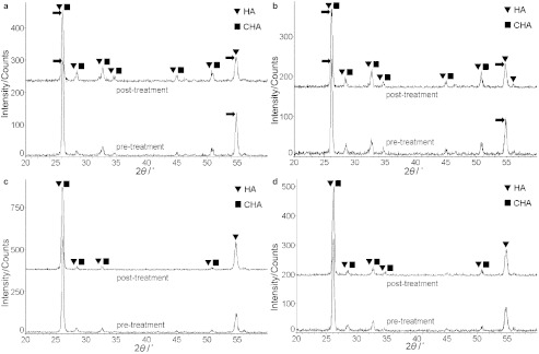
The XRD patterns of specimens from the four groups are shown in Figure 2 (Figure 2a for Group LP; Figure 2b for Group P; Figure 2c for Group NP; and Figure 2d for Group L). The pre-treatment and post-treatment patterns are displayed in one figure for comparison. As the XRD patterns shows, the major phases of the specimens are hydroxyapatite (HA) and carbonated hydroxyapatite (CHA). Comparing the pre-treatment pattern with that of the post-treatment in Groups LP and P, the pre-treatment peaks were sharper than the post-treatment peaks. Meanwhile, some peaks clearly visible in the pre-treatment pattern became weak or even disappeared at the same site in the post-treatment pattern. However, in Groups NP and L, the changes in peaks were not significant.
Figure 2.

Pre-treatment and post-treatment XRD patterns of specimen. (a) Group LP. ‘HA' and ‘CHA' refer to ‘hydroxyapatite' and ‘carbonated hydroxyapatite', respectively. Arrows indicated the obvious difference between the pre-treatment and post-treatment XRD patterns. (b) Group P. ‘HA' and ‘CHA' refer to ‘hydroxyapatite' and ‘carbonated hydroxyapatite', respectively. Arrows indicated the obvious difference between the pre-treatment and post-treatment XRD patterns. (c) Group NP. ‘HA' and ‘CHA' refer to ‘hydroxyapatite' and ‘carbonated hydroxyapatite', respectively. (d) Group L. ‘HA' and ‘CHA' refer to ‘hydroxyapatite' and ‘carbonated hydroxyapatite', respectively. XRD, X-ray diffraction.
Enamel crystal size and crystallinity
The crystal size and crystallinity of the specimens were calculated from the XRD data for three times and the changes of each after different treatment are shown in Table 2. Generally, the values of crystal size and crystallinity decreased after treatment in Group LP, Group P and Group NP. The changes were most distinct in Group LP. The means (and s.d.) of the changes in the crystal size and crystallinity of Group LP were −2.78 (2.35) nm and −2.548% (0.021 73%), respectively. The results of the Student's t-test showed significant decrease in crystal size and crystallinity after treatments in Group LP (P<0.005) and Group P (P<0.01). However, the changes in Group NP and Group L had no statistically significance (P>0.05). The results of the ANOVA indicated a statistically significant difference in the changes of crystal size and crystallinity in Group LP vs. Group NP, Group LP vs. Group L, Group P vs. Group NP and Group P vs. Group L, respectively (P<0.01). However, the ANOVA indicated no statistically significant difference in Group LP vs. Group P, as well as Group NP vs. Group L (P>0.05). It is known that crystals with higher crystallinity have sharper diffraction peaks and, conversely, low crystallinity results in weak or even no diffraction peaks.27 Thus, the results of crystallinity were consistent with the XRD patterns shown in Figure 2.
Table 2. Mean (s.d.) of crystal size and crystallinity changes after treatment in specimens of Group LP, Group P, Group NP and Group L.
| Groups | Changes in crystal size/nm | Changes in crystallinity/% | Agent pH |
|---|---|---|---|
| LP | −2.78 (2.35)**,a,b,c | −2.548 (0.021 73)**,a,b,c | 5.0 |
| P | −2.23 (2.05)*,d,e | −2.105 (0.019 14)*,d,e | 5.0 |
| NP | −0.86 (1.21)f,g | −0.647 (0.009 12)f,g | 5.9 |
| L | −0.10 (0.15)f | −0.013 (0.000 22)f | — |
**,*Student's t-test indicated a statistically significant decrease in post-treatment vs. pre-treatment measure (**P<0.005, *P<0.01).
Student's t-test indicated no statistically significant difference in pre-treatment vs. post-treatment measure (P>0.05).
ANOVA indicated a statistically significant difference in Group LP vs. Group NP, Group LP vs. Group L, Group P vs. Group NP and Group P vs. Group L, respectively (P<0.01).
ANOVA indicated no statistically significant difference in Group LP vs. Group P and Group NP vs. Group L, respectively (P>0.05).
Enamel surface morphology
The high-resolution SEM micrographs of specimens are shown in Figure 1 (Figure 1a for post-treatment specimens of Group LP; Figure 1b for post-treatment specimens of Group P; Figure 1c for post-treatment specimens of Group NP; Figure 1d for post-treatment specimens of Group L; and Figure 1e for pre-treatment specimens). As Figure 1 shows, the surface morphology of pre-treatment specimens was similar to that of post-treatment specimens of Group L, which appeared as a smooth flat surface. However, the surface morphology of post-treatment specimens of Group LP, Group P and Group NP was no longer smooth. Instead, a rough surface was observed, and a clear spherical enamel crystal structure can be identified. These changes were most evident in Group LP among the four groups.
Discussion
The significant work in our study was the application of micro-area XRD to evaluate the changes after tooth bleaching. This approach has never before been applied in evaluating the effects of a bleaching procedure on enamel. As is widely known, dental enamel is the hardest and most highly mineralized biological tissue. It is composed of approximately 96% minerals, 3% water and 1% organic matter by weight.28,29 The 96% mineral component, always referred to as enamel crystal, is the primary constituent of dental enamel. Micro-area XRD is a common approach used in materials science to analyze crystals, so it also can be applied in analyzing dental enamel. It enables the enamel crystal to be investigated in a non-destructive way. Furthermore, no special pre-treatment of specimens is necessary prior to micro-area XRD analysis, unlike others, more common methods, such as micro-hardness measurements. In micro-hardness measurements, it is necessary to flatten the upper surface layer of enamel to provide a more uniform surface, thereby improving the precision of the indentations.12,13,14,15,16 This procedure will lead to unreliable results in evaluating the adverse effects of peroxide-containing bleaching products on dental enamel,9 because the removed upper surface layer, the chief structure of which is prismless enamel, is generally more highly mineralized than the subsurface and is thus more resistant to demineralization.30 In the present study, the depth of X-ray penetration was less than 10 µm. At this depth, the enamel is prismless enamel because the average thickness of prismless is approximately 30 µm.30 In our study, to keep the enamel intact and improve the reliability of the results, no polishing or flattening measures were applied on the enamel surface before the bleaching treatment.
The primary mineral in tooth enamel is HA.31 The XRD pattern of the specimens in the current study was identified as HA and CHA, which is in agreement with the results of a previous study.32 However, the observed peaks did not agree with the diffraction patterns of standard, artificially synthesized HA or CHA. In the XRD pattern of the specimens, some peaks were lower or weaker and some peaks had disappeared altogether compared with the standard HA or CHA diffraction patterns. This phenomenon could be explained by the low crystallinity of natural enamel crystals and the immaturity of the enamel specimens applied in the present study. The enamel specimens were obtained from 10 premolars, which were extracted for orthodontic reasons from young people aged 12–15 years. It is known that after eruption, the tooth enamel will continue the mineralization in saliva before it reaches maturity.
The results from XRD patterns, enamel crystal size and crystallinity, as well as enamel surface SEM morphology, verified that changes occurred in enamel surface after cold-light tooth bleaching. These observed changes actually involved demineralization. There were three factors which may have influenced the enamel surface after bleaching, namely cold-light, peroxide in the bleaching agent and components of the bleaching agent other than peroxide. The cold-light, by itself, cannot cause demineralization. Thus, either peroxide or other components of the bleaching agent would be the causes of demineralization. The results in our study verified that 35% hydrogen peroxide solution had a major demineralization effect on enamel surface and the cold-light had no significant increase or decrease effect on this demineralization. The 35% hydrogen peroxide solution is acidic (pH=6.0), and its pH decreased further (pH=5.0) when the bleaching agents were prepared by mixing the 35% hydrogen peroxide solution with silicon dioxide catalyst. The critical pH for the occurrence of enamel demineralization is between 5.2 and 5.8.33 Obviously, the pH of mixed bleaching agent was lower than 5.2, and it can easily initiate the dissolution of enamel crystals. As the pH value of whitening agent fell, hydrogen ions started to attack the enamel crystals, freeing calcium and phosphate ions from enamel surface. Additionally, the substitution of carbonate ions in the HA crystal lattice would cause disturbances in the regular array of ions in the crystal lattice, making the crystal susceptible to attack by hydrogen ions.34 Because the 96% component of enamel is inorganic mineral, the acid pH takes the primary demineralization effects on the enamel. The oxidative effects are more likely to occur deeper in the tooth, where more organic materials are present.17
Conclusion
We can conclude from this study that the cold-light bleaching treatment resulted in enamel surface demineralization in vitro. It reduced crystal size and crystallinity and led to obvious changes in the surface morphology of enamel surface. The acidic peroxide-containing bleaching agent was the major factor that caused enamel demineralization, whereas cold-light alone had no significant increase or decrease effect on this demineralization.
Acknowledgments
The authors thank Mr Chao-liang Zhang (State Key Laboratory of Oral Diseases, Sichuan University) for his assistance in the SEM analysis and Ms Ru Shen (Key Laboratory of Advanced Technologies of Materials, Ministry of Education, Southwest Jiaotong University) for her help with the micro-area XRD experiment.
References
- Joiner A. The bleaching of teeth: a review of the literature. J Dent. 2006;34 7:412–419. doi: 10.1016/j.jdent.2006.02.002. [DOI] [PubMed] [Google Scholar]
- Dahl JE, Pallesen U. Tooth bleaching—a critical review of the biological aspects. Crit Rev Oral Biol Med. 2003;14 4:292–304. doi: 10.1177/154411130301400406. [DOI] [PubMed] [Google Scholar]
- Deliperi S, Bardwell DN. Two-year clinical evaluation of non-vital tooth whitening and resin composite restorations. J Esthet Restor Dent. 2005;17 6:369–378. doi: 10.1111/j.1708-8240.2005.tb00472.x. [DOI] [PubMed] [Google Scholar]
- Hannig C, Lindner D, Attin T. Efficacy and tolerability of two home bleaching systems having different peroxide delivery. Clin Oral Invest. 2007;11 4:321–329. doi: 10.1007/s00784-007-0128-x. [DOI] [PubMed] [Google Scholar]
- Wiegand A, Drebenstedt S, Roos M, et al. 12-month color stability of enamel, dentine, and enamel-dentine samples after bleaching. Clin Oral Invest. 2008;12 4:303–310. doi: 10.1007/s00784-008-0195-7. [DOI] [PubMed] [Google Scholar]
- Joiner A, Thakker G, Cooper Y. Evaluation of a 6% hydrogen peroxide tooth whitening gel on enamel and dentine microhardness in vitro. . J Dent. 2004;32 Suppl:27–34. doi: 10.1016/j.jdent.2003.10.010. [DOI] [PubMed] [Google Scholar]
- Justino LM, Tames DR, Demarco FF. In situ and in vitro effects of bleaching with carbamide peroxide on human enamel. Oper Dent. 2004;29 2:219–225. [PubMed] [Google Scholar]
- Götz H, Duschner H, White DJ, et al. Effects of elevated hydrogen peroxide ‘strip' bleaching on surface and subsurface enamel including subsurface histomorphology, micro-chemical composition and fluorescence changes. J Dent. 2007;35 6:457–466. doi: 10.1016/j.jdent.2007.01.004. [DOI] [PubMed] [Google Scholar]
- Joiner A. Review of the effects of peroxide on enamel and dentine properties. J Dent. 2007;35 12:889–896. doi: 10.1016/j.jdent.2007.09.008. [DOI] [PubMed] [Google Scholar]
- Cadenaro M, Breschi L, Nucci C, et al. Effect of two in-office whitening agents on the enamel surface in vivo: a morphological and non-contact profilometric study. Oper Dent. 2008;33 2:127–134. doi: 10.2341/07-89. [DOI] [PubMed] [Google Scholar]
- Maia E, Baratieri LN, Caldeira de Andrada MA, et al. The influence of two home-applied bleaching agents on enamel microhardness: an in situ study. J Dent. 2008;36 1:2–7. doi: 10.1016/j.jdent.2007.09.011. [DOI] [PubMed] [Google Scholar]
- Lewinstein I, Fuhrer N, Churaru N, et al. Effect of different peroxide bleaching regimens and subsequent fluoridation on the hardness of human enamel and dentin. J Prosthet Dent. 2004;92 4:337–342. doi: 10.1016/j.prosdent.2004.07.019. [DOI] [PubMed] [Google Scholar]
- Rodrigues JA, March GM, Ambrosano GM, et al. Microhardness evaluation of in situ vital bleaching on human dental enamel using a novel study design. Dent Mater. 2005;21 11:1059–1067. doi: 10.1016/j.dental.2005.03.011. [DOI] [PubMed] [Google Scholar]
- Chen HP, Chang CH, Liu JK, et al. Effect of fluoride containing bleaching agents on enamel surface properties. J Dent. 2008;36 9:718–725. doi: 10.1016/j.jdent.2008.05.003. [DOI] [PubMed] [Google Scholar]
- Jiang T, Ma X, Wang ZJ, et al. Beneficial effects of hydroxyapatite on enamel subjected to 30% hydrogen peroxide. J Dent. 2008;36 11:907–914. doi: 10.1016/j.jdent.2008.07.005. [DOI] [PubMed] [Google Scholar]
- Azer SS, Machado C, Sanchez E, et al. Effect of home bleaching systems on enamel nanohardness and elastic modulus. J Dent. 2009;37 3:185–190. doi: 10.1016/j.jdent.2008.11.005. [DOI] [PubMed] [Google Scholar]
- Hegedüs C, Bistey T, Flόra-Nagy E, et al. An atomic force microscopy study on the effect of bleaching agents on enamel surface. J Dent. 1999;27 7:509–515. doi: 10.1016/s0300-5712(99)00006-8. [DOI] [PubMed] [Google Scholar]
- Spalding M, Taveira LA, Assis GF. Scanning electron microscopy study of dental enamel surface exposed to 35% hydrogen peroxide: alone, with saliva, and with 10% carbamide peroxide. J Esthet Restor Dent. 2003;15 3:154–165. doi: 10.1111/j.1708-8240.2003.tb00185.x. [DOI] [PubMed] [Google Scholar]
- Al-Salehi SK, Wood DJ, Hatton PV. The effect of 24 h nonstop hydrogen peroxide concentration on bovine enamel and dentine mineral content and microhardness. J Dent. 2007;35 11:845–850. doi: 10.1016/j.jdent.2007.08.001. [DOI] [PubMed] [Google Scholar]
- Bistey T, Nagy IP, Sim A, et al. In vitro FT-IR study of the effects of hydrogen peroxide on superficial tooth enamel. J Dent. 2007;35 4:325–330. doi: 10.1016/j.jdent.2006.10.004. [DOI] [PubMed] [Google Scholar]
- Severcan F, Gokduman K, Dogan A, et al. Effects of in-office and at-home bleaching on human enamel and dentin: an in vitro application of Fourier transform infrared study. Appl Spectrosc. 2008;62 11:1274–1279. doi: 10.1366/000370208786401554. [DOI] [PubMed] [Google Scholar]
- Tezel H, Ertas OS, Ozata F, et al. Effect of bleaching agents on calcium loss from the enamel surface. Quintessence Int. 2007;38 4:339–347. [PubMed] [Google Scholar]
- Cavalli V, Giannini M, Carvalho RM. Effect of carbamide peroxide bleaching agents on tensile strength of human enamel. Dent Mater. 2004;20 8:733–739. doi: 10.1016/j.dental.2003.10.007. [DOI] [PubMed] [Google Scholar]
- Pobbe POS, Viapiana R, Souza-Gabriel AE, et al. Coronal resistance to fracture of endodontically treated teeth submitted to light-activated bleaching. J Dent. 2008;36 11:935–939. doi: 10.1016/j.jdent.2008.07.007. [DOI] [PubMed] [Google Scholar]
- Wang B, Liang JP.Observation of the bleaching effect of Beyond whitening accelerator Chin J Conserv Dent 200616(6337–339.Chinese. [Google Scholar]
- Zhang Y, Kang YY, Sun Y.The effect of Beyond cold light whitening kit on bleaching pulpless teeth Chin J Conserv Dent 200818(11615–617.Chinese.
- Huang JW.Manual of MDI JadeAvailable at http://wenku.baidu.com/view/857703778e9951e79b892700.html (accessed 10 February 2012). Chinese.
- Deakins M, Volker JF. Amount of organic matter in enamel from several types of human teeth. J Dent Res. 1941;20 2:117–121. [Google Scholar]
- Cuy JL, Mann AB, Livi KJ, et al. Nanoindentation mapping of the mechanical properties of human molar tooth enamel. Arch Oral Biol. 2002;47 4:281–291. doi: 10.1016/s0003-9969(02)00006-7. [DOI] [PubMed] [Google Scholar]
- Whittaker DK. Structural variations in the surface zone of human tooth enamel observed by scanning electron microscopy. Archs Oral Biol. 1982;27 5:383–392. doi: 10.1016/0003-9969(82)90147-9. [DOI] [PubMed] [Google Scholar]
- White T, Ferraris C, Kim J, et al. Apatite: an adaptive framework structure. Rev Mineral Geochem. 2005;57 1:307–401. [Google Scholar]
- Sydney-Zax M, Mayer I, Deutsch D. Carbonate content in developing human and bovine enamel. J Dent Res. 1991;70 5:913–916. doi: 10.1177/00220345910700051001. [DOI] [PubMed] [Google Scholar]
- Driessens FC, Theuns HM, Borggreven JM, et al. Solubility behavior of whole human enamel. Caries Res. 1986;20 2:103–110. doi: 10.1159/000260928. [DOI] [PubMed] [Google Scholar]
- Featherstone JDB. The science and practice of caries prevention. J Am Dent Assoc. 2000;131 7:887–899. doi: 10.14219/jada.archive.2000.0307. [DOI] [PubMed] [Google Scholar]


