Abstract
Much information about the coupling of presynaptic ionic currents with the release of neurotransmitter has been obtained from invertebrate preparations, most notably the squid giant synapse1. However, except for the preparation described here, few vertebrate preparations exist in which it is possible to make simultaneous measurements of neurotransmitter release and presynaptic ionic currents. Embryonic Xenopus motoneurons and muscle cells can be grown together in simple culture medium at room temperature; they will form functional synapses within twelve to twenty-four hours, and can be used to study nerve and muscle cell development and synaptic interactions for several days (until overgrowth occurs). Some advantages of these co-cultures over other vertebrate preparations include the simplicity of preparation, the ability to maintain the cultures and work at room temperature, and the ready accessibility of the synapses formed2-4. The preparation has been used widely to study the biophysical properties of presynaptic ion channels and the regulation of transmitter release5-8. In addition, the preparation has lent itself to other uses including the study of neurite outgrowth and synaptogenesis9-12, molecular mechanisms of neurotransmitter release13-15, the role of diffusible messengers in neuromodulation16,17, and in vitro synaptic plasticity18-19.
Keywords: Neuroscience, Issue 73, Physiology, Biophysics, Neurobiology, Developmental Biology, Cellular Biology, Anatomy, Electrophysiology, Neurophysiology, Xenopus, patch clamp, primary culture, embryo, synapses, synaptogenesis, synaptic currents, neurotransmitter release, varicosity, neurite guidance, neurons, motoneurons, cell culture, microdisection, animal model
Protocol
1. Pre-experimental Preparation
Prepare the following solutions (see Table 1 for compositions): (a) 1 liter NFR (Normal Frog Ringer), (b) 100 ml 10% saline, (c) 100 ml CMF (Ca2+ /Mg2+-free solution), (d) 1 ml ITS (Insulin-Transferrin-Selenium, Sigma I1884), (e) 100 ml L-15 culture medium, (f) 10 ml HCG (human chorionic gonadotropin Sigma CG-10), (g) 100 ml K+-internal solution, (h) 100 ml K+-internal solution for Amphotericin B. The osmotic strength of solution 1 should be checked to ensure that it is approximately 260 mOsM using a vapor pressure osmometer (e.g. Wescor model #5100C).
Filter sterilize solutions (b) and (c). Solutions (a) (b) and (c) can be stored up to three months at 4 °C. Prepare solution (d) by adding 1 ml deionized H2O to the vial containing the lyophilized powder via a 25 gauge syringe needle and syringe filter. This final solution can be stored at 4 °C for 30 days.
Prepare solution (e) in a laminar flow hood by combining all components in a beaker or flask. Filter sterilize the final solution and transfer 10 ml aliquots into sterile centrifuge tubes. Store at -20 °C.
Prepare the HCG (solution (f)) by adding 10 ml deionized H20 to the vial containing the lyophilized powder via a 25 gauge syringe needle and syringe filter. This final solution can be stored at 4 °C for 30 days.
Fabricate at least two microdissection tools by gluing a Minutien pin (26002-10, Fine Science Tools) to the end of a glass Pasteur pipette with cyanoacrylate glue. Allow the sharp end of the pin to extend past the end of the pipette approximately 0.5 cm.
Two days before preparing cultures, induce breeding with the following procedure. Identify a breeding-ready pair of Xenopus by observing a prominent, reddish cloaca on the female and dark pigmentation on the planar surface of the front paws of the male. One at a time, net each frog and hold it ventral side down on a sink with the net so that it cannot escape. Inject 1 ml of solution (f) through the net and subcutaneously into one of the dorsal lymph sacs.
Place the breeding pair together in a covered ten gallon tank of water. To ensure that the animals will not trample the newly laid and fertilized eggs, install a screened floor with a mesh size of approximately ½" fitted approximately 1-2 inches above the bottom of the tank (Figure 1A).
Leave frogs undisturbed for 12-48 hr until the animals are in amplexus and fertilized eggs are observed on the floor below the screen (Figure 1B).
Remove the animals from the tank, but leave the eggs undisturbed for at least 24 hr longer. This pair of frogs can be re-bred after six weeks.
Loosen the embryos from the bottom of the tank and transfer them to four or five 60x15 mm culture dishes containing 10% saline (solution b). Sort the embryos by stage according to the scheme of Niewkoop and Faber (ref. 20). Useful embryos will be those at stage 22-24. Figure 2A shows an embryo at approximately stage 22 while Figure 2B shows an embryo that is too far along in development to be useful (approximately stage 28). It is important to choose embryos that are healthy: those that are smooth in appearance with light brown and white mottling are ideal. Embryos that have large black or white patches are generally unhealthy and unusable.
2. Microdissection of Xenopus Embryos
Inside a laminar flow hood, label and fill approximately halfway three 60x15 mm sterile culture dishes with 10 % saline; and one with CMF.
Using a sterile, glass Pasteur pipette transfer five to ten stage 22-24 embryos into one of the dishes containing 10% saline. With the aid of a stereo zoom dissecting microscope inside the hood (0.6-5x with 10 x eyepieces), remove the jelly coat and vitelline membrane from each embryo using two pairs of sterile #5 forceps (11251-30, Fine Science Tools). (See Figures 3A and 3B.)
Wash the bare embryos by passing them, one at a time, through the remaining two dishes of 10% saline and finally into the dish containing CMF. Use a new, sterile pipette for each transfer and minimize the volume of solution transferred from dish to dish.
In turn, hold each embryo gently but firmly with a pair of forceps and, using the microdissection tool fashioned in step 1.5, remove the neural tube and associated myotomes which are located at the most dorsal aspect of the animal. Do this by making three slices, one at either end of the location of the neural tube and a third just ventral to it (Figure 4A). Move each dissected neural tube/myotome to a clean part of the dish away from the yolk granules and other debris (Figure 4B). The dorsal-ventral axis, and the locations of the neural tube and myotomes are indicated in Figure 4.
After about fifteen minutes in this solution (CMF) use forceps to lift the pigmented skin free of the dissected tissue and discard. After an additional 30-60 min, the cells will form a "sand pile" as they become dissociated from one another (Figure 4C).
3. Preparation of Nerve-muscle Co-cultures
Thaw a 10 ml tube of culture medium and aseptically add 70 μl ITS and 35 ng/ml BDNF (Sigma B3795).
Label and fill sterile 35 mm culture dishes (FD35-100 World Precision Instruments) approximately halfway with L-15 culture medium (solution (e)), one for each embryo dissected.
Fabricate a plating pipette by grasping each end of a glass Pasteur pipette while holding the tapered portion over a flame. Pull the ends apart to about 10 cm and then break off the end to yield a tip of approximately 0.2 mm.
Use the plating pipette to suck up the "sand pile" from one embryo minimizing the amount of solution drawn. Expel the cells onto the bottom of a culture dish. Plate the cells into several lines. Observe plated undifferentiated cells (Figure 5A and 5B).
Leave the dishes of plated cells undisturbed for at least fifteen minutes to allow the cells time to attach to the dish.
4. Patch Clamping Nerve-muscle Synapses
After 12-24 hr in culture, plated cells take on distinct morphological characteristics: muscle cells become spindle-shaped and spinal neurons extend long processes (Figure 6). Functional synaptic contact between neurite varicosities and muscle cells can be confirmed with simultaneous paired electrophysiological recordings. "M", "S" and "V" refer respectively to the muscle cell, neuronal soma and presynaptic varicosity.
Prepare two patch electrodes for recording: one for the presynaptic varicosity and one for the muscle cell.
For the presynaptic electrode, prepare an amphotericin-containing solution as follows: Add 100 μl DMSO to a 1.5 ml microcentrifuge tube containing 5 mg amphotericin B (Sigma A4888). Vortex this solution for 10 sec. Next, add 10 μl of this solution to a second microcentrifuge tube that contains 625 μl K+-internal solution for Amphotericin B (solution (h)). Vortex as above.
Fill the pre-synaptic electrode with the amphotericin-containing K+-internal solution prepared in step 4.3 and the post-synaptic electrode with K+-internal solution (solution (g)).
Replace media in the culture dish with NFR and relocate it to an inverted microscope fitted with phase contrast optics.
Position both electrodes just above their respective targets. Obtain the whole-cell configuration with the muscle cell electrode before the perforated patch configuration with the varicosity electrode.
To confirm functionality of the synapse, depolarize the varicosity with a patch clamp amplifier (e.g. Axopatch 200B, Molecular Devices) under software control (e.g. by pCLAMP 9, Molecular Devices) and observe the simultaneous presynaptic and postsynaptic currents. Figure 7 shows the currents seen in response to depolarizing steps of voltage given to the presynaptic varicosity in 10 mV increments from -30 mV to +40 mV. The inward currents seen in the presynaptic cell are carried by Na+ and Ca2+ and the outward currents by K+. Currents recorded in the postsynaptic cell are responses to neurotransmitter released from the varicosity and are carried mostly by Na+ through nicotinic acetylcholine receptor channels.
Representative Results
Figure 4B shows the dorsal view of an isolated spinal cord/myotome immediately after its removal from a stage 22 Xenopus embryo. Figure 4C shows that, after skin removal and incubation in Ca2+-Mg2+-free solution (CMF, solution (c)), cells dissociate into a "sand pile" and are ready for plating. Immediately after plating into culture medium cells exhibit little morphological variability (Figure 5B) but take on distinct shapes after twenty-four hours in culture. Muscle cells become spindle-shaped while neurons remain spherical whilst extending neurites that elaborate varicosites at synapses with muscle cells (Figure 6). Simultaneous paired patch recording from the presynaptic varicosity and postsynaptic muscle cell (Figure 7) reveals the presynaptic inward and outward currents associated with the release of neurotransmitter and the resultant excitatory postsynaptic currents.
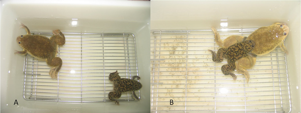 Figure 1. A: Breeding pair of frogs in mating bucket atop screen. B: Frogs in amplexus with fertilized eggs.
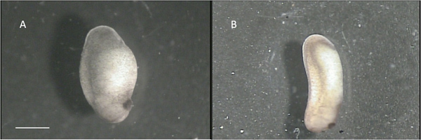 Figure 2. A: "suitable" stage 22 embryo. B: "unsuitable" stage 28 embryo. Scale bar represents 1 mm.
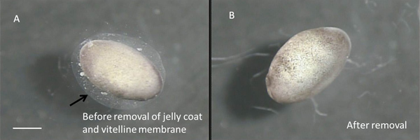 Figure 3. A: Stage 22 embryo before removal of vitelline membrane and jelly coat. The arrow points to the outer edge of the jelly coat. The vitelline membrane adheres closely to the embryo and is virtually invisible. B: Bare embryo. Scale bar represents 1 mm.
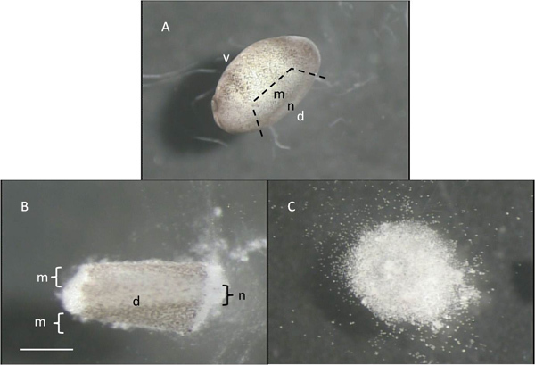 Figure 4. A: Stage 22 embryo with a dashed line indicating to-be dissected portion. B: Acutely isolated dorsal portion of embryo; "d" and "v" refer to the dorsal and ventral aspects of the embryos, while "m" and "n" indicate the approximate locations of the mytomes and neural tube. C: "Sand pile" of cells after 60 min in CMF. Scale bar represents 1 mm for A, and 0.5 mm for B and C.
 Figure 5. A: 5x power view of cells in culture immediately after plating. B: Same culture at 40x. Scale bar represents 40 μm for A, and 5 μm for B.
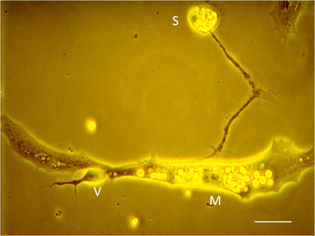 Figure 6. Neuromuscular junction in culture with identification of neuronal soma (S), presynaptic varicosity (V), and postsynaptic muscle cell (M). Scale bar: 5 μm.
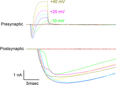 Figure 7. Presynaptic (top) and postsynaptic (bottom) currents seen in response to application of 10 mV incremental voltage steps presented to the presynaptic varicosity from -30 mV to +40 mV. Voltage steps are indicated next to some of the presynaptic traces. The holding potential for both cells was -70 mV.
Discussion
Key steps in the successful co-culturing of motoneurons and muscle cells are the use of appropriately staged embryos produced from the induced breeding of Xenopus frogs, and the careful aseptic dissection of the undifferentiated spinal neurons and myoblasts. Fertilized eggs should be left undisturbed until they reach approximately stage 10 or so as moving them sooner often halts their development. Healthy embryos are identified by a smooth appearance and a brown and white mottled coloring. Stage 22-24 embryos are the most useful because this is the point during development just after the neural tube closes and before the myocytes have differentiated significantly. In addition, cells from older embryos fail to dissociate well in CMF. Care should be taken when removing the vitelline membrane as it adheres strongly to the embryo. The membrane should be torn apart using sharp forceps so that the embryo emerges intact. Another important precaution is to carefully place the cells onto the bottom of the culture dish (rather than letting them settle there). This method is preferred as this increases the likelihood that the cells will adhere to the culture dish.
Functional neurotransmission between nerve and muscle can be determined with just postsynaptic recording and often can be ascertained by the observation of spontaneous muscle contraction after innervation. Un-innervated muscle cells do not twitch in culture. Postsynaptic recording alone is useful for recording miniature endplate currents or potentials but measurements of evoked release requires presynaptic stimulation, and correlation of pre- and postsynaptic currents requires double patch-clamp.
Besides the paired-patch recording method described here, this preparation offers the opportunity to introduce a third pipette at the neuronal soma5 This allows the generation of an action potential that can propagate to the presynaptic varicosity and lead to the release of neurotransmitter. In addition, putative agents that are thought to mediate or modulate synaptic transmission can be introduced to either side of the synapse: via the muscle cell pipette or through diffusion from a third pipette placed at the soma.
Disclosures
No conflicts of interest declared.
Acknowledgments
Funded by the NSF (0854551).
References
- Augustine GJ, Eckert R. Divalent cations differentially support transmitter release at the squid giant synapse. J. Physiol. 1984;346:257–271. doi: 10.1113/jphysiol.1984.sp015020. [DOI] [PMC free article] [PubMed] [Google Scholar]
- Spitzer NC, Lamborghini JE. The development of the action potential mechanism of amphibian neurons isolated in culture. Proc. Natl. Acad. Sci. U.S.A. 1976;73:1641. doi: 10.1073/pnas.73.5.1641. [DOI] [PMC free article] [PubMed] [Google Scholar]
- Weldon PR, Cohen MW. Development of synaptic ultrastructure at neuromuscular contacts in an amphibian cell culture system. J. Neurocytol. 1979;8:239–259. doi: 10.1007/BF01175564. [DOI] [PubMed] [Google Scholar]
- Tabti N, Poo M-M. In: Culturing Nerve Cells. Banker G, Goslin K, editors. Cambridge, Massachusetts: MIT Press; 1991. pp. 137–153. [Google Scholar]
- Yazejian B, DiGregorio DA, Vergara JL, Poage RE, Meriney SD, Grinnell AD. Direct measurements of presynaptic calcium and calcium-activated potassium currents regulating neurotransmitter release at cultured Xenopus nerve-muscle synapses. J. Neurosci. 1997;17:2990. doi: 10.1523/JNEUROSCI.17-09-02990.1997. [DOI] [PMC free article] [PubMed] [Google Scholar]
- DiGregorio DA, Peskoff A, Vergara JL. Measurement of action potential-induced presynaptic calcium domains at a cultured neuromuscular junction. J. Neurosci. 1999;19:7846. doi: 10.1523/JNEUROSCI.19-18-07846.1999. [DOI] [PMC free article] [PubMed] [Google Scholar]
- Yazejian B, Sun XP, Grinnell AD. Tracking presynaptic Ca2+ dynamics during neurotransmitter release with Ca2+-activated K+ channels. Nat. Neurosci. 2000;3:566. doi: 10.1038/75737. [DOI] [PubMed] [Google Scholar]
- Sun XP, Chen BM, Sand O, Kidokoro Y, Grinnell AD. Depolarization-induced Ca2+ entry preferentially evokes release of large quanta in the developing Xenopus neuromuscular junction. J. Neurophysiol. 2010;104(5):2730–2740. doi: 10.1152/jn.01041.2009. [DOI] [PMC free article] [PubMed] [Google Scholar]
- Xie SP, Poo MM. Initial events in the formation of neuromuscular synapse: rapid induction of acetylcholine release from embryonic neurons. Proc. Natl. Acad. Sci. U.S.A. 1986;83:7069. doi: 10.1073/pnas.83.18.7069. [DOI] [PMC free article] [PubMed] [Google Scholar]
- Li PP, Chen C, Lee CW, Madhavan R, Peng HB. Axonal filopodial asymmetry induced by synaptic target. Mol. Biol Cell. 2011;22(14):2480–2490. doi: 10.1091/mbc.E11-03-0198. [DOI] [PMC free article] [PubMed] [Google Scholar]
- Feng Z, Ko CP. Schwann cells promote synaptogenesis at the neuromuscular junction via transforming growth factor-beta1. J. Neurosci. 2008;28(39):9599–9609. doi: 10.1523/JNEUROSCI.2589-08.2008. [DOI] [PMC free article] [PubMed] [Google Scholar]
- Song HJ, Ming GL, Poo MM. cAMP-induced switching in turning direction of nerve growth cones. Nature. 1997;17(6639):275–279. doi: 10.1038/40864. [DOI] [PubMed] [Google Scholar]
- Morimoto T, Wang XH, Poo MM. Overexpression of synaptotagmin modulates short-term synaptic plasticity at developing neuromuscular junctions. Neuroscience. 1998;82(4):969–978. doi: 10.1016/s0306-4522(97)00343-6. [DOI] [PubMed] [Google Scholar]
- Lu B, Czernik AJ, Popov S, Wang T, Poo MM, Greengard P. Expression of synapsin I correlates with maturation of the neuromuscular synapse. Neuroscience. 1996;74(4):1087–1097. doi: 10.1016/0306-4522(96)00187-x. [DOI] [PubMed] [Google Scholar]
- Schaeffer E, Alder J, Greengard P, Poo MM. Synapsin IIa accelerates functional development of neuromuscular synapses. Proc. Natl. Acad. Sci. U.S.A. 1994;26(9):3882–3886. doi: 10.1073/pnas.91.9.3882. [DOI] [PMC free article] [PubMed] [Google Scholar]
- Liou JC, Tsai FZ, Ho SY. Potentiation of quantal secretion by insulin-like growth factor-1 at developing motoneurons in Xenopus cell culture. J. Physiol. 2003;553(Pt. 3):719–7128. doi: 10.1113/jphysiol.2003.050955. [DOI] [PMC free article] [PubMed] [Google Scholar]
- Peng HB, Yang JF, Dai Z, Lee CW, Hung HW, Feng ZH, Ko CP. Differential effects of neurotrophins and schwann cell-derived signals on neuronal survival/growth and synaptogenesis. J. Neurosci. 2003;23(12):5050–5060. doi: 10.1523/JNEUROSCI.23-12-05050.2003. [DOI] [PMC free article] [PubMed] [Google Scholar]
- Dan Y, Poo MM. Hebbian depression of isolated neuromuscular synapses in vitro. Science. 1992;12(5063):1570–1573. doi: 10.1126/science.1317971. [DOI] [PubMed] [Google Scholar]
- Xiao Q, Xu L, Spitzer NC. Target-dependent regulation of neurotransmitter specification and embryonic neuronal calcium spike activity. J. Neurosci. 2010;30(16):5792–5801. doi: 10.1523/JNEUROSCI.5659-09.2010. [DOI] [PMC free article] [PubMed] [Google Scholar]
- Niewkoop PD, Faber J. Normal table of Xenopus laevis (Daudin) North-Holland, Amsterdam: 1967. [Google Scholar]


