



Abstract
Oral myiasis is a common parasitic infestation of live human and animals caused by species of dipteran fly larvae known as maggots which may be secondary to medical disease. This case involves a 51-year-old female, poorly debilitated with advanced periodontal disease infected by the dipteral larvae in the anterior maxillary region which belonged to the family Calliphoridae and Chrysomya bezziana species. This lady was neglected from her family and presented oral myiasis with the previous history of filariasis and Hansen's disease. Secondary infestations may occur in cancrum oris, oral extraction wounds, jaw bone wounds, oral leprosy lesion, filariasis, and carcinoma. Hansen's disease (leprosy) is bacterial in origin whereas filariasis (elephantiasis) is parasitic in origin like-myiasis. The treatment consisted of manual removal of the larvae by topical application of turpentine oil, oral therapy, and surgical debridement of the oral wound.
Keywords: Filariasis, flies larvae, Hansen's disease, maggots, oral myiasis
INTRODUCTION
Myiasis comes from the Latin word ‘myia’ meaning fly and ‘iasis’ means disease. Parasitic invasion of the tissues in the oral cavity by larvae of flies are called oral myiasis. Oral myiasis was first described by Laurence in 1909 which occur in the skin and mucosa.[1] The incidence of oral myiasis is less when compared to cutaneous myiasis as oral tissues are not permanently exposed to the external environment. The predisposing factors include low socioeconomic status, immunocompromised state, debilitated, and unhygienic living conditions. Suppurative lesions, facial trauma, mouth-breathers, extraction wounds, fungating carcinomas, and other conditions are considered to be the risk factors.[2] Human myiasis can occur in any part of the globe, but is more common in regions with warm and humid climate. Myiasis can be classified based on the substrate, degree of host dependence, mode of infestation, and anatomic sites Table 1. Flies causing myiasis belong to the order Diptera. Chrysomya bezziana, the old world screw worm fly, belongs to genera Calliphoridae which is an obligatory parasite.[3]
Table 1.
Classification of myiasis

CASE REPORT
A 51-year-old woman came to the Department of Oral and Maxillofacial Surgery with a chief complaint of pain and mobility in the upper front teeth, and in the month, it was noticed that couple of insects emerging out from her gums [Figure 1]. She gives a history of leprosy and filariasis for which she had undergone treatment. Extra oral examination revealed no active infection in hands (leprosy) [Figure 2] and legs (elephantiasis) [Figure 3]. On intraoral examination, she had poor oral hygiene with periodontally compromised grade III mobile teeth. The patient had anterior open bite and was a habitual mouth breather. Exploration of the labial gingival revealed deepening of the gingival sulcus with formation of the deep periodontal pocket in the region of the upper left lateral incisor and canine teeth. The surrounding mucosa was inflamed and tender on palpation with pus discharge [Figure 1a and b]. A provisional diagnosis of oral myiasis was made. The hematological report was normal. The patient was sent for a medical opinion regarding leprosy of the hand and filariasis of the right leg, and no signs of active infections with healed scar were mentioned in the report obtained and no contraindication to carry out the dental procedure was sought from them. Orthopantamography revealed mild bone destruction in relation to the mobile anterior maxillary teeth. The treatment plan consisted of removal of the maggots, debridement, and irrigation. The wound was debrided under local anesthesia and roller gauze impregnated with turpentine oil was inserted into the cavity present deep to the gingival sulcus. A live larva was removed with the help of tweezer. Copious irrigation with normal saline and povidine iodine was performed. Under antibiotic coverage with oral penicillin, the patient underwent debridement for three consecutive days and the wound healed by secondary intention.
Figure 1.
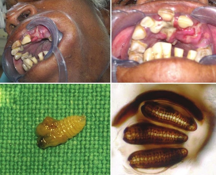
(a and b) Intraoral appearance of the wound showing pus discharge at the perforated mucosa in the anterior maxilla. (c) The extracted maggot from the wound. (d) Microscopic examination revealing five fingers like process in the anterior spiracle, incomplete peritreme in the posterior spiracle, and straight spiracular slits
Figure 2.

Photograph of hands affected by Hansen's disease
Figure 3.

Photograph of left leg affected by filariasis
The larvae recovered from the wound were preserved in 40% formaldehyde. It appeared tapered in shape and creamy white in color, their segments giving the appearance of transverse rows, with a brown black tip anteriorly [Figure 1c] and was identified as larvae of Chrysomya bezziana by an entomologist. Microscopic examination [Figure 1d] revealed five fingers like process in the anterior spiracle, incomplete peritreme in the posterior spiracle, and straight spiracular slits which belong to the family Calliphoridae.
DISCUSSION
The patient in the present case was of low socio-economic status having poor living conditions and neglected from family. Persistent mouth opening due to incompetent lips, class II malocclusion with poor oral hygiene, and advanced periodontal disease as seen in our case are common predisposing factors for oral myiasis. Periodontal pockets provide a perfect environment for the eggs to hatch and for the larvae to grow in the warm and moist conditions. In addition, the patient had previous bacterial lepromatous infection as well as parasitic elephantiasis infection which could be a cause to get neglected from her family and moreover, the patient was physically dependent on her relatives for day-to-day routine activities which could be a contributing factor to her neglected oral hygiene. It is usually the special people with mental or physical disability who are affected because of the difficulties in maintaining good oral hygiene due to poor manual dexterity and negligence of parents/guardians.[4]
Infestation of the nose with larvae of certain flies can occur in leprosy patients. Granulomatous infiltration of nasal mucosa and consequent sensory loss may result in atrophic rhinitis and/or painless ulceration, which is usually asymptomatic except for later complications of epistaxis, septal perforation, inflammation, and/or myiasis in long neglected patients. This results in severe distress and agony and can cause extensive tissue damage.[5]
In India, filariasis is predominantly caused by a species of nematode called Wuchereria bancrofti. The disease is transmitted through the bite of blood sucking mosquitoes. The salient clinical feature of the disease is lymphangitis leading to elephantiasis of the legs, arms, scrotum, and breast. Oral or perioral involvement is very rare.[6] Patients with lymphoedema belong to socially neglected groups; however, nosocomically acquired myiasis are not necessarily associated with patient negligence.[7] Extensive wounds occur when infested by larvae of the screwworm fly. Although the fly larvae do not carry infectious agents themselves, there is, however, an increased risk for complications due to secondary bacterial infection. There are few reports of myiasis in edema/lymphoedema patients living in areas endemic with bancroftian filariasis.[8] Oral myiasis with active filariasis patients can be treated with a single annual dose of albendazole 400 mg along with diethylcarbamazine 6 mg/kg or ivermectin 200-400 μg/kg body weight.[9]
Poor oral hygiene, ulcerated lesions, severe halitosis, and alcoholism are the pathological factors associated with oral myiasis. Weak, senile, pediatric, or mentally handicapped patients who are unable to defend themselves and who often present with a lack of labial sealing are attacked by flies and consequently by the disease.[10] The prevention of human myiasis is important, and involves the control of fly population, general cleanliness of dwelling areas, and provision for basic sanitation and health education. In our case, the patient represented with healed Hansen's disease and elephantiasis with infestation of oral myiasis, which, according to our knowledge is the first report.
Footnotes
Source of Support: Nil
Conflict of Interest: None declared
REFERENCES
- 1.Laurence SM. Dipterous larvae infection. Br Med J. 1909;9:88. [Google Scholar]
- 2.Sankari LS, Ramakrishnan K. Oral myiasis caused by Chrysomya bezziana. J Oral Maxillofac Pathol. 2010;14:16–8. doi: 10.4103/0973-029X.64304. [DOI] [PMC free article] [PubMed] [Google Scholar]
- 3.Sheikh S, Pallagatti S, Singla I, Kalucha A, Aggarwal A, Kaur H. Oral Myiasis: A review. J Clin Exp Dent. 2011;3:e465–8. [Google Scholar]
- 4.Ramli R, Rahman AR. Oral myiasis: Case report. Malays J Med Sci. 2002;9:47–50. [PMC free article] [PubMed] [Google Scholar]
- 5.Thami GP, Baurah MC, Sharmce SC, Behera NK. Nasal myiasis in leprosy leading to unusual tissue destruction. J Dermatol. 1995;22:348–50. doi: 10.1111/j.1346-8138.1995.tb03401.x. [DOI] [PubMed] [Google Scholar]
- 6.Baliga M, Ramanathan A, Uppal N. Oral filariasis: A case report. Br J Oral Maxillofac Surg. 2010;48:143–4. doi: 10.1016/j.bjoms.2009.04.020. [DOI] [PubMed] [Google Scholar]
- 7.Sherman RA. Wound myiasis in urban and suburban United States. Arch Intern Med. 2004;160:14. doi: 10.1001/archinte.160.13.2004. [DOI] [PubMed] [Google Scholar]
- 8.Srinivasan R, Pani SP. Myiasis in human filarial lymphedema. Southeast Asian J Trop Med Public Health. 1992;23:807–8. [PubMed] [Google Scholar]
- 9.Palumbo E. Filariasis: Diagnosis, treatment and prevention. Acta Biomed. 2008;79:106–9. [PubMed] [Google Scholar]
- 10.Gealh WC, Ferreira GM, Farah GJ, Teodoro U, Camarini ET. Treatment of oral myiasis caused by Cochliomyia hominivorax: Two cases treated with ivermectin. Br J Oral Maxillofac Surg. 2009;47:23–6. doi: 10.1016/j.bjoms.2008.04.009. [DOI] [PubMed] [Google Scholar]