Abstract
Advanced abdominal pregnancies with live twin fetuses are extremely rare and are misdiagnosed in up to 60% of the cases. Such a case is presented here, highlighting the diagnostic and management challenges encountered. A high index of suspicion in making the diagnosis of this rare variety of ectopic pregnancy, emphasizing adherence to basic imaging principles, and appropriate placental management is very important in reducing the associated morbidity and mortality.
Keywords: Abdominal pregnancy, Advanced, Live, Placenta, Twin gestation
Introduction
Advanced abdominal pregnancy of twin pregnancy is very rare, and most of the case reports in the literature originate primarily in the developed world.[1] A literature search did not reveal any similar cases in sub-Saharan Africa. This difference is likely related to the ability to detect abdominal pregnancy using sophisticated imaging especially in the community.[1] Generally, abdominal pregnancy is difficult to diagnose clinically and missed in 60% of cases due to absence of distinguishing clinical features.[2] Here, we present a case of live advanced abdominal twin pregnancy misdiagnosed as abruptio placenta, emphasizing adherence to basic imaging principles to ensure an early ultrasound diagnosis of this rare variety of ectopic pregnancy and appropriate management of placenta, particularly in the developing world.
Case Report
A 24-year-old Tanzanian woman, primigravida at the gestational age of 26 weeks, was referred from another nearby facility with a one-month history of persistent abdominal pain associated with long standing recurrent vaginal bleeding during the antenatal period. She had been admitted repeatedly at a local community hospital due to abdominal pain. She denied any history that would suggest previous pelvic inflammatory disease, intrauterine contraceptive device use or pelvic surgery. During her last admission, abruptio placenta was suspected, and the patient was unsuccessfully induced twice with oxytocin without knowing the status of the fetuses. She was subsequently transferred to our hospital for further evaluation and treatment.
On arrival, physical examination revealed a conscious woman in severe pain, pale, afebrile. Her abdominal examination was notable for distended abdomen consisted with uterine fundus of 30 cm, generalized tenderness was elicited on palpation and fetal heartbeats could not be detected by fetoscope. Vaginal examination revealed a closed, uneffaced, firm and posterior cervix, and the presenting part was not found. Transabdominal ultrasound revealed live intrauterine diamniotic–dichorionic twins, estimated gestational age was 26 weeks and there were enlarged cystic placentas suggestive of retro-placental clots.
Laboratory investigation revealed hemoglobin level of 7.0g/dl, the blood group A, and Rhesus positive.
The diagnosis of concealed abruptio placenta with live twin pregnancy was reached, and the patient was counseled and an emergency abdominal delivery was performed.
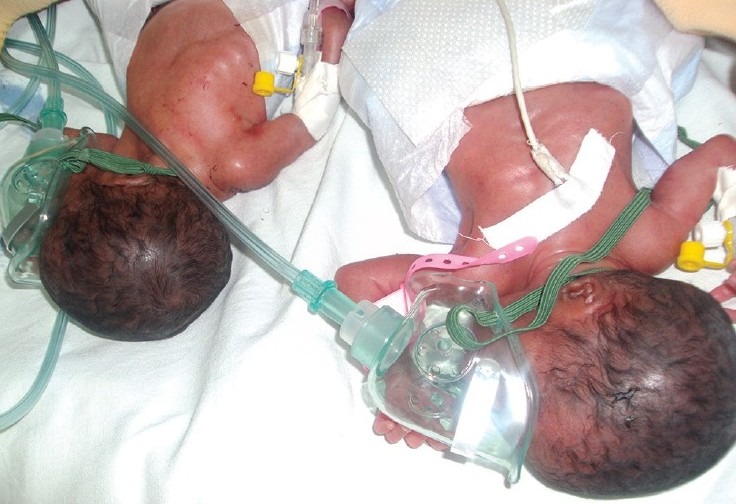
At laparotomy, the findings were: A thickened peritoneum with area of hematoma resembling a cystic mass, and a normal-sized intact uterus separate from the pregnant sac which was engulfed by thickened omentum. Both fallopian tubes and ovaries were grossly normal. Live female twins were extracted from the gestational sacs. Two placentas were seen, one attached to the ceacum and ascending colon and the other firmly adhered to the posterior aspect of the omentum, the transverse and the sigmoid colon. The placenta attached to the ceacum and ascending colon developed profuse bleeding during the delivery of the babies, requiring ligation of placental blood vessels to control bleeding. Both placentas were left in situ with their umbilical cords cut to 2cm long. Estimated blood loss was 2000ml; the patient was subsequently transfused 4 units of whole blood. The first twin weighed 700 g and the other 800g, each with Apgar score of 5 at first minutes and 6 at fifth minute. Grossly, both fetuses had no congenital abnormalities and were admitted to the neonatal intensive care unit due to prematurity [Figure 1]. Unfortunately, both babies died within the first week of life.
Figure 1.

Diamniotic–dichorionic twins; first day post delivery admitted at Bugando neonatal unit
Postoperatively, the patient was covered with broad spectrum antibiotics and admitted to the intensive care unit. Three days later she developed septicemia which was successful treated with meropenem. The patient improved and was discharged on postoperative day 14, with plan for outpatient follow-up in one month.
Fourteen days after discharge, the patient was readmitted with peritonitis which necessitated repeat laparotomy and attempt to remove the placentas provoked massive hemorrhage which was controlled by intra-abdominal packing and the patient was transfused with 3 units of blood. The abdominal packs were removed after 48 hours with no active bleeding from placental site. Eight days after removing the packing, the patient developed fascial dehiscence and was taken to operating room for exploration, where both placentas were easily detached without provoking bleeding. Her postoperative recovery thereafter was unremarkable, and she was discharged to attend outpatient clinic. After three visits of 11 months, she was discharged from the clinic in a good condition.
Discussion
Abdominal pregnancy is a rare form of ectopic pregnancy that has seldom been described in the developing world.[1,3] This case was unique in that there were live twins without gross malformations at an advanced gestational age.
Predisposing factors for abdominal twin gestation are the same as for other forms of ectopic pregnancy. Of note, this patient had additional family history of twin pregnancy on maternal side.
Despite the remarkable improvements in sophisticated imaging such as ultrasound scan, magnetic resonance imaging (MRI) and computerized tomography (CT) scan,[2] diagnosis of abdominal pregnancy often requires a high index of suspicion based on clinical presentations such as persistent abdominal pain, painful fetal movements, weight loss, abnormal presentations, uneffaced and displaced cervix, vaginal bleeding, fetal death, failed induction of labor and palpation of an abdominal mass distinct from the uterus.[1–3] There were many challenges in diagnosis and management of this case. First, it was difficult to reach a correct diagnosis prior to surgery because clinical features mimicked that of abruptio placenta and transabdominal ultrasound failed to detect the extra uterine pregnancy.[3,4] In a limited resource area like Tanzania, one should pay close attention to patient's presentation and adherence to the basic imaging principles preferably with the use of trans-vaginal ultrasound to improve the likelihood of identifying abdominal pregnancy.[3]
The second challenge occurred during management in the operating room. Leaving the placenta in situ is the best way to deal with abdominal pregnancy, based on previous experience and review of the literature.[5] In the present case, an attempt to remove the placentas during the repeat laparotomy provoked massive bleeding requiring blood transfusion. We were uncertain about the role of methotrexate in post-operative handling of placenta. Some authors have reported sepsis as a feared complication following use of this drug.[5]
Advanced abdominal pregnancy is associated with significant fetal and maternal morbidity and mortality.[1,2] These complications may be worse in extra uterine multiple gestations, as two or more placentas occupy a large area, and attach to vital organs. One should be prepared for possible complications such as hemorrhage and sepsis when managing these patients.[1]
A high index of suspicion in making the diagnosis of this rare variety of ectopic pregnancy, emphasizing adherence to basic imaging principles by using transvaginal ultrasound and appropriate placental management is very important in reducing the associated morbidity and mortality.
Footnotes
Source of Support: Nil.
Conflict of Interest: None declared.
References
- 1.da Silva BB, de Araujo EP, Cronemberger JN, dos Santos AR, Lopes-Costa PV. Primary twin omental pregnancy: Report of a rare case and literature review. Fertil Steril. 2008;90:2006e13–5. doi: 10.1016/j.fertnstert.2008.03.038. [DOI] [PubMed] [Google Scholar]
- 2.Worley KC, Hnat MD, Cunningham FG. Advanced extrauterine pregnancy: Diagnostic and therapeutic challenges. Am J Obstet Gynecol. 2008;198:297e1–7. doi: 10.1016/j.ajog.2007.09.044. [DOI] [PubMed] [Google Scholar]
- 3.Dassah ET, Odoi AT, Opoku BK. Advanced twin abdominal pregnancy: Diagnostic and therapeutic challenges. Acta Obstet Gynecol Scand. 2009;88:1291–3. doi: 10.3109/00016340903281006. [DOI] [PubMed] [Google Scholar]
- 4.Ikechebelu JI, Onwusulu DN, Chukwugbo CN. Term abdominal pregnancy misdiagnosed as abruptio placenta. Niger J Clin Pract. 2005;8:43–5. [PubMed] [Google Scholar]
- 5.Oneko O, Petru E, Masenga G, Ulrich D, Obure J, Zeck W. Management of the placenta in advanced abdominal pregnancies at an East-African tertiary referral center. J Womens Health (Larchmt) 2010;19:1369–75. doi: 10.1089/jwh.2009.1704. [DOI] [PubMed] [Google Scholar]


