SUMMARY
Characterizing dopaminergic neuronal development and function in novel genetic animal models might uncover strategies for researchers to develop disease-modifying treatments for neurologic disorders. Id2 is a transcription factor expressed in the developing central nervous system. Id2−/− mice have fewer dopaminergic neurons in the olfactory bulb and reduced olfactory discrimination, a pre-clinical marker of Parkinson’s disease. Here, we summarize behavioral, histological and in vitro molecular biological analyses to determine whether midbrain dopaminergic neurons are affected by Id2 loss. Id2−/− mice were hyperactive at 1 and 3 months of age, but by 6 months showed reduced activity. Id2−/− mice showed age-dependent histological alterations in dopaminergic neurons of the substantia nigra pars compacta (SNpC) associated with changes in locomotor activity. Reduced dopamine transporter (DAT) expression was observed at early ages in Id2−/− mice and DAT expression was dependent on Id2 expression in an in vitro dopaminergic differentiation model. Evidence of neurodegeneration, including activated caspase-3 and glial infiltration, were noted in the SNpC of older Id2−/− mice. These findings document a novel role for Id2 in the maintenance of midbrain dopamine neurons. The Id2−/− mouse should provide unique opportunities to study the progression of neurodegenerative disorders involving the dopamine system.
INTRODUCTION
Dopamine (DA) is a catecholaminergic neurotransmitter of the central nervous system (CNS) that plays a prominent role in cognition as well as motor and endocrine function (Iversen and Iversen, 2007). Most DA neurons in the adult mammalian brain are found in the substantia nigra pars compacta (SNpC) and the ventral tegmental area (VTA) of the ventral midbrain (Prakash and Wurst, 2006a). Age-dependent neurodegeneration of midbrain dopaminergic (mDA) neurons of the SNpC underlies altered motor function in patients with Parkinson’s disease (PD) (Dauer and Przedborski, 2003). Dysfunction of mDA neurons is also a prominent characteristic of neuropsychiatric and addictive disorders (Chao and Nestler, 2004). Understanding the molecular pathways mediating the development and survival of mDA neurons and how perturbations of these pathways affect mature DA neurons should provide insights into the prevention and treatment of these disorders.
mDA neurons in the mouse arise from mDA progenitors between embryonic days e9 and e14. The induction, specification and maturation of mDA neurons can be monitored by evaluation of the temporal pattern of expression of genes associated with differentiation of DA neurons. Expression of DAT and establishment of DA re-uptake are among the last events in the maturation of mDA neurons, and thus indicative of terminal differentiation (for reviews, see Prakash and Wurst, 2006b; Prakash and Wurst, 2006a). DAT is a highly specific Na+/Cl−-dependent neuronal membrane transporter thought to be the major regulator of synaptic DA levels. DAT is the functional target of drugs of abuse, notably cocaine and amphetamine (Pifl et al., 1996). Loss of DAT function in mice results in behavioral abnormalities, including increased locomotor activity and reduced avoidance behaviors (Gainetdinov et al., 1998). DAT dysfunction detected by advanced imaging is a biomarker of PD (Spiegel et al., 2006), an observation that has prompted numerous studies investigating the possibility that DAT polymorphisms would be predicative of increased risk for developing PD. These studies have yielded mixed results (see, PD GENE, http://www.pdgene.org/); however, recent studies provide compelling evidence of a gene-environment interaction between DAT polymorphisms and occupational pesticide exposure underlying an increased risk of developing PD (Ritz et al., 2009).
During adult neurogenesis in the mouse, Id2 loss leads to a diminished olfactory bulb (OB) size associated with a specific decrease in periglomerular DA neurons (Havrda et al., 2008). Id proteins (Id1-Id4) are helix-loop-helix (HLH) transcription factors highly expressed in the developing CNS (Andres-Barquin et al., 1997). Id proteins inhibit the function of class I basic helix-loop-helix (bHLH) transcription factors (Kee, 2009) and class II tissue-specific bHLH proteins (Chen et al., 2012) by heterodimerization that results in the bHLH factors being unable to bind DNA (Benezra et al., 1990; Andres-Barquin et al., 2000). Transcription factors of the bHLH family direct the proliferation, specification and maturation of multiple neuronal lineages (Atchley and Fitch, 1997; Guillemot, 1999; Ross et al., 2003). However, a role for Id2 during the development and maintenance of mDA neurons has not been described.
Previously, we reported decreased olfactory discrimination in Id2−/− mice associated with a reduction in OB DA neurons (Havrda et al., 2008). Olfactory dysfunction is under investigation as a potential preclinical biomarker of PD (Wu et al., 2011) and is a feature of multiple neurologic disorders (Doty 2012). In this study, we continued our characterization of Id2−/− mice to determine whether Id2 is required for the establishment, maintenance and functioning of mDA neurons. Histological and behavioral changes occurring in Id2−/− mice, comparable to those found in human diseases involving the DA system, would provide strong evidence for the importance of Id2 in the maintenance and function of midbrain DA neurons. In this regard, Id2−/− mice might provide opportunities for studying the genesis, progression and clinical course of important neurologic disorders.
TRANSLATIONAL IMPACT.
Clinical issue
Medical advances have allowed individuals to live longer but age-related neurological disorders have become more prevalent. Parkinson’s disease (PD) is a complex neurodegenerative disorder characterized by severely debilitating cognitive and motor symptoms that can occur sporadically. The disease is difficult to detect before an incurable loss of midbrain dopaminergic (DA) neurons has already taken place. The development of disease-modifying treatment strategies for age-related neurodegenerative diseases such as PD will require a broader understanding of disease initiation and progression. In this context, the value of new genetic animal models that recapitulate features of PD cannot be understated.
Results
These authors’ ongoing characterization of neurological defects in the Id2−/− mouse has revealed key features mimicking PD. In previous work, they reported that Id2−/− mice have olfactory functional defects reminiscent of those associated with PD in humans. Here, they report age-dependent degeneration of midbrain DA tissues in these mice. They show that histological abnormalities that arise in Id2−/− mice occur in association with reduced activity, following a period of early life hyperactivity. Behavioral and histological changes occur along with early life suppression of dopamine transporter (DAT), a finding validated using an in vitro model of DA neuron differentiation. These observations suggest that inactivation of Id2 can affect the motor system, alter the maintenance of DA neurons and contribute to age-dependent neurodegeneration.
Implications and future directions
It is expected that novel genetic models of PD will provide insights into disease progression and identify pathways that are important in the pathobiology of the disease. Behavioral and histological characterization of the Id2−/− mouse indicates an unexpected role for this transcription factor in midbrain DA tissues that, when lost, results in histological changes similar to those that occur in human PD. This discovery opens the door for future studies that examine the molecular mechanisms of Id2 activity in the maintenance of aging DA neurons. Such studies have a high likelihood of informing methods to detect and treat PD and other neurologic disorders involving the DA system such as attention deficit hyperactivity disorder, schizophrenia, pathologic gambling and substance abuse.
RESULTS
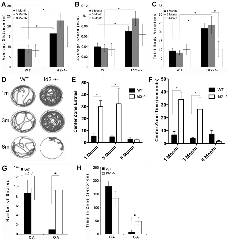
Id2−/− mice displayed increased home cage activity compared with wild-type littermates housed in the same cages (our unpublished observations). Locomotor activity in Id2−/− mice was therefore quantified using the open-field test. At 1 and 3 months of age, Id2−/− mice demonstrated increases in average distance travelled (Fig. 1A) and average speed (Fig. 1B). As shown in Fig. 1C, Id2−/− mice also exhibited significantly increased bi-directional rotation, a behavior mediated by the DA system (Richter et al., 1999) and associated with hemi-Parkinsonism in humans (Bracha et al., 1987). By contrast, when tested at 6 months of age, the same Id2−/− mice did not have a significantly increased locomotor activity compared with wild-type mice and had significantly reduced rotational activity compared with measures at 3 months of age (Fig. 1A–C), suggesting that these changes were age-dependent.
Fig. 1.
Biphasic ambulatory activity and reduced avoidance in Id2−/− mice. (A–C) Spontaneous locomotor activity in wild-type (WT) and Id2−/− mice (n=8 mice per group) was analyzed using the open-field test at 1, 3 and 6 months of age (video-tracking, analyzed using Anymaze software). Changes in distance travelled (A), average speed (B) and total body rotations (C) were analyzed using two-way repeated measure ANOVA. Changes at individual time points were analyzed using post-hoc Bonferroni-corrected t-test for multiple comparisons; number of comparisons, 3. (D) Representative track plot tracings generated by Anymaze software demonstrate ambulatory activity in wild-type and Id2−/− mice at 1, 3 and 6 months of age. (E,F) Crossings into, and time spent in, an identically defined center zone were quantified from the same cohort of animals analyzed in A–C. Total entries (E) and time spent in the center zone (F) were analyzed as for A–C. (G,H) Avoidance behaviors were analyzed in a naïve cohort of wild-type and Id2−/− mice (n=6 mice per group) using the elevated plus maze paradigm. Entries (G) and time spent (H) in open arms (OA) and closed arms (CA) were calculated from track plots using Anymaze software. All data are expressed as the average of each group; error bars represent s.e.m. *P<0.05 analyzed using a two-tailed t-test for effect resulting from genotype in each measure.
Close inspection of track plots obtained from open-field studies revealed a distinct change in activity in the majority of Id2−/− mice as compared with wild-type mice, and recordings obtained from the same mice at earlier time points (Fig. 1D). During this inspection, we also noted a particular change in thigmotaxis. Id2−/− mice appeared to have reduced center zone avoidance at earlier time points compared with wild-type mice. Quantification of center zone crossing and time spent in the center zone indicate that Id2−/− mice had reduced avoidance behaviors at young ages and that this behavioral phenotype was lost at 6 months of age (Fig. 1E,F). To extend the finding of reduced avoidance in younger Id2−/− animals, plus-maze analysis was conducted in a naïve cohort of 3-month-old Id2−/− mice. Plus-maze analysis revealed significant increases in total maze open arm entries (Fig. 1G) and time spent in open arms (Fig. 1H). Elevated plus-maze tests can indicate changes in avoidance behavior; however, they are potentially confounded by the highly penetrant hyperactive behavior of Id2−/− mice at 3 months of age. Because Id2−/− mice travelled much greater distances in the elevated plus maze (data not shown), we were unable to rule out the alternative interpretation that changes observed in the plus-maze test were the result of increased activity.
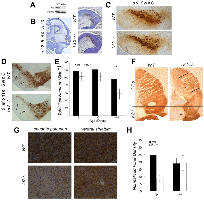
Alterations in locomotor activity are associated with mDA dysfunction in animal models of addiction and neurodegeneration (Giros et al., 1996; Nestler, 2000; Kurosaki et al., 2003). Because Id2 is highly expressed during CNS development and is an important regulator of proliferation and cell-fate specification (Andres-Barquin et al., 2000), we examined the developing midbrain to determine whether Id2 loss affected the establishment of mDA tissues. The Id2 transcript has been detected in the mesencephalic flexure (Lein et al., 2007), known to be rich in midbrain mDA progenitors (Prakash and Wurst, 2006a). We used immunoblotting to verify Id2 protein expression in tissues derived from the ventral midbrain of wild-type embryos at e12.5 (Fig. 2A). Using immunohistochemistry, we evaluated tyrosine hydroxylase (TH)-positive mDA neurons (Fig. 2B) in wild-type and Id2−/− tissues and found that they were histologically indistinguishable at e12.5. Similarly, histological examination of TH-stained mDA neurons of the SNpC obtained from Id2−/− mice at postnatal day 6 (p6) revealed that these tissues were also indistinguishable from wild-type animals (Fig. 2C). These data indicated that Id2 does not have a major role in the establishment of normal mDA neuronal tissues.
Fig. 2.
Age-dependent histological alterations in Id2−/− mice. (A) Protein extracts obtained from micro-dissected midbrain tissue from wild-type (WT) and Id2−/− littermates at e12.5. SDS-PAGE and immunoblotting demonstrate Id2 expression in the developing midbrain, absent in Id2−/− animals. (B) Histological sections obtained from e12.5 embryos cut on the sagittal plane and stained for TH (brown) and counterstained with hematoxylin. Left: low magnification image of wild-type TH-positive progenitors in the mesencephalic flexure. Right: maximized images of boxed zone show TH-positive progenitors in wild-type and Id2−/− mice. (C) Representative coronal sections obtained from 6-day-old wild-type and Id2−/− mice stained for TH (brown) and counterstained with hematoxylin. (D) Morphometrically aligned sections from wild-type and Id2−/− mice at 6 months of age stained with anti-TH antibody (brown) and counterstained with hematoxylin. TH staining of the SNpC is indicated by black arrows (original magnification 10×). (E) Total SNpC cell number manually counted in serial histological sections (6 sections per animal) in 6, 30 and 180-day-old mice stained for TH. Serial sections were cut at 5 μM and counted sections were spaced at 25 μM apart; only nucleated cells were counted; n=8 animals per group; *P=0.023, analyzed using a two-tailed t-test. (F) Sagittal sections obtained from 6-month-old wild-type and Id2−/− mice stained with anti-TH antibodies (brown). Arrows indicate TH-staining intensity within the caudate putamen (CPu). Black line designates the dorsal boundary of the ventral striatum (Vst) (original magnification 10×). (G) Representative brightfield photomicrographs of the TH-stained (brown) histological sections of the CPu and ventral striatum obtained from wild-type and Id2−/− mice counterstained with hematoxylin (original magnification 40×). (H) Densitometric measurements of TH-positive fiber density obtained from the CPu and the ventral striatum (vst) of wild-type and Id2−/− mice.
Photomicrographs obtained at 40× magnification were batch converted to binary, grayscale images. TH-positive fiber density was calculated using ImageJ software. Average density was normalized to background staining in the cortex (n=3 animals per group, three representative areas sampled per individual) *P=0.004, analyzed using a two-tailed t-test. Error bars represent s.e.m.
Despite hyperactivity at 1 and 3 months of age, movement of Id2−/− mice decreased with age (Fig. 1), a hallmark symptom associated with mDA neurodegeneration in PD (Lees et al., 2009). To determine whether the reduced locomotor activity of older Id2−/− mice correlated with alterations in the histology of mDA neurons, we evaluated TH-positive mDA neurons in the SNpC and the VTA in cohorts of wild-type and Id2−/− mice at 6 and 30 days of age, as well as in tissues obtained from the same group of 6-month-old wild-type and Id2−/− mice whose activity was observed to have decreased at later ages. In contrast to younger animals (Fig. 2B,C), we found a readily observable decrease in TH-positive fibers in the SNpC of Id2−/− individuals at 6 months of age (Fig. 2D), correlating with their reduced physical activity (Fig. 1). Although we were unable to rigorously quantify cell numbers using stereologic techniques, manual cell counts in serial sections provided strong evidence of a reduction in TH-positive cells in the SNpC but not within the VTA (Fig. 2D,E and data not shown) observable only at 6 months of age. This observation, suggesting reduced SNpC TH-positive neurons with an apparent sparing of VTA TH-positive neurons, was evaluated within the striatum. TH-positive fibers appeared reduced in the caudate putamen (CPu) of Id2−/− mice but appeared normal within the ventral striatum (Fig. 2F). Densitometric analysis of TH-positive fibers within these regions indicated that reduced fibers were present within the CPu, a region innervated by the SNpC, in Id2−/− mice, but that fiber density was normal within the VTA innervated ventral striatum (Fig. 2G,H). Of particular importance in this regard, we observe that at 6 months of age, which is the most advanced age that these out-bred Id2−/− mice attain before dying with extensive neurologic dysfunction (Figs 1, 2), rare Id2−/− animals that did not exhibit changes in activity also had histologically normal tissues of the SNpC and striatum (data not shown).
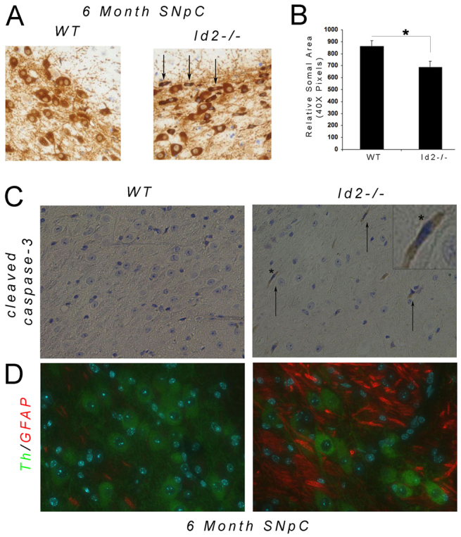
Age-dependent loss of mDA neurons is a defining feature of important neurodegenerative diseases including PD, and therefore we analyzed tissues obtained from Id2−/− mice for additional histological characteristics indicative of neurodegeneration. Analysis of SNpC cytology identified an obvious alteration in the cytoplasmic morphology of TH-positive neurons in Id2−/− mice, characterized by numerous small, dark-staining cell bodies (Fig. 3A, arrows). This cytologic abnormality was associated with a significant overall reduction in average somal area of SNpC TH-positive Id2−/− neurons, as compared with SNpC neurons from wild-type littermates (Fig. 3B). Immunohistochemical staining of adjacent sections for cleaved caspase-3, a marker of apoptosis, revealed cleaved caspase-3-positive cells in the SNpC of Id2−/− mice that had morphologies similar to that of cells identified as having reduced somal areas. These cells did not appear in wild-type mice (Fig. 3C). Areas of noticeably diminished TH-positive fibers and reduced numbers of cell bodies in the SNpC of Id2−/− mice also displayed an increased density of GFAP-positive fibers, a histological characteristic of degenerating neuronal tissue (Fig. 3D) (Przedborski, 2004). Cytologic changes in DA neurons and immunohistological features of neurodegeneration were observed only in Id2−/− animals at 6 months of age and correlate temporally with decreased locomotor activity (Fig. 1).
Fig. 3.

Histological evidence of neurodegeneration in the SNpC of Id2−/− mice. (A) TH-stained histological sections of the SNpC in wild-type and Id2−/− mice at 6 months of age. Black arrows indicate neurons of the SNpC with increased staining intensity and reduced somal volume in Id2−/− mice. (B) Average TH-stained somal area, excluding nuclei, was calculated from 100 neurons per section, three sections per animal (n=3 animals per group). Values indicate means and s.e.m.; *P<0.05, calculated using a two-tailed t-test. (C) Adjacent histological SNpC sections to A, stained for cleaved caspase-3 (brown) and counterstained with hematoxylin. Arrows indicate cleaved caspase-3-positive cells visible in the SNpC of Id2−/− but not wild-type mice. Inset: maximized image of a cleaved caspase-3-positive cell (asterisk) within the SNpC of a representative Id2−/− mouse. (D) Adjacent histological sections to A of the SNpC in wild-type and Id2−/− mice, stained for Th (green) and GFAP (red). Images indicate increased glial fibers present within degenerating tissues of the SNpC in Id2−/− mice.
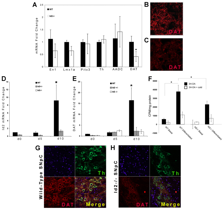
The observation that Id2−/− had a biphasic behavioral phenotype characterized by early-life hyperactivity and decreased activity later led us to look for evidence of molecular changes in younger Id2−/− mice that might affect the functioning and maintenance of mDA tissues. To detect subtle changes in mDA neuron development, we used real-time PCR analysis to study DA lineage-associated gene expression in mRNA obtained from e12.5 ventral midbrain tissues. No differences in early mDA differentiation markers, including En-1 and Lmx1A, or later markers, including Th and Pitx3, were detected (Fig. 4A). However, even at this early age there was decreased expression of DAT (Fig. 4A), a late marker identifying mature mDA neurons (Prakash and Wurst, 2006a). Using immunohistochemistry and confocal microscopy, we observed reduced DAT-positive fibers in the CPu of 6-month-old Id2−/− mice (Fig. 4B,C), a well-characterized feature of PD (Spiegel et al., 2006).
Fig. 4.
Id2 is required for normal levels of DAT expression. (A) Total mRNA was obtained from micro-dissected midbrain tissues of wild-type and Id2−/− mice at e12.5. Real-time PCR analysis was performed in triplicate to determine the expression levels of the DA markers En1, Lmx1a, Pitx3, Th, AADC and DAT. Data was normalized to the expression of β-actin and expressed as relative fold change compared with the average wild-type expression level (n=4 per group); *P<0.05 analyzed using a Bonferroni-corrected t-test for multiple comparisons. (B,C) Representative images of striatal cryosections from wild-type (B) and Id2−/− (C) mice stained with anti-DAT antibody. Fluorescent conjugated secondary antibodies were used for visualization and the images examined by confocal microscopy. The images are representative of an evaluation of 6 animals of each genotype. (D,E) Id2 (D) and DAT (E) expression was examined in wild-type, Id2+/− and Id2−/− NPCs at 0, 5 and 10 days after the induction of DA-directed differentiation. The values shown are from a representative experiment conducted in triplicate; *P<0.04 analyzed using a Bonferroni-corrected t-test for multiple comparisons. (F) DA uptake was assessed in wild-type and Id2−/− cells immediately prior to and following DA-directed differentiation (day 10) as described for D and E. Black bars represent [3H]DA uptake and white bars represent identical experiments conducted in the presence of unlabeled DA competitor. Data were normalized to data obtained from identical assays conducted at 4°C. Three independent experiments were conducted in triplicate; *P<0.05 analyzed using a two-tailed t-test. (G,H) Confocal images of histological sections of the SNpC of 1-month-old wild-type (F) and Id2−/− (G) mice co-immunolabeled for TH (green) and DAT (red) and counterstained with Hoechst 33342 dye (blue). All values indicate means ± s.e.m.
Because the observed reduction in DAT expression at late ages (Fig. 4B) could simply be the result of overall mDA neuronal cell loss (Fig. 2), we sought to determine whether Id2 was important for DAT expression and whether changes in DAT expression preceded the changes in activity or SNpC histology that we observed in Id2−/− mice. We isolated primary neural progenitor cells (NPCs) from wild-type, Id2+/− and Id2−/− animals and analyzed them using well-characterized culture conditions known to support DA differentiation (Lee et al., 2000). We analyzed Id2 and DAT expression prior to the start of differentiation (day 0), following an induction period during which NPCs were exposed to the cytokines sonic hedgehog (Shh) and FGF8b (day 5) and at the end of a maturation period (day 10), as described by others (Yan et al., 2005; Papanikolaou et al., 2008). We found that Id2 expression in wild-type cells, although maintained at basal levels during 5 days of induction, increased more than ninefold following the maturation period of 10 days (Fig. 4D). As expected, under these conditions the expression of DAT mRNA increased almost 20-fold above basal expression levels following 10 days of maturation (Fig. 4E). This expected induction of DAT mRNA at late stages of differentiation was not seen in cells isolated from Id2+/− or Id2−/− mice differentiated under identical conditions (Fig. 4E). We next sought to determine whether the observed reduction in DAT expression was associated with measurable differences in DA uptake. We assayed NPCs isolated from wild-type and Id2−/− mice for the uptake of DA before and after 10 days of DA-directed differentiation. We observed a significant increase in DA uptake in wild-type cells at day 10 of DA-directed differentiation, as compared with undifferentiated cells (Fig. 4F), at the same time point where we observed significant increases in the expression of DAT (Fig. 4E). A significant increase in DA uptake was not observed in Id2−/− NPCs cultured under identical conditions as compared with undifferentiated Id2−/− cells (Fig. 4F). These data provide strong evidence suggesting that Id2 is required for normal levels of DAT expression and function in vitro.
Having observed reduced DAT mRNA during embryonic development in Id2−/− mice (Fig. 4A), and during in vitro differentiation assays (Fig. 4E), we examined the tissue of 30-day-old Id2−/− mice to determine whether DAT expression was suppressed in SNpC DA neurons (Fig. 2) at early ages. Although we consistently observed a histologically normal SNpC in wild-type and Id2−/− mice at 30 days of age (Fig. 2), we noted a reduction in DAT staining intensity in TH-positive SNpC neurons in 30-day-old Id2−/− mice (Fig. 4G,H). These in vitro and in vivo findings indicate that DAT expression is repressed in younger, hyperactive Id2−/− mice prior to observable histological changes in the SNpC.
DISCUSSION
Genetic models of mDA neuron dysfunction should enhance our understanding of the pathology underlying neuropsychiatric disorders, substance abuse and neurodegenerative diseases as well as informing their treatment. Our characterization of the Id2−/− mouse has revealed several important characteristics indicative of alterations in the function of the dopamine system. We previously reported that Id2−/− mice have reduced numbers of DA neurons in the OB, accompanied by reduced olfactory discrimination (Havrda et al., 2008). Here, we report that Id2−/− mice are hyperactive at a young age and undergo age-dependent SNpC cell loss with a concomitant reduction in locomotor activity. This neurodegenerative phenotype occurs subsequent to early-life suppression of DAT, suggesting that inefficient DA re-uptake might contribute to these behavioral and histological changes.
Our findings support a model by which Id2 functions beyond the level of mDA progenitor cells to regulate the maturation and maintenance of DA neurons. Id proteins are highly expressed in the developing CNS and a proliferative function of Id proteins in proliferative, undifferentiated cells has been well described (for reviews see Andres-Barquin et al., 2000; Perk et al., 2005). We previously reported that during adult neurogenesis, Id2 loss diminished the numbers of migrating neuroblasts in the rostral migratory stream, resulting in a highly specific reduction of periglomerular DA neurons. These changes, associated with a diminished OB in Id2−/− mice, occurred in the absence of any detectable loss of SVZ progenitors (Havrda et al., 2008). We interpreted these findings to indicate that Id2 functioned either at later stages of DA neuronal differentiation or in tissue maintenance. Analogous to our previous findings in the adult mouse forebrain, we now find that although Id2 is highly expressed in the developing midbrain there are no obvious defects in the establishment of mDA progenitors. We did find, however, that Id2 loss results in reduced expression of DAT, a late marker of mDA differentiation both in vivo and in vitro, and that Id2 was required for the maintenance of SNpC tissues at advanced ages.
Along with DAT expression being a marker of mature DA neurons, DAT dysfunction is emerging as potential predictor of neurodegenerative and neuropsychiatric disorders. Genetic variations in the DAT locus have been reported to increase the risk of developing PD, in particular following exposure to environmental toxins (Ritz et al., 2009). Attention deficit hyperactivity disorder (ADHD) (Barkley et al., 1990) is characterized by enhanced DA tone and hyperactivity, and an association between DAT polymorphisms and ADHD has been reported (Cook et al., 1995). By dramatically reducing DA reuptake, the DAT−/− mouse recapitulates this pathology of enhanced DA tone and thus is a potential model for understanding the biological underpinnings of these disorders (Gainetdinov et al., 1998). Phenotypic similarities between the Id2−/− mouse and the DAT−/− mouse are easily discernible: olfactory dysfunction (Tillerson et al., 2006; Havrda et al., 2008), increased locomotor activity (Fig. 1) (Giros et al., 1996) and a defect in lactation (Mori et al., 2000; Morice et al., 2004). Because Id2−/− mice model the symptoms of neurologic disorders associated with DAT dysfunction and they share features with the DAT−/− mouse, they represent a novel animal model in which to identify potential pathways of interest underlying neurodegenerative and neuropsychiatric pathologies.
Neurodegeneration occurring in association with DAT inhibition is thought to arise from DA toxicity (Nassogne et al., 1997; Hattoria et al., 2009), a concept likely to be recapitulated by models of repressed DAT expression such as the Id2−/− mouse. Our ongoing characterization of the Id2−/− mouse has identified characteristics shared with PD patients and several different animal models of PD (Dauer and Przedborski, 2003). For example, Id2−/− mice exhibit defects in olfaction (Havrda et al., 2008) and have histopathologic changes associated with degeneration of DA neurons in the midbrain. Specifically, Id2−/− mice have reduced numbers of DA neurons in the SNpC (Fig. 2) and exhibit age-dependent reductions in locomotor activity (Fig. 1), a behavior identified by others (Bracha et al., 1987; Giros et al., 1996; Smits et al., 2008) to reflect these changes. Neurodegenerative diseases, particularly PD, are often associated with bradykinesia (Lees et al., 2009), a symptom manifested in mouse models of DA neuronal loss (Li et al., 2009). Paradoxically, in certain mouse models that exhibit DA neuronal loss such as weaver (Girk2−/−) and Pitx3−/−, the mice are hyperactive (Schmidt et al., 1982; Hwang et al., 2003; Smits et al., 2008), raising the question of whether early-life hyperactivity is a predicator of PD (Walitza et al., 2007).
The early-life hyperactivity that we observed in Id2−/− mice is reminiscent of certain α-synuclein transgenic mouse models of PD. Similar early-life hyperactivity behaviors to those observed in Id2−/− mice are also observed in A53T human α-synuclein transgenic mice in the open-field and elevated plus maze tests (Fig. 1) (Giasson et al., 2002). Also, the hyperactivity observed in the A53T human α-synuclein mouse is associated with DAT repression (Unger et al., 2006). Importantly, others have demonstrated that in these mice overexpressing human α-synuclein, increased locomotor activity is associated with dopamine synaptic dysfunction and precedes the loss of striatal dopamine (Lam et al., 2011). Our findings that in Id2−/− mice the biphasic changes in locomotor activity are associated with early-life DAT repression and later neurodegeneration are consistent with these findings.
Characterization of the function of Id2 in our murine model at advanced ages is compromised by phenotypic co-morbidities including immunologic deficiencies and lactation defects (Yokota et al., 1999; Mori et al., 2000). The defects in lactation could be relevant to a defect in the DA system because prolactin secretion is known to be negatively regulated by DA (Voogt et al., 2001). Analysis of mammary tissues in Id2−/− mice, however, indicates that this defect is unlikely to be related to CNS regulation of prolactin secretion, but rather results from reduced cellular proliferation and viability during lobulo-alveolar development (Mori et al., 2000). Id2−/− mice are also impaired in the development of peripheral lymphoid organs and natural killer cells of the immune system (Yokota et al., 1999). This characteristic of Id2−/− mice is of interest given the potential role of neuroinflammation and innate immunity in the development of PD (Mihara et al., 2008). The histological and neurochemical alterations, consistent with the behavioral changes we observe, are the predominant phenotype of Id2−/− mice at the ages we have analyzed and we reason that symptoms would be increasingly evident at even later ages.
Although we cannot isolate the neurologic effects of Id2 loss at advanced ages using this model system, germ-line deletion of Id2 provides a platform for studying the maintenance of DA neurons as well as potential cell non-autonomous factors contributing to PD, such as defects in innate immunity. Analysis of the Id2−/− mouse will thus be useful in providing insights into the pathological development of movement disorders such as PD, and should contribute to the identification of early biomarkers of disease associated with complex changes such as early-life hyperactivity or preclinical symptoms such as olfactory dysfunction (Wu et al., 2011). Based on these studies, it can be expected that the development of tissue-specific deletion models of Id2, in which confounding non-neurologic alterations are eliminated, will further our understanding of the molecular mechanisms underlying maintenance of mDA neurons and the progression of neurodegenerative disorders.
MATERIALS AND METHODS
Research animals
The development and maintenance of Id2−/− mice has been previously reported (Havrda et al., 2008). Briefly, Id2+/− animals were maintained and bred to produce the desired wild-type and Id2−/− animals. All experimental cohorts were made up of pairs of Id2−/− animals along with gender-matched littermates. Pairs of either gender were analyzed in open-field assays and males were analyzed in the elevated plus maze assay. All animal studies we describe were conducted in accordance with federal guidelines and approved by the Dartmouth Institutional Animal Care and Use Committee.
Behavioral analysis
Spontaneous locomotor activity of wild-type and Id2−/− mice was assayed using the open-field test (Walsh and Cummins, 1976). Mice locomotion was recorded using a digital video camera and analyzed using video tracking software (Anymaze, Stoelting, IL). For each trial, mice were placed in a 1.2-m2 open enclosure for 5 minutes. An identically defined center zone of approximately 0.4 m2 was used for center zone measurements. Mice were tested at 1, 3 and 6 months of age (n=8 per group). Two trials per animal were conducted during each session and overall activity, average speed and rotation were analyzed.
For elevated plus maze analysis, mice were placed into the maze (Columbus Instruments, Columbus, OH) facing the same open arm for each trial, and 5-minute runs were conducted. Naïve, age-matched, male animals were tested once in a cross-sectional manner to eliminate potential conditioned responses resulting from multiple exposures of each mouse to the apparatus. Behavior was captured using digital video recording and tracking as described above. Maze arms were designated zones; zone entries and total time spent in each zone were defined by movement of the entire animal into a zone.
Tissue harvest, immunochemistry, cell counting and densitometry
Embryos were harvested from CO2-euthanized adult wild-type and Id2−/− mice at e12.5 and fixed overnight in buffered formalin. Following behavioral analysis at 6 months of age, adult mice were perfusion-fixed with buffered formalin and their brains dissected and post-fixed overnight. Similar tissue isolation was used for animals at 6 and 30 days of age. Embryos and tissues were either paraffin-embedded or prepared for cryosectioning using standard techniques. Immunostains were conducted using mouse monoclonal anti-tyrosine hydroxylase (anti-TH) antibody (Millipore, Billerica, MA), rat polyclonal anti-DAT (Millipore, Billerica, MA), rabbit polyclonal anti-GFAP (Millipore), and rabbit polyclonal cleaved-caspase-3 (Cell Signaling Technology, Beverly, MA) with antigen-matched blocking peptide (Cell Signaling Technology). For cell counting, serial histological sections throughout the SNpC were obtained from Id2−/− and wild-type mice. Sections cut at 5 μM were spaced at 25 μM intervals, exceeding the diameter of the nucleus in these cells, to eliminate the potential of counting individual cells in multiple sections. Computer-assisted cell counts (ImagePro, MediaCybernetics, Bethesda, MD) of total nucleated TH-positive cells were made in both hemispheres. Three sections of the SNpC and ventral tegmental area (VTA) morphometrically aligned between wild-type and Id2−/− animals were analyzed at 6, 30 and 180 days of age. For somal size measurements, images were captured at 40× magnification and the TH-positive cytoplasm of SNpC neurons was quantified using ImagePro analysis software. Somal measurements were made of TH-positive cells from photomicrographs taken from three individual aligned sections throughout the SNpC, with >150 cells examined per animal. Densitometric measurements were made on photomicrographs obtained from histological sections at 40× magnification. Identically obtained images were batch-converted to binary, grayscale images resulting in TH-positive fiber density appearing as dark gray. Total TH-fiber area, excluding striatal nuclei, was defined using identical computer-assisted image thresholding. TH-positive fiber density was calculated as mean area fraction per image using ImageJ software. Average density was normalized to background staining in photomicrographs obtained from the cortex of each animal examined, with three representative stained areas sampled per individual.
Cell culture and in vitro DA differentiation
NPCs were explanted from the brains of postnatal wild-type, Id2+/− and Id2−/− mice and propagated as described (Reynolds and Weiss, 1992). DA-directed differentiation was induced as previously described (Yan et al., 2005; Papanikolaou et al., 2008). Briefly, cells were plated on poly-lysine and laminin surfaces in an induction medium consisting of DMEM:F12 (50:50) supplemented with N2 (Gibco), laminin (Sigma), 10 ng/ml FGF-2, 100 ng/ml FGF-8b and 500 ng/ml sonic hedgehog (Shh) ligand (R&D Systems). Following induction (5 days), the medium was removed and replaced with maturation medium consisting of the same basal medium lacking growth factors and supplemented with 200 μM ascorbic acid for an additional 5 days.
RNA isolation and real-time PCR analysis
Total RNA was collected from wild-type, Id2+/− and Id2−/− NPCs or tissues using the RNAeasy Kit (Qiagen, Valencia, CA). PCR amplification was monitored in real-time by incorporation of SYBR green (Bio-Rad, Hercules, CA) and authenticated by examination of the PCR product melting profile and appearance following size fractionation by gel electrophoresis. Threshold cycle numbers during the log-phase of amplifications were normalized to the expression of β-actin and cyclophilin. Relative gene expression between experimental groups was determined using the comparative CT (2−ΔΔCT) methods (Schmittgen and Livak, 2008). Primer sequences are available upon request.
[3H]DA uptake assays
DA-uptake assays were conducted using NPCs isolated from wild-type and Id2−/− mice. NPCs were plated in triplicate in 24-well plates (50,000 cells/well) and DA-uptake was assayed under proliferation conditions, or following 10 days of DA-directed differentiation. Cells were rinsed thoroughly with Kreb’s phosphate buffer (KB) and incubated in medium containing either 40 nM [3H]DA or 40 nM [3H]DA and 4 μM unlabeled ‘cold’ DA (Perkin-Elmer, Sigma-Aldrich) and incubated for 5 minutes at either 37°C or 4°C. Following incubation, cells were rinsed thoroughly with KB and lysed in 500 μl of 1% SDS. For protein analysis, 50 μl of total cell lysate was removed from each sample and the remaining 450 μl was added to 5 ml scintillation fluid and the emission read using a liquid scintillation counter. [3H]DA measured at 4°C was subtracted from [3H]DA measured at 37°C and the resultant [3H]DA measurements were normalized to protein content and expressed as counts per minute per mg protein.
Statistical analysis
Paired comparisons were conducted using a two-tailed t-test. For longitudinal studies, comparisons between genotype over time were made using repeated measure ANOVA, with changes at specific time points analyzed using multiple post-hoc Bonferroni-corrected t-tests for multiple comparisons. P-values of less than 0.05 were considered significant.
Acknowledgments
We thank Pierre-Pascale J. Lenck-Santini and Nicholas Shworak (Geisel School of Medicine at Dartmouth, Lebanon, NH) for assistance with behavioral assays. For technical assistance we thank Audrey Nailor, Alison Young, Sarah Purdy-Gilman, Rebecca O’Meara, Eric York and Eve Kemble. For editing, we thank Hermes Yeh, Brian Luikart, Mark Israel, Christopher Dant and Tabatha Richardson.
Footnotes
COMPETING INTERESTS
The authors declare that they do not have any competing or financial interests.
AUTHOR CONTRIBUTIONS
M.C.H. conceived, designed and performed experiments, and wrote the paper. B.R.P., N.M.W. and K.B.H. performed experiments.
FUNDING
This work was supported by an award from Dartmouth SYNERGY: The Center for Clinical and Translational Science ( to M.C.H.), a research grant from The Hitchcock Foundation (to M.C.H.) and a fellowship from the National Institutes of Health (F31NS064634 to B.R.P.).
REFERENCES
- Andres-Barquin P. J., Hernandez M. C., Hayes T. E., McKay R. D., Israel M. A. (1997). Id genes encoding inhibitors of transcription are expressed during in vitro astrocyte differentiation and in cell lines derived from astrocytic tumors. Cancer Res. 57, 215–220 [PubMed] [Google Scholar]
- Andres-Barquin P. J., Hernandez M. C., Israel M. A. (2000). Id genes in nervous system development. Histol. Histopathol. 15, 603–618 [DOI] [PubMed] [Google Scholar]
- Atchley W. R., Fitch W. M. (1997). A natural classification of the basic helix-loop-helix class of transcription factors. Proc. Natl. Acad. Sci. USA 94, 5172–5176 [DOI] [PMC free article] [PubMed] [Google Scholar]
- Barkley R. A., DuPaul G. J., McMurray M. B. (1990). Comprehensive evaluation of attention deficit disorder with and without hyperactivity as defined by research criteria. J. Consult. Clin. Psychol. 58, 775–789 [DOI] [PubMed] [Google Scholar]
- Benezra R., Davis R. L., Lockshon D., Turner D. L., Weintraub H. (1990). The protein Id: a negative regulator of helix-loop-helix DNA binding proteins. Cell 61, 49–59 [DOI] [PubMed] [Google Scholar]
- Bracha H. S., Shults C., Glick S. D., Kleinman J. E. (1987). Spontaneous asymmetric circling behavior in hemi-parkinsonism; a human equivalent of the lesioned-circling rodent behavior. Life Sci. 40, 1127–1130 [DOI] [PubMed] [Google Scholar]
- Chao J., Nestler E. J. (2004). Molecular neurobiology of drug addiction. Annu. Rev. Med. 55, 113–132 [DOI] [PubMed] [Google Scholar]
- Chen X. S., Zhang Y. H., Cai Q. Y., Yao Z. X. (2012). ID2: A negative transcription factor regulating oligodendroglia differentiation. J. Neurosci. Res. 90, 925–932 [DOI] [PubMed] [Google Scholar]
- Cook E. H., Jr, Stein M. A., Krasowski M. D., Cox N. J., Olkon D. M., Kieffer J. E., Leventhal B. L. (1995). Association of attention-deficit disorder and the dopamine transporter gene. Am. J. Hum. Genet. 56, 993–998 [PMC free article] [PubMed] [Google Scholar]
- Dauer W., Przedborski S. (2003). Parkinson’s disease: mechanisms and models. Neuron 39, 889–909 [DOI] [PubMed] [Google Scholar]
- Doty R. L. (2012). Olfaction in Parkinson’s disease and related disorders. Neurobiol. Dis. 46, 527–552 [DOI] [PMC free article] [PubMed] [Google Scholar]
- Gainetdinov R. R., Jones S. R., Fumagalli F., Wightman R. M., Caron M. G. (1998). Re-evaluation of the role of the dopamine transporter in dopamine system homeostasis. Brain Res. Brain Res. Rev. 26, 148–153 [DOI] [PubMed] [Google Scholar]
- Giasson B. I., Duda J. E., Quinn S. M., Zhang B., Trojanowski J. Q., Lee V. M. (2002). Neuronal alpha-synucleinopathy with severe movement disorder in mice expressing A53T human alpha-synuclein. Neuron 34, 521–533 [DOI] [PubMed] [Google Scholar]
- Giros B., Jaber M., Jones S. R., Wightman R. M., Caron M. G. (1996). Hyperlocomotion and indifference to cocaine and amphetamine in mice lacking the dopamine transporter. Nature 379, 606–612 [DOI] [PubMed] [Google Scholar]
- Guillemot F. (1999). Vertebrate bHLH genes and the determination of neuronal fates. Exp. Cell Res. 253, 357–364 [DOI] [PubMed] [Google Scholar]
- Hattoria N., Wanga M., Taka H., Fujimura T., Yoritaka A., Kubo S., Mochizuki H. (2009). Toxic effects of dopamine metabolism in Parkinson’s disease. Parkinsonism Relat. Disord. 15 Suppl. 1, S35–S38 [DOI] [PubMed] [Google Scholar]
- Havrda M. C., Harris B. T., Mantani A., Ward N. M., Paolella B. R., Cuzon V. C., Yeh H. H., Israel M. A. (2008). Id2 is required for specification of dopaminergic neurons during adult olfactory neurogenesis. J. Neurosci. 28, 14074–14086 [DOI] [PMC free article] [PubMed] [Google Scholar]
- Hwang D. Y., Ardayfio P., Kang U. J., Semina E. V., Kim K. S. (2003). Selective loss of dopaminergic neurons in the substantia nigra of Pitx3-deficient aphakia mice. Brain Res. Mol. Brain Res. 114, 123–131 [DOI] [PubMed] [Google Scholar]
- Iversen S. D., Iversen L. L. (2007). Dopamine: 50 years in perspective. Trends Neurosci. 30, 188–193 [DOI] [PubMed] [Google Scholar]
- Kee B. L. (2009). E and ID proteins branch out. Nat. Rev. Immunol. 9, 175–184 [DOI] [PubMed] [Google Scholar]
- Kurosaki R., Muramatsu Y., Watanabe H., Michimata M., Matsubara M., Imai Y., Araki T. (2003). Role of dopamine transporter against MPTP (1-methyl-4-phenyl-1,2,3,6-tetrahydropyridine) neurotoxicity in mice. Metab. Brain Dis. 18, 139–146 [DOI] [PubMed] [Google Scholar]
- Lam H. A., Wu N., Cely I., Kelly R. L., Hean S., Richter F., Magen I., Cepeda C., Ackerson L. C., Walwyn W., et al. (2011). Elevated tonic extracellular dopamine concentration and altered dopamine modulation of synaptic activity precede dopamine loss in the striatum of mice overexpressing human α-synuclein. J. Neurosci. Res. 89, 1091–1102 [DOI] [PMC free article] [PubMed] [Google Scholar]
- Lee S. H., Lumelsky N., Studer L., Auerbach J. M., McKay R. D. (2000). Efficient generation of midbrain and hindbrain neurons from mouse embryonic stem cells. Nat. Biotechnol. 18, 675–679 [DOI] [PubMed] [Google Scholar]
- Lees A. J., Hardy J., Revesz T. (2009). Parkinson’s disease. Lancet 373, 2055–2066 [DOI] [PubMed] [Google Scholar]
- Lein E. S., Hawrylycz M. J., Ao N., Ayres M., Bensinger A., Bernard A., Boe A. F., Boguski M. S., Brockway K. S., Byrnes E. J., et al. (2007). Genome-wide atlas of gene expression in the adult mouse brain. Nature 445, 168–176 [DOI] [PubMed] [Google Scholar]
- Li Y., Liu W., Oo T. F., Wang L., Tang Y., Jackson-Lewis V., Zhou C., Geghman K., Bogdanov M., Przedborski S., et al. (2009). Mutant LRRK2(R1441G) BAC transgenic mice recapitulate cardinal features of Parkinson’s disease. Nat. Neurosci. 12, 826–828 [DOI] [PMC free article] [PubMed] [Google Scholar]
- Mihara T., Nakashima M., Kuroiwa A., Akitake Y., Ono K., Hosokawa M., Yamada T., Takahashi M. (2008). Natural killer cells of Parkinson’s disease patients are set up for activation: a possible role for innate immunity in the pathogenesis of this disease. Parkinsonism Relat. Disord. 14, 46–51 [DOI] [PubMed] [Google Scholar]
- Mori S., Nishikawa S. I., Yokota Y. (2000). Lactation defect in mice lacking the helix-loop-helix inhibitor Id2. EMBO J. 19, 5772–5781 [DOI] [PMC free article] [PubMed] [Google Scholar]
- Morice E., Denis C., Giros B., Nosten-Bertrand M. (2004). Phenotypic expression of the targeted null-mutation in the dopamine transporter gene varies as a function of the genetic background. Eur. J. Neurosci. 20, 120–126 [DOI] [PubMed] [Google Scholar]
- Nassogne M. C., Louahed J., Evrard P., Courtoy P. J. (1997). Cocaine induces apoptosis in cortical neurons of fetal mice. J. Neurochem. 68, 2442–2450 [DOI] [PubMed] [Google Scholar]
- Nestler E. J. (2000). Genes and addiction. Nat. Genet. 26, 277–281 [DOI] [PubMed] [Google Scholar]
- Papanikolaou T., Lennington J. B., Betz A., Figueiredo C., Salamone J. D., Conover J. C. (2008). In vitro generation of dopaminergic neurons from adult subventricular zone neural progenitor cells. Stem Cells Dev. 17, 157–172 [DOI] [PubMed] [Google Scholar]
- Perk J., Iavarone A., Benezra R. (2005). Id family of helix-loop-helix proteins in cancer. Nat. Rev. Cancer 5, 603–614 [DOI] [PubMed] [Google Scholar]
- Pifl C., Giros B., Caron M. G. (1996). The dopamine transporter. The cloned target site of parkinsonism-inducing toxins and of drugs of abuse. Adv. Neurol. 69, 235–238 [PubMed] [Google Scholar]
- Prakash N., Wurst W. (2006a). Development of dopaminergic neurons in the mammalian brain. Cell. Mol. Life Sci. 63, 187–206 [DOI] [PMC free article] [PubMed] [Google Scholar]
- Prakash N., Wurst W. (2006b). Genetic networks controlling the development of midbrain dopaminergic neurons. J. Physiol. 575, 403–410 [DOI] [PMC free article] [PubMed] [Google Scholar]
- Przedborski S. (2004). Clarification: Pathogenic role of glial cells in Parkinson’s disease. Mov. Disord. 19, 118. [DOI] [PubMed] [Google Scholar]
- Reynolds B. A., Weiss S. (1992). Generation of neurons and astrocytes from isolated cells of the adult mammalian central nervous system. Science 255, 1707–1710 [DOI] [PubMed] [Google Scholar]
- Richter A., Ebert U., Nobrega J. N., Vallbacka J. J., Fedrowitz M., Löscher W. (1999). Immunohistochemical and neurochemical studies on nigral and striatal functions in the circling (ci) rat, a genetic animal model with spontaneous rotational behavior. Neuroscience 89, 461–471 [DOI] [PubMed] [Google Scholar]
- Ritz B. R., Manthripragada A. D., Costello S., Lincoln S. J., Farrer M. J., Cockburn M., Bronstein J. (2009). Dopamine transporter genetic variants and pesticides in Parkinson’s disease. Environ. Health Perspect. 117, 964–969 [DOI] [PMC free article] [PubMed] [Google Scholar]
- Ross S. E., Greenberg M. E., Stiles C. D. (2003). Basic helix-loop-helix factors in cortical development. Neuron 39, 13–25 [DOI] [PubMed] [Google Scholar]
- Schmidt M. J., Sawyer B. D., Perry K. W., Fuller R. W., Foreman M. M., Ghetti B. (1982). Dopamine deficiency in the weaver mutant mouse. J. Neurosci. 2, 376–380 [DOI] [PMC free article] [PubMed] [Google Scholar]
- Schmittgen T. D., Livak K. J. (2008). Analyzing real-time PCR data by the comparative C(T) method. Nat. Protoc. 3, 1101–1108 [DOI] [PubMed] [Google Scholar]
- Smits S. M., Noorlander C. W., Kas M. J., Ramakers G. M., Smidt M. P. (2008). Alterations in serotonin signalling are involved in the hyperactivity of Pitx3-deficient mice. Eur. J. Neurosci. 27, 388–395 [DOI] [PubMed] [Google Scholar]
- Spiegel J., Storch A., Jost W. H. (2006). Early diagnosis of Parkinson’s disease. J. Neurol. 253 Suppl. 4, iv2–iv7 [DOI] [PubMed] [Google Scholar]
- Tillerson J. L., Caudle W. M., Parent J. M., Gong C., Schallert T., Miller G. W. (2006). Olfactory discrimination deficits in mice lacking the dopamine transporter or the D2 dopamine receptor. Behav. Brain Res. 172, 97–105 [DOI] [PubMed] [Google Scholar]
- Unger E. L., Eve D. J., Perez X. A., Reichenbach D. K., Xu Y., Lee M. K., Andrews A. M. (2006). Locomotor hyperactivity and alterations in dopamine neurotransmission are associated with overexpression of A53T mutant human alpha-synuclein in mice. Neurobiol. Dis. 21, 431–443 [DOI] [PubMed] [Google Scholar]
- Voogt J. L., Lee Y., Yang S., Arbogast L. (2001). Regulation of prolactin secretion during pregnancy and lactation. Prog. Brain Res. 133, 173–185 [DOI] [PubMed] [Google Scholar]
- Walitza S., Melfsen S., Herhaus G., Scheuerpflug P., Warnke A., Müller T., Lange K. W., Gerlach M. (2007). Association of Parkinson’s disease with symptoms of attention deficit hyperactivity disorder in childhood. J. Neural Transm. Suppl. 2007, 311–315 [DOI] [PubMed] [Google Scholar]
- Walsh R. N., Cummins R. A. (1976). The open-field test: a critical review. Psychol. Bull. 83, 482–504 [PubMed] [Google Scholar]
- Wu Y., Le W., Jankovic J. (2011). Preclinical biomarkers of Parkinson disease. Arch. Neurol. 68, 22–30 [DOI] [PubMed] [Google Scholar]
- Yan Y., Yang D., Zarnowska E. D., Du Z., Werbel B., Valliere C., Pearce R. A., Thomson J. A., Zhang S. C. (2005). Directed differentiation of dopaminergic neuronal subtypes from human embryonic stem cells. Stem Cells 23, 781–790 [DOI] [PMC free article] [PubMed] [Google Scholar]
- Yokota Y., Mansouri A., Mori S., Sugawara S., Adachi S., Nishikawa S., Gruss P. (1999). Development of peripheral lymphoid organs and natural killer cells depends on the helix-loop-helix inhibitor Id2. Nature 397, 702–706 [DOI] [PubMed] [Google Scholar]


