Abstract
Glucose variability predicts hypoglycemia in both type 1 and type 2 diabetes and has consistently been related to mortality in nondiabetic patients in the intensive care unit. SD and mean amplitude of glycemic excursions have historically been very popular measures of glucose variability. For reasons outlined in this counterpoint, I propose to use coefficient of variation and the mean absolute glucose change as preferred measures of glucose variability.
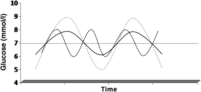
There is little consensus on the importance of glucose variability (1). In order to recognize glucose variability as an independent risk factor or a causal factor in the development of microvascular and macrovascular complications or mortality, glucose variability should be deleterious above the harmful effects of both hyperglycemia and hypoglycemia. In other words, the question is whether larger or more frequent glucose excursions within the euglycemic range are more harmful than small, less frequent glucose excursions, which are also seen in healthy individuals and are therefore likely to be of no consequence (or could theoretically even be beneficial) (Fig. 1). Correction for mean glucose of any relation between glucose variability and a given outcome is important, as high correlations between glucose variability and mean glucose have been demonstrated (2,3). This also makes sense from a clinical point of view, as higher glucose values open up space for more glucose variability.
FIG. 1.
Visualization of glucose variability. Solid line: a given excursion. Dashed line: higher glucose variability due to a higher frequency of oscillation. Dotted line: higher glucose variability due to a larger amplitude. Note that the mean and area under the curve are identical in the three situations.
The importance of glucose variability
Glucose variability has been identified as a predictor of hypoglycemia and has been found to be related to intensive care unit mortality. Other putative relations are between glucose variability and oxidative stress, as well as microvascular and macrovascular complications of diabetes. With regard to prediction of hypoglycemia, glucose variability has been shown predictive of severe hypoglycemia in type 1 diabetes (4,5) and of nonsevere hypoglycemia in type 2 diabetes (6–8). The relation of glucose variability to intensive care mortality was shown in nondiabetic patients with stress hyperglycemia in at least 12 independent cohorts, which have been meta-analyzed (9). The relation between glucose variability and oxidative stress, quantified as urinary 8-isoprostanes by radioimmunoassay, was first reported in patients with type 2 diabetes in poor control on oral agents (r = 0.86, P < 0.001) (10). Surprisingly (3), no relation between mean amplitude of glycemic excursions (MAGE) and mean glucose was found in this study. Our group could not confirm a relation between glucose variability and 8-isoprostanes, now measured by tandem mass spectroscopy, in patients with type 2 diabetes on oral agents with good control (11). Subsequently, Monnier et al. (12) extended their investigations to a larger sample of patients with type 2 diabetes with poor glycemic control, finding a weaker relationship (r = 0.386). Of note, this relation was restricted to those with an HbA1c above the median of the cohort, 8.2%. Again, no relation between MAGE and mean glucose was seen. Neither Monnier et al. nor we found a relation between oxidative stress and glucose variability in type 1 diabetic patients, although glucose variability is larger in this patient group (12,13).
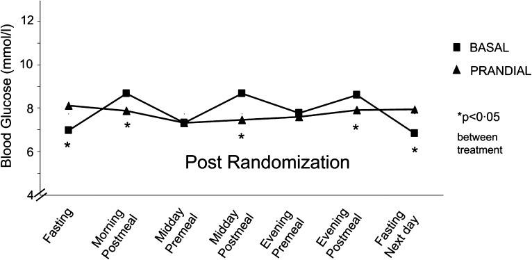
As to microvascular complications in type 1 diabetes, analyses of the Diabetes Control and Complications Trial (DCCT) dataset did not reveal any relation between microvascular complications and glucose variability above mean glucose, for retinopathy, neuropathy, or nephropathy (14,15). Other, much smaller datasets reported relationships between glucose variability and microvascular complications (16) and coronary artery content (17), although a larger study found no relation between glucose variability and a composite score for cardiovascular risk (18). There is a lack of intervention studies specifically aiming at reducing glucose variability without affecting mean glucose. In a reanalysis of the HEART2D (Hyperglycemia and Its Effect After Acute Myocardial Infarction on Cardiovascular Outcomes in Patients With Type 2 Diabetes Mellitus) study (19), in which glucose variability is notably lower in the mealtime-related insulin arm than in the basal insulin arm (Fig. 2), we could find a difference in one measure of glucose variability, but this did not translate into a difference in mortality rates between the two groups (20).
FIG. 2.
Glucose profiles from the prandial (triangles) and basal (squares) insulin groups in HEART2D. Reproduced with permission from Siegelaar et al. (20).
How to measure glucose variability?
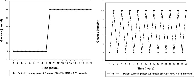
There are many measures of variability, reviewed elsewhere (21). Rather than commenting on all of them, I would like to make a few salient points. The most widely used method to assess variability is SD. Actually, this is a measure of dispersion rather than a measure of glucose variability (Fig. 3). It shows a linear relationship with mean glucose, with an r of 0.56 (3). When corrected for the mean by dividing it through the mean, the coefficient of variation is obtained and the linear relationship with mean glucose largely disappears (3). Although glucose values in a typical dataset are usually not completely normally distributed, SD remains a fairly robust measure because a linear relation has been established between interquartile range and SD (22). Therefore transformation of the data to improve precision can be done but doesn’t seem obligatory.
FIG. 3.
Two fictitious patients with identical mean and SD of glucose throughout a 20-h period, but markedly different glucose variability. The MAG change of both patients differs by a factor 19. Reproduced with permission from Hermanides et al. (27).
MAGE was designed to capture mealtime-related glucose excursions. To separate mealtime-induced from other glucose excursions, investigations were done in healthy volunteers, and it was found that excursions larger than 1.0 SD of the glucose measurements taken were consistently mealtime related. From this, it follows that postprandial excursions and glucose variability are closely related, the former contributing to the latter, but also that they are not identical. MAGE has been criticized on five main points. First, with the advent of continuous glucose monitoring, postprandial excursions can be assessed much more precisely by using the area under the curve as assessed by the trapezoidal method. Second, calculation of MAGE is operator dependent (3,21,23,24) and not defined unambiguously. The criterion that both the ascending and descending limb of an excursion must be larger than 1 SD to qualify as an excursion, while then only the ascending or the descending limb is used for calculation of MAGE, depending on the direction of the first excursion, has especially confused investigators and resulted in erroneous calculations. Computerized determination, rather than using ruler and pencil, has been recommended (23), but different methods do not give the same outcome (M. Sechterberger, unpublished results). Third, the outcome differs depending on whether ascending or descending limbs are used for calculation of MAGE, the difference being ∼7% (23,25). Fourth, there is a high correlation with SD—in one study up to 0.9 (21). Fifth and last, it is questionable whether only mealtime excursions or excursions larger than 1.0 SD would have clinical importance (23). Indeed, some excursions that would seem quantitatively relevant are not taken in to account. An example is seen in Fig. 2 of the accompanying point article (25), when the excursion following the inflection at 156 mg/dL does not qualify.
With the availability of continuous glucose monitoring, investigators were confronted with much more variability than previously surmised. Rather than seeing variability as predominantly related to postprandial glucose excursions, they sought to measure glucose variability in a mathematically sound way, taking all variability into account. Continuous overall net glycemic action is an example of such a new measure. Starting at the intensive care unit, where glucose variability is also less related to meals, we proposed the mean absolute glucose (MAG) change (26,27). This is a simple summation of all absolute changes in glucose, divided by the time over which measurements were taken (Fig. 3). In this way, two excursions of identical extent but differing in duration contribute differently to the overall sum of variability. These new measures did not start with healthy populations, but of course reference values in healthy populations could easily be obtained. A downside of variability measures that take all variation into account is that noise, a particular problem in continuous glucose monitoring, is hard to separate from the signal, although efforts in this direction are being made (28).
Is it possible to make a statement on which measure of variability is to be preferred? To do so, we can look at which measures show the strongest relation to a given outcome, provided the relationship between glucose variability and that particular outcome is well established. As explained above, this can only be said of the relation between glucose variability and intensive care unit mortality and of glucose variability as a predictor of hypoglycemia. We showed that MAG had a stronger relation to intensive care unit mortality than SD, with a 5.4% larger area under the receiver operator curve (27), although another study found SD to have a stronger relation with mortality than MAG (29). We recently used a general boosted model to investigate the predictive power of different measures of glucose variability for hypoglycemia. Coefficient of variation showed the highest relative influence on hypoglycemia rate, >40%, followed by MAG contributing with somewhat more than 10%, although not statistically significant, and contributions below 5% of average daily risk range (ADRR), SD, and MAGE (8). In HEART2D, where the difference in variability between the two study arms was apparent to the naked eye (Fig. 2), this could only be confirmed with statistical significance by MAG, whereas MAGE and SD, although numerically higher in the basal insulin arm, did not indicate a significant difference.
In conclusion, when it comes to assessing postprandial excursions, the area under the curve is the preferred method. When diabetes investigators want to assess glucose variability, I would recommend coefficient of variation and MAG.
ACKNOWLEDGMENTS
No potential conflicts of interest relevant to this article were reported.
Footnotes
See accompanying article, p. 1398.
REFERENCES
- 1.Siegelaar SE, Holleman F, Hoekstra JB, DeVries JH. Glucose variability; does it matter? Endocr Rev 2010;31:171–182 [DOI] [PubMed] [Google Scholar]
- 2.Borg R, Kuenen JC, Carstensen B, et al. ; ADAG Study Group Associations between features of glucose exposure and A1C: the A1C-Derived Average Glucose (ADAG) study. Diabetes 2010;59:1585–1590 [DOI] [PMC free article] [PubMed] [Google Scholar]
- 3.Rodbard D. Clinical interpretation of indices of quality of glycemic control and glycemic variability. Postgrad Med 2011;123:107–118 [DOI] [PubMed] [Google Scholar]
- 4.Cox DJ, Kovatchev BP, Julian DM, et al. Frequency of severe hypoglycemia in insulin-dependent diabetes mellitus can be predicted from self-monitoring blood glucose data. J Clin Endocrinol Metab 1994;79:1659–1662 [DOI] [PubMed] [Google Scholar]
- 5.Kilpatrick ES, Rigby AS, Goode K, Atkin SL. Relating mean blood glucose and glucose variability to the risk of multiple episodes of hypoglycaemia in type 1 diabetes. Diabetologia 2007;50:2553–2561 [DOI] [PubMed] [Google Scholar]
- 6.Murata GH, Hoffman RM, Shah JH, Wendel CS, Duckworth WC. A probabilistic model for predicting hypoglycemia in type 2 diabetes mellitus: The Diabetes Outcomes in Veterans Study (DOVES). Arch Intern Med 2004;164:1445–1450 [DOI] [PubMed] [Google Scholar]
- 7.Monnier L, Wojtusciszyn A, Colette C, Owens D. The contribution of glucose variability to asymptomatic hypoglycemia in persons with type 2 diabetes. Diabetes Technol Ther 2011;13:813–818 [DOI] [PubMed] [Google Scholar]
- 8.Qu Y, Jacober SJ, Zhang Q, Wolka LL, DeVries JH. Rate of hypoglycemia in insulin-treated patients with type 2 diabetes can be predicted from glycemic variability data. Diabetes Technol Ther 2012;14:1008–1012 [DOI] [PubMed] [Google Scholar]
- 9.Eslami S, Taherzadeh Z, Schultz MJ, Abu-Hanna A. Glucose variability measures and their effect on mortality: a systematic review. Intensive Care Med 2011;37:583–593 [DOI] [PMC free article] [PubMed] [Google Scholar]
- 10.Monnier L, Mas E, Ginet C, et al. Activation of oxidative stress by acute glucose fluctuations compared with sustained chronic hyperglycemia in patients with type 2 diabetes. JAMA 2006;295:1681–1687 [DOI] [PubMed] [Google Scholar]
- 11.Siegelaar SE, Barwari T, Kulik W, Hoekstra JB, DeVries JH. No relevant relationship between glucose variability and oxidative stress in well-regulated type 2 diabetes patients. J Diabetes Sci Tech 2011;5:86–92 [DOI] [PMC free article] [PubMed] [Google Scholar]
- 12.Monnier L, Colette C, Mas E, et al. Regulation of oxidative stress by glycaemic control: evidence for an independent inhibitory effect of insulin therapy. Diabetologia 2010;53:562–571 [DOI] [PubMed] [Google Scholar]
- 13.Wentholt IM, Kulik W, Michels RP, Hoekstra JB, DeVries JH. Glucose fluctuations and activation of oxidative stress in patients with type 1 diabetes. Diabetologia 2008;51:183–190 [DOI] [PubMed] [Google Scholar]
- 14.Kilpatrick ES, Rigby AS, Atkin SL. The effect of glucose variability on the risk of microvascular complications in type 1 diabetes. Diabetes Care 2006;29:1486–1490 [DOI] [PubMed] [Google Scholar]
- 15.Siegelaar SE, Kilpatrick ES, Rigby AS, Atkin SL, Hoekstra JB, DeVries JH. Glucose variability does not contribute to the development of peripheral and autonomic neuropathy in type 1 diabetes: data from the DCCT. Diabetologia 2009;52:2229–2232 [DOI] [PMC free article] [PubMed] [Google Scholar]
- 16.Bragd J, Adamson U, Bäcklund LB, Lins PE, Moberg E, Oskarsson P. Can glycaemic variability, as calculated from blood glucose self-monitoring, predict the development of complications in type 1 diabetes over a decade? Diabetes Metab 2008;34:612–616 [DOI] [PubMed] [Google Scholar]
- 17.Snell-Bergeon JK, Roman R, Rodbard D, et al. Glycaemic variability is associated with coronary artery calcium in men with Type 1 diabetes: the Coronary Artery Calcification in Type 1 Diabetes study. Diabet Med 2010;27:1436–1442 [DOI] [PMC free article] [PubMed] [Google Scholar]
- 18.Borg R, Kuenen JC, Carstensen B, et al. ; ADAG Study Group HbA₁(c) and mean blood glucose show stronger associations with cardiovascular disease risk factors than do postprandial glycaemia or glucose variability in persons with diabetes: the A1C-Derived Average Glucose (ADAG) study. Diabetologia 2011;54:69–72 [DOI] [PMC free article] [PubMed] [Google Scholar]
- 19.Raz I, Wilson PW, Strojek K, et al. Effects of prandial versus fasting glycemia on cardiovascular outcomes in type 2 diabetes: the HEART2D trial. Diabetes Care 2009;32:381–386 [DOI] [PMC free article] [PubMed] [Google Scholar]
- 20.Siegelaar SE, Kerr L, Jacober SJ, DeVries JH. A decrease in glucose variability does not reduce cardiovascular event rates in type 2 diabetic patients after acute myocardial infarction: a reanalysis of the HEART2D study. Diabetes Care 2011;34:855–857 [DOI] [PMC free article] [PubMed] [Google Scholar]
- 21.Rodbard D. Interpretation of continuous glucose monitoring data: glycemic variability and quality of glycemic control. Diabetes Technol Ther 2009;11(Suppl. 1):S55–S67 [DOI] [PubMed] [Google Scholar]
- 22.Rodbard D. Optimizing display, analysis, interpretation and utility of self-monitoring of blood glucose (SMBG) data for management of patients with diabetes. J Diabetes Sci Tech 2007;1:62–71 [DOI] [PMC free article] [PubMed] [Google Scholar]
- 23.Baghurst PA. Calculating the mean amplitude of glycemic excursion from continuous glucose monitoring data: an automated algorithm. Diabetes Technol Ther 2011;13:296–302 [DOI] [PubMed] [Google Scholar]
- 24.Rodbard D. New and improved methods to characterize glycemic variability using continuous glucose monitoring. Diabetes Technol Ther 2009;11:551–565 [DOI] [PubMed] [Google Scholar]
- 25.Service FJ. Glucose variability. Diabetes 2013;62:1398–1404 [DOI] [PMC free article] [PubMed] [Google Scholar]
- 26.McDonnell CM, Donath SM, Vidmar SI, Werther GA, Cameron FJ. A novel approach to continuous glucose analysis utilizing glycemic variation. Diabetes Technol Ther 2005;7:253–263 [DOI] [PubMed] [Google Scholar]
- 27.Hermanides J, Vriesendorp TM, Bosman RJ, Zandstra DF, Hoekstra JB, DeVries JH. Glucose variability is associated with intensive care unit mortality. Crit Care Med 2010;38:838–842 [DOI] [PubMed] [Google Scholar]
- 28.Facchinetti A, Sparacino G, Guerra S, et al. Real-time improvement of continuous glucose monitoring accuracy: the smart sensor concept. Diabetes Care 2013;36:793–800 [DOI] [PMC free article] [PubMed] [Google Scholar]
- 29.Meynaar IA, Eslami S, Abu-Hanna A, van der Voort P, de Lange DW, de Keizer N. Blood glucose amplitude variability as predictor for mortality in surgical and medical intensive care unit patients: a multicenter cohort study. J Crit Care 2012;27:119–124 [DOI] [PubMed] [Google Scholar]


