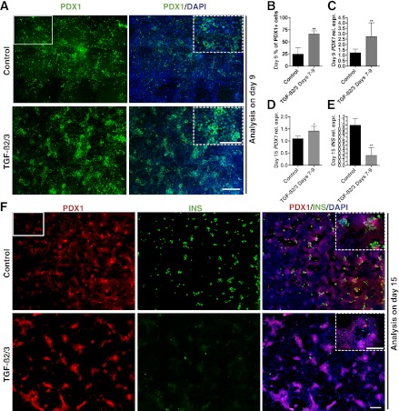
FIG. 4.
TGF-β signaling is important for efficient induction of pancreatic progenitors but prohibits endocrine differentiation. A: Immunostaining of PDX1 reveals induction of pancreatic progenitor cells in control and TGF-β2/3–treated samples. Each image is a composite of nine individual images at original magnification ×10 (representative ×10 image is shown as white rectangle) to cover a representative area of the culture (higher magnification pictures is marked by dotted lines). B: Quantification of PDX1-positive cells in day 9 samples with and without TGF-β2/3 treatment. Total number of cells counted for each condition is ≥10,000. C: Q-PCR showing day 9 PDX1 expression levels in control and TGF-β2/3-treated samples (n = 2 independent experiments). D: Q-PCR analysis of day 15 PDX1 expression levels in control and TGF-β2/3-treated samples (n = 3 independent experiments). E: Q-PCR analysis of day 15 INS expression levels in control and TGF-β2/3–treated samples (n = 3 independent experiments). F: Immunostaining of PDX1 and insulin in day 15 samples shows induction of endocrine cells in control but not in TGF-β2/3–treated samples. Each image is a composite of 20 individual original magnification ×10 images to cover a representative area of the culture (representative ×10 image is shown as white rectangle). Insets show higher magnification view. B–E: Statistical analysis was performed using the Student t test. *P < 0.05; **P < 0.01. A–F: Scale bars = 400 μm for all images. A and F insets: Scale bars = 200 μm.

