Abstract
Lipoma is the commonest benign tumor occurring at any anatomical site, where fat is present. In oral cavity and oropharynx, it is a relatively uncommon neoplasm. Tongue, which is totally devoid of fat cell is also a site for lipoma but very rarely. We report one such rare case of the universal tumor, of 20 years of duration and 9 cm in size, presenting at the lateral margin, dorsal and ventral surface of the tongue, for which complete tumor excision was done.
Keywords: Lipoma, tongue, duration of tumor
Introduction
The benign fatty tumor, the lipoma, is composed of adult fat cells that are subdivided into lobules by septae of fibrous connective tissue. It appears most frequently in the subcutis of adults and is histologically indistinguishable from normal adipose tissue. The metabolism of the lipoma differs from that of the normal adipose tissue. It has been shown that the fat of lipoma is not used for energy production during starvation periods as happens with normal adipose tissue. Although lipomas are common in many parts of the body, they are infrequently found in the oral cavity.
Case Report
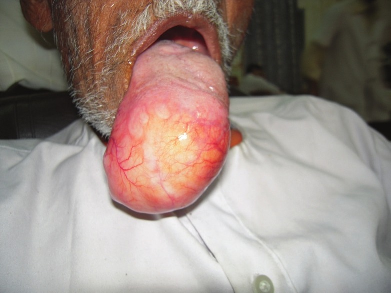
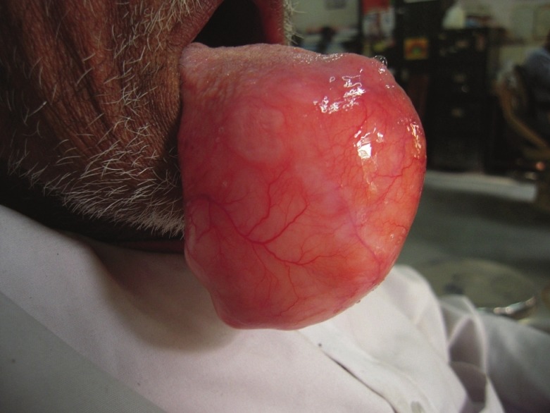
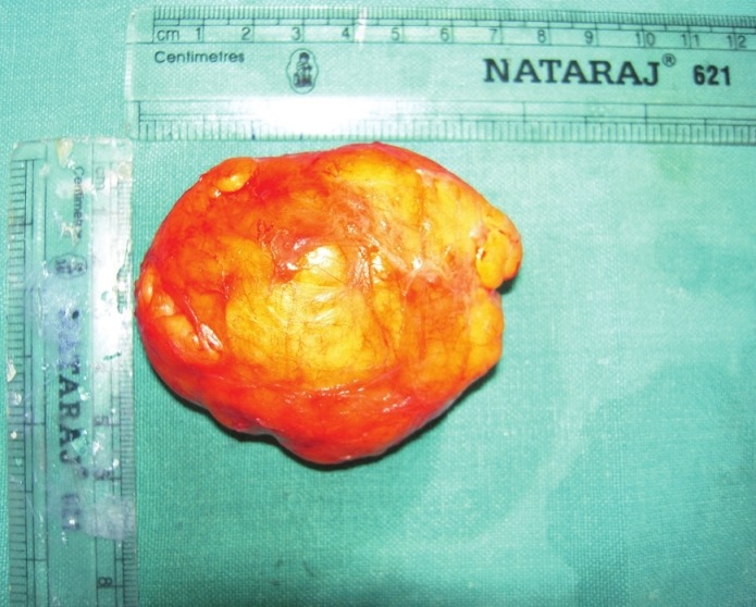
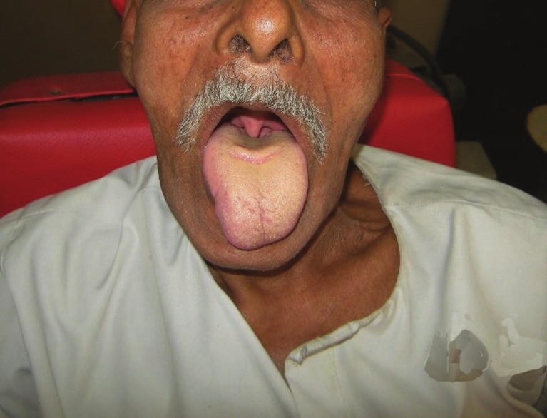
A 75-year-old man was referred to our department with a tumor tongue, which he had first noticed approximately 16 years earlier. His medical history was non-contributory. The mass measured 9 × 8 cm and was extending from dorsal to ventral surface supero-inferiorly and right to left lateral border of anterior third of tongue [Figures 1 and 2]. The mass was painless, firm, yellowish in color with multiple engorged blood vessels over its surface. Patient had difficulty in mastication, swallowing. He frequently used to get up from sleep because of obstruction in airway. The lesion was excised under general anesthesia. The tumor was well encapsulated [Figure 3]. The mucosal layers were closed together with absorbable sutures obliterating the dead space. He made an uneventful recovery from the surgery [Figure 4].
Figure 1.

Front view of patient
Figure 2.

Profile view of patient showing tumor from the tip and ventral surface of the tongue
Figure 3.

Surgical specimen
Figure 4.

Appearance of tongue 6 months postoperatively
Macroscopic examination showed a single mass of tissue covered with adipose tissue, 9 × 8 × 6 cm and weighting 56 g. The cut surface showed fatty tissue. Microscopically the sections showed sheets of mature adipocytes and extravasated red blood cells. The tumor cells were arranged in lobules. The features were consistent with a lipoma. The patient has since regained normal speech and feeding capacity with no loss of sensory or motor functions of the tongue. The tongue now fits comfortably in the mouth without obstructing the airway.
Discussion
Lipomas are common tumors in the human body, but are less frequent in the oral cavity, comprising no more than 1-5% of all the neoplasms.[1] They commonly present as slow growing asymptomatic lesions with a characteristic yellowish color and soft, doughy feel, in the buccal mucosa, floor of the mouth and tongue in the fourth and fifth decade generally without gender.
Predilection.[2–4] Some studies, however, have shown a male preponderance.[4] They may present as solitary or multiple lesions, for instance, as in Gardner's or Bournville's syndrome[5] or as macroglosia.[5–9] Their clinical course is usually asymptomatic until they grow to large sizes.[5] The majority remain unulcerated. When they are ulcerated, they present diagnostic problems. This was noted by a report of a lipoma that presented as a non-healing ulcer. In the present case, the large size interfered with speech and mastication, similar to a case reported by Gray and Barker[5] and also difficulty in respiration. The average duration of the lipoma before excision is 3.2 years with a range of 6 weeks to 15 years.[4] The usual range in size is 0.5-8 cm.[4] The present case was 10 cm in diameter.
The differential diagnosis includes ranula, dermoid cyst, thyroglossal duct cyst, ectopic thyroid tissue, pleomorphic adenoma and mucoepidermoid carcinoma, angiolipoma, fibrolipoma and malignant lymphoma.[6–9] The definitive diagnosis is by microscopic examination, which shows adult fat tissue cells embedded in a stroma of connective tissue and surrounded by a fibrous capsule.[9] Lipoma has a characteristic radiographic appearance. On Computed Tomography (CT) scan it shows a high density from 83 to 143 Hounsfield units with well or poorly defined margins depending on the capsule.
Ultrasonography shows a lesion, which is around or elliptical in shape with intact or mostly intact capsule. Most lipomas are hypoechoic with echogenic lines or spots.[10] Surgical excision is the mainstay of treatment.[4] Recurrence is reduced by wide surgical excision at the same time preserving the surrounding structures. Well encapsulated lipomas, as the present case, easily shell out with no possibility of recurrence or damage to the surrounding structures. It is still advisable to excise them with a little cuff of surrounding normal tissue to prevent recurrence but still conserving surrounding structures.[3] Infiltrating lipomas are difficult to extirpate and when multiple are liable to recurrence due to difficulty in adequate excision. Recurrence rate of as high as 62.5% has been recorded.[2] Simple lipomas regardless of their size are easily extirpated without recurrence. This unusually huge tongue lipoma was surgically excised uneventfully. The present case demonstrates the size lipomas can grow to, if untreated.
Footnotes
Source of Support: Nil,
Conflict of Interest: None declared
References
- 1.Capodiferro S, Scully C, Maiorano E, Lo Muzio L, Favia G. Liposarcoma circumscriptum (lipoma-like) of the tongue: Report of a case. Oral Dis. 2004;10:398–400. doi: 10.1111/j.1601-0825.2004.01040.x. [DOI] [PubMed] [Google Scholar]
- 2.Dattilo DJ, Ige T, Nwana EJ. Intraoral lipoma of the tongue and submandibular space: Report of a case. J Oral Maxillofac Surg. 1996;54:915–7. doi: 10.1016/s0278-2391(96)90549-2. [DOI] [PubMed] [Google Scholar]
- 3.Fregnani ER, Pires FR, Falzoni R, Lopes MA, Vargas PA. Lipomas of the oral cavity: Clinical findings, histological classification and proliferative activity of 46 cases. Int J Oral Maxillofac Surg. 2003;32:49–53. doi: 10.1054/ijom.2002.0317. [DOI] [PubMed] [Google Scholar]
- 4.Furlong MA, Fanburg-Smith JC, Childers EL. Lipoma of the oral and maxillofacial region: Site and subclassification of 125 cases. Oral Surg Oral Med Oral Pathol Oral Radiol Endod. 2004;98:441–50. doi: 10.1016/j.tripleo.2004.02.071. [DOI] [PubMed] [Google Scholar]
- 5.Favia G, Maiorano E, Orsini G, Piattelli A. Myxoid liposarcoma of the oral cavity with involvement of the periodontal tissues. J Clin Periodontol. 2001;28:109–12. doi: 10.1034/j.1600-051x.2001.028002109.x. [DOI] [PubMed] [Google Scholar]
- 6.Gray AR, Barker GR. Sublingual lipoma: Report of an unusually large lesion. J Oral Maxillofac Surg. 1991;49:747–50. doi: 10.1016/s0278-2391(10)80241-1. [DOI] [PubMed] [Google Scholar]
- 7.Moore PL, Goede A, Phillips DE, Carr R. Atypical lipoma of the tongue. J Laryngol Otol. 2001;115:859–61. doi: 10.1258/0022215011909198. [DOI] [PubMed] [Google Scholar]
- 8.Nunes FD, Loducca SV, de Oliveira EM, de Araujo VC. Well-differentiated liposarcoma of the tongue. Oral Oncol. 2002;38:117–9. doi: 10.1016/s1368-8375(01)00030-6. [DOI] [PubMed] [Google Scholar]
- 9.Piattelli A, Rubini C, Fioroni M, Iezzi G. Spindle-cell lipoma of the cheek: A case report. Oral Oncol. 2000;36:495–6. doi: 10.1016/s1368-8375(00)00035-x. [DOI] [PubMed] [Google Scholar]
- 10.Zhong LP, Zhao SF, Chen GF, Ping FY. Ultrasonographic appearance of lipoma in the oral and maxillofacial region. Oral Surg Oral Med Oral Pathol Oral Radiol Endod. 2004;98:738–40. doi: 10.1016/j.tripleo.2004.04.022. [DOI] [PubMed] [Google Scholar]


