Abstract
Frenal attachments are thin folds of mucous membrane with enclosed muscle fibers that attach the lips to the alveolar mucosa and underlying periosteum. Most often, during the oral examination of the patient the dentist gives very little importance to the frenum, for assessing its morpholology and attachment. However, it has been seen that an abnormal frenum can be an indicator of a syndrome. This paper highlights the different frenal attachments seen in association with various syndromic as well as non-syndromic conditions.
Keywords: Aberrant frena, frenulum, syndromes
INTRODUCTION
One of the more interesting yet often misunderstood anatomic structures in the oral cavity is the frenum- a mucosal attachment of a loose part to a more rigid part.
A frenulum is a small frenum.[1] There are several frena that are usually present in a normal oral cavity, most notably the maxillary labial frenum, the mandibular labial frenum, and the lingual frenum.
Their primary function is to provide stability of the upper and lower lip and the tongue.[1] The extent of their involvement in mastication is in dispute. Labial frenal attachments are thin folds of mucous membrane with enclosed muscle fibers originating from orbicularis oris muscle of upper lip that attach at the lips to the alveolar mucosa and underlying periosteum.[2] It extends over the alveolar process in infants and forms a raphe that reaches the palatal papilla. Through the growth of alveolar process as the teeth erupt, this attachment generally changes to assume the adult configuration.
CLASSIFICATION
Depending upon the extension of attachment of fibers, frena have been classified as:[3]
Mucosal – when the frenal fibers are attached up to mucogingival junction [Figure 1]
Gingival – when fibres are inserted within attached gingiva [Figure 2]
Papillary – when fibres are extending into inter dental papilla; [Figure 3] and
Papilla penetrating – when the frenal fibres cross the alveolar process and extend up to palatine papilla [Figure 4].
Figure 1.
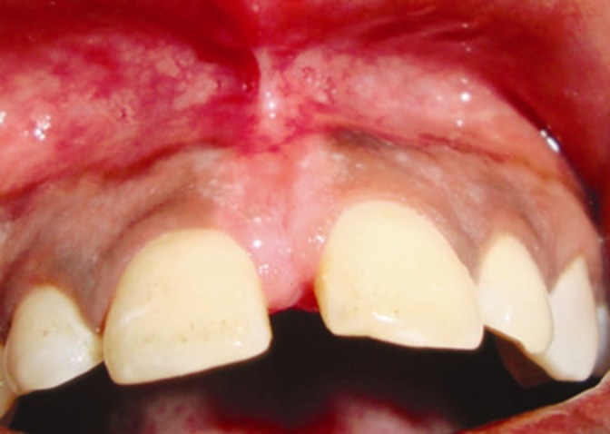
Mucosal frenal attachment
Figure 2.
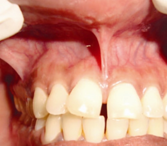
Gingival frenal attachment
Figure 3.
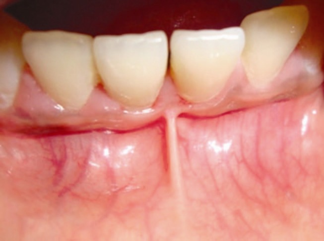
Papillary frenal attachment
Figure 4.
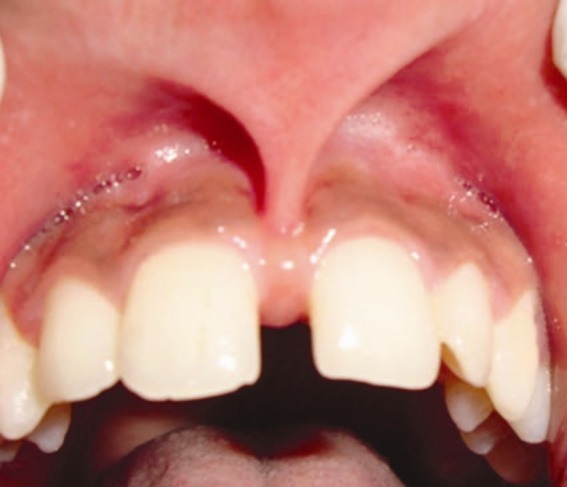
Papilla penetrating frenal attachment
Other variations of normal frenal attachment include:[4]
Simple frenum with anodule [Figure 5]
Simple frenum with appendix [Figure 6]
Simple frenum with nichum [Figure 7]
Bifid labial frenum
Persistent tectolabial frenum
Double frenum
Wider frenum [Figure 8]
Figure 5.
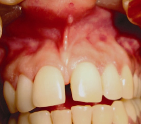
Simple frenum with a nodule
Figure 6.
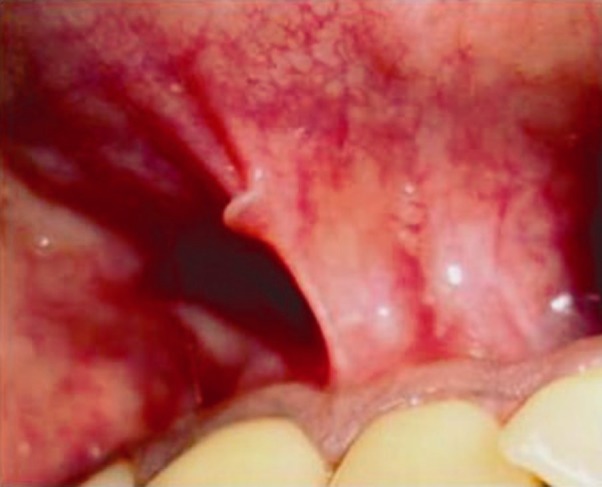
Simple frenum with appendix
Figure 7.
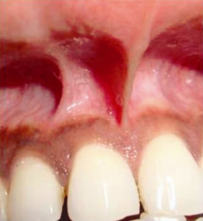
Simple frenum with nichum
Figure 8.
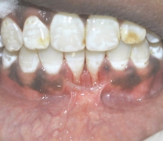
Wider frenal attachment
Abnormal or aberrant frena are detected visually, by applying tension over it to see the movement of papillary tip or blanching produced due to ischemia of the region. Clinically, papillary and papilla penetrating frena are considered as pathological and have been found to be associated with loss of papilla, recession, diastema, difficulty in brushing, malalignment of teeth and it may also prejudice the denture fit or retention leading to psychological disturbances to the individual.[5]
A frenum can become a significant problem if tension from lip movement pulls the gingival margin away from the tooth, or if the tissue inhibits the closure of a diastema during orthodontic treatment.
Frenal attachment that encroach on the marginal gingiva distend the gingival sulcus, fostering plaque accumulation, increasing the rate of progression of periodontal recession and thereby leading to recurrence after treatment.
Syndromes associated with different frenal attachments:
Ehlers-Danlos syndrome
Infantile hypertrophic pyloric stenosis,
Holoprosencephaly,
Ellis-van Creveld syndrome, and
Oro-facial-digital syndrome.
Each syndrome exhibits relatively specific frenal abnormalities, ranging from multiple, hyper plastic, hypoplastic, or an absence of frena.
Oro-facial-digital syndrome
Oro-facial-digital syndrome arises as the result of a single gene malformation showing X-linked dominant inheritance.[6]
Clinical features within the oral cavity:
The tongue is lobulated with hamartomata between lobules, hypertrophied lingual frenum which is incompletely differentiated from the floor of the mouth
Gums are clefted by abnormal supernumerary frenula
Cleft often in soft palate, might be bilateral or asymmetrical
Teeth are malpositioned, often and may have enamel hypoplasia
Midline notch (pseudocleft) may be seen in the upper lip.
Infantile hypertrophic pyloric stenosis
Occurs commonly in males at a ratio of 4.5 to 1 with an unknown etiology. There is a disturbance in the frenum formation. The absence or hypoplasia of mandibular frenum represents an important diagnostic tool in detection of this disease.[7]
Holoprosencephaly
It is an autosomal dominant condition characterized by a brain malformation due to defects in prosencephalon. It is characterized by defects including cyclopia, single nostril, single central incisor and premaxillary agenesis. Absence of labial maxillary frenum is one of the characteristic features of this condition.[8]
Ellis-van Creveld syndrome
Ellis-van Creveld (EvC) syndrome is an autosomal recessive disorder, mainly affecting the ectodermal components such as enamel, nail, and hair. The gene for EvC syndrome is located on chromosome 4p16. Patients with EvC syndrome characteristically presents with congenitally missing teeth, abnormal frenal attachment, microdontia, and hexadactyly.[9]
Oral manifestations in the EvC syndrome are characteristic and constant. The most constant finding is fusion of the anterior portion of the upper lip to the maxillary gingival margin, as a result of which no mucobuccal fold exists, causing the upper lip to present a slight V-shaped notch in the middle. Changes in the upper lip can be addressed by various names such as partial hare-lip or lip tie. The anterior portion of the lower ridge is often serrated and presents with multiple small labial frenula. The maxillary and mandibular alveolar process presents with notching or submucous clefts and continuous or broad labial frenula with dystrophic philtrum.[9]
Ehlerdanlos syndrome
It is a genetic disorder characterized by hyper extensive skin and hyper mobile joints with no gender predilection. Absence of the inferior labial and lingual frena has been described in this disorder.[10]
Pallister-hall syndrome
Inherited as an autosomal dominant pattern. The gene responsible for this disorder has been mapped to 7p13 and is identified as GL13.Clinical features include short mid face and nose with a flat nasal bridge and anteverted nostrils. Oral manifestations include micrognathia, microglossia and abnormal supernumerary frena extending from the buccal mucosa to the alveolar ridge.[11]
Opitz C syndrome
Exhibits similar frenal abnormalities as Pallister-Hall syndrome.[12]
Nonsyndromic conditions
In addition to abnormal oral frena observed in syndromic conditions, anomalous frena are encountered without other associated phenotypic features of genetic or chromosomal states. For example ankylosis of superior labial frena may show a familiar pattern of occurrence.[1]
Aberrant frenal attachments may be seen after orthognathic surgeries. Problems are probably caused by errors in the surgical technique. The design of the soft tissue incisions is critical, vertical incisions in the area of osteotomy will predictably create periodontal problems.[13]
MANAGEMENT
Three surgical techniques are effective in removal of frenal attachments
The simple excision technique
The Z-plasty technique
A localized vestibuloplasty with secondary epithelialization.
The first two techniques are effective when the mucosal and fibrous tissue band is relatively narrow; the third technique is often preferred when the frenal attachment has a wide base.
Simple excision technique
A narrow elliptic incision around the frenal area down to the periosteum is given
The fibrous frenum is then sharply dissected from the underlying periosteum and soft tissue, and the margins of the wound are gently undermined and re-approximated.
Z-plasty technique
After excision of the fibrous tissue, two oblique incisions are made in a Z fashion, one at each end of the previous area of excision
Two pointed flaps are then gently undermined and rotated to close the initial vertical incision horizontally.
Localized vestibuloplasty with secondary epithelialization:
An incision is made through mucosal tissue and underlying submucosal tissue, without perforating the periosteum
A supraperiosteal dissection is completed by undermining the mucosal and submucosal tissue with scissors
After a clean periosteal layer is identified, the edge of the mucosal flap is sutured to the periosteum at the maximal depth of the vestibule and the exposed periosteum is allowed to heal by secondary epithelialization.
CONCLUSION
Frenum may not regularly draw close scrutiny on routine dental examination. However, the detection of abnormal frenum may represent a highly useful indicator in the diagnosis of a wide array of syndromic and non-syndromic conditions. However, the presence of any abnormal frenal attachments can be corrected with a wide variety of surgical techniques available.
Footnotes
Source of Support: Nil
Conflict of Interest: None declared.
REFERENCES
- 1.Mintz SM, Siegel MA, Seider PJ. An overview of oral frena and their association with multiple syndromes and nonsyndromic conditions. Oral Surg Oral Med Oral Pathol Oral RadiolEndod. 2005;99:321–4. doi: 10.1016/j.tripleo.2004.08.008. [DOI] [PubMed] [Google Scholar]
- 2.Kotlow LA. Oral diagnosis of abnormal frenum attachments in neonates and infants: Evaluation and treatment of maxillary frenum using the Erbium YAG Laser. J Pediatr Dent Care. 2004c;10:11–4. [Google Scholar]
- 3.Mirko P, Miroslav S, Lubor M. Significance of the labial frenum attachment in periodontal disease in man. Part I. Classification and epidemiology of the labial frenum attachment. J Periodontol. 1974;45:891–4. doi: 10.1902/jop.1974.45.12.891. [DOI] [PubMed] [Google Scholar]
- 4.Kakodkar PV, Patel TN, Patel SV, Patel SH. Clinical assessment of diverse frenum morphology in Permanent dentition. Internet J Dent Sci. 2009:7. [Google Scholar]
- 5.Anubha N, Chaubey KK, Arora VK, Narula IS. Frenectomy combined with a laterally displaced pedicle graft. Indian J Dent Sci. 2010;2:47–51. [Google Scholar]
- 6.Dodge JA, Kernohan DC. Oral facial digital syndrome. Arch Dis Child. 1967;42:214–9. doi: 10.1136/adc.42.222.214. [DOI] [PMC free article] [PubMed] [Google Scholar]
- 7.Jenista JA. Madibular frenulum as a sign of infantile hypertrophic pyloric stenosis. 2001;138:447. J Pediatr. 2001;138:447–7. doi: 10.1067/mpd.2001.109192. [DOI] [PubMed] [Google Scholar]
- 8.Martin RA, Jones KL. Absence of the superior labial frenulum in holoprosencephaly: A new diagnostic sign. J Pediatr. 1998;133:151–3. doi: 10.1016/s0022-3476(98)70198-2. [DOI] [PubMed] [Google Scholar]
- 9.Babaji P. Oral abnormalities in the Ellis-van creveldsyndrome. Indian J Dent Res. 2010;21:143–5. doi: 10.4103/0970-9290.62791. [DOI] [PubMed] [Google Scholar]
- 10.De Felice C, Toti P, Di Maggio G, Parinni S, Bagnoli F. Absence of the inferior labial and lingual frenula in Ehlers-Danlos syndrome. 2001;357:1500-2. Lancet. 2001;357:1500–2. doi: 10.1016/S0140-6736(00)04661-4. 10. [DOI] [PubMed] [Google Scholar]
- 11.Hall JG, Pallister PD, Clarren SK, Beck with JB. Congenital hypothalamic hamartoblastoma, hypopituitarism, imperforate anus and postaxial polydactyly: A new syndrome? Part I: Clinical, causal and pathogenic considerations. Am J Med Genet. 1980;7:47–74. doi: 10.1002/ajmg.1320070110. [DOI] [PubMed] [Google Scholar]
- 12.Brooks JK, Leonard CO, Coccaro PJ., Jr (Opitz (BBG/G) syndrome: Oral manifestations. Am J Med Genet. 1992;43:595–601. doi: 10.1002/ajmg.1320430318. [DOI] [PubMed] [Google Scholar]
- 13.Panula K, Finne K, Oikarinen K. Incidence of complications and problems related to orthognathic surgery: A review of 655 patients. J Oral MaxillofacSurg. 2001;59:1128–36. doi: 10.1053/joms.2001.26704. [DOI] [PubMed] [Google Scholar]


