Abstract
Background:
Neurosurgeons are frequently among the first physicians asked to evaluate patients with papilledema, and the patient is often referred with the implication that they may require shunting. After an initial evaluation to exclude potential neurosurgical emergencies, the physician should carefully consider various etiologies of papilledema to prevent unnecessary neurosurgical operations.
Case Description:
The authors report two illustrative cases of unusual causes of papilledema: Anemia and leukemic infiltration of the central nervous system. In each case, a complete blood count provided clues for the diagnosis. A review of the literature is also included.
Conclusions:
Both patients responded to medical management/treatment of the underlying disease and did not require neurosurgical operative intervention. Papilledema may be caused by other etiologies besides increased intracranial pressure. The authors present two unusual cases leading to papilledema and provide an outline for the workup of these conditions.
Keywords: Anemia, cerebrospinal fluid shunt, idiopathic intracranial hypertension, papilledema
INTRODUCTION
Papilledema is clinically defined as optic disc swelling secondary to increased intracranial pressure (ICP).[23] Unilateral or bilateral optic disc swelling requires the clinician to obtain a thorough history and perform a detailed general, neurologic, and ophthalmologic exam to develop a broad differential diagnosis. If papilledema is confirmed, the cause of increased ICP needs to be identified. Commonly encountered causes of papilledema include mass lesions, cerebral edema, hydrocephalus, shunt failure, and idiopathic intracranial hypertension (IIH). Less frequently encountered etiologies of papilledema include systemic disease processes and medications. It is important for the clinician to consider these other medically treatable etiologies before attributing papilledema to IIH/pseudotumor cerebri (PTC) and considering an invasive procedure such as cerebrospinal fluid (CSF) shunting. The list of potential etiologies for papilledema is extensive, but includes optic neuritis, anterior ischemic optic neuropathy, anterior toxic neuropathy, uveitis, and hypotony.
We report two unusual etiologies of papilledema—one due to anemia and the other due to leukemic infiltration of the central nervous system. Both patients had abnormal bloodwork results shown on an initial complete blood count. Both patients responded to medical management and treatment of the underlying disease and operative intervention was not required. We also conducted a literature review to highlight the work-up for papilledema in an attempt to create a roadmap for diagnosis of papilledema.
CASE REPORTS
Case 1
A 14-year-old boy with Crohn's disease complained of “spots” in his vision for a 24-hour period and severe headaches during the prior several weeks. An initial evaluation revealed bilateral retinal hemorrhages and disc edema. Laboratory studies demonstrated severe anemia with hemoglobin of 3.0 mg/dL, white blood cells (WBC) of 1.8 × 10³/mm3, and platelets of 101 × 103/mm3. He was transfused with packed red blood cells (RBC) and referred to our facility.
One to two days before admission, the headaches were described as retro-orbital and bilateral, accompanied by multiple spots in both visual fields. On admission, the spots had consolidated into one large spot in the center of his vision. The patient also admitted to intermittent diplopia, pulsatile tinnitus, and several episode of emesis over the previous 2 weeks. He had noted bloody stools the previous day. His medications before admission included 6-mercaptopurine (6-MP), mesalamine, and vitamin D supplements. He had been taking 6-MP to treat Crohn's disease for several months. On retrospective chart review, we noted that the patient's blood counts had gradually declined since 6-MP therapy had been initiated.
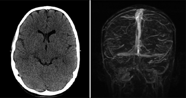
An ophthalmologic assessment at our institution demonstrated visual acuity of 20/800 right eye and 20/400 left eye, bilateral 4+ optic disc edema, bilateral central scotomas, nerve fiber layer hemorrhages, and bilateral scattered peripheral intraretinal hemorrhages. Computed tomography (CT) scan and magnetic resonance imaging/magnetic resonance venography (MRI/MRV) were unremarkable [Figure 1]. The patient underwent a lumbar puncture, which revealed an opening pressure in excess of 55 cm H2O. Other CSF studies were also unrevealing.
Figure 1.

Patient in Case 1. A normal head CT (left) and a normal MRV
We postulated that the patient's severe anemia may have precipitated the elevated ICP, causing his symptoms. He was placed on oral acetazolamide 5 mg/kg/dose daily to reduce ICP. His 6-MP was discontinued as this was thought to be the proximate cause of anemia, and he received a transfusion of packed RBC to increase his hemoglobin to 8.5 g/dL.
The patient's clinical course showed improvement by hospital day 3 [Table 1]. His visual acuity improved to 20/40 right eye and 20/40 left eye with a central bitemporal field loss; papilledema was reduced to 2-3 + OD, 2 + OS; and confrontation to visual fields still showed persistent but incomplete binasal central scotomas. His headaches gradually improved during hospitalization. His blood counts slowly increased after 6-MP was discontinued. One week after admission, his visual acuity remained 20/40 bilaterally with resolution of his partial central scotoma while disc edema improved 1.5-2 + OD, 1.5 + OS. A follow-up visit 2 weeks later revealed flat optic discs with good color, no hemorrhages, and visual acuity 20/40 right eye, 20/50 left eye.
Table 1.
Hgb, visual acuity, and disc edema trends for patient in case 1 report

Case 2
A 16-year-old obese girl presented with a 2-week history of jaw pain, headaches, sore throat, and tactile fevers. One week after the onset of symptoms, she was diagnosed with acute sinusitis and treated with amoxicillin-clavulinate. Due to persistent symptoms, further tests were performed that revealed anemia (Hgb of 7.8 mg/dL) and thrombocytopenia (platelets of 15 × 103/mm3). Blasts were noted on the smear. A monospot test was negative. Leukemia was suspected and she was referred to our facility for further management.
A review of the patient's systems revealed diffuse headaches, which were moderated by acetaminophen, mild neck pain, nausea with three episodes of emesis for 2 days, and bleeding when brushing her teeth. Bone marrow biopsy results were consistent with acute promyelocytic leukemia. She underwent induction therapy that consisted of all-transretinoic acid (ATRA) on days 1 through 28, daunomycin on days 3 through 6, and cytarabine (Ara-C) intravenously on days 3 through 9, and she also received four doses of intrathecal Ara-C. Her blood cell counts were monitored daily and transfusions of packed RBC and platelets were provided as needed.
Two days into her chemotherapy, the patient complained of progressive vision loss. An ophthalmology evaluation revealed visual acuity of 20/200 right eye and 20/200 left eye, bilateral 4+ optic nerve edema, and macular exudates. Intravenous acetazolamide (500 mg twice daily) was administered.
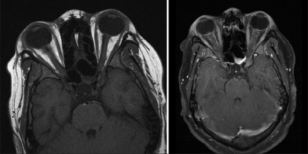
A “high” opening pressure during lumbar puncture was reported. CSF studies revealed RBC of 16/mm3, WBC of 1/mm3, bands of 18%, neutrophils of 59%, lymphocytes of 14%, monocytes of 4%, and other cells of 2% (myelocytes, metamyelocytes). Myeloid cells were also noted in the cytopathology specimen consistent with the diagnosis of leukemia. MRI demonstrated multifocal irregular nodules, dural thickening, and gyral enhancement in the posterior fossa. In addition, the patient's optic nerve sheaths were dilated and demonstrated thick nodular enhancement diffusely [Figure 2], consistent with leukemic infiltration of the optic nerve sheaths. Infiltration of the leptomeninges, and scalp were also noted. There was no evidence of cerebral venous sinus thrombosis or narrowing. An MRI of the spine was negative. We postulated that the patient's papilledema was secondary to leukemic infiltration of her optic sheaths.
Figure 2.

Patient in Case 2. Axial T1 MRI without (left) and with Gadolinium demonstrating thick nodular enhancement of the bilateral optic nerves
Acetazolamide did not lead to any improvement in the patient's condition. She urgently underwent a course of palliative craniospinal radiation therapy (12 daily treatments of 2160 cGy). Notably, her headaches and vision improved by the end of radiation treatment as evident by interval improvement in her papilledema with ongoing resolution of nerve hemorrhages and macula exudates. The patient continued taking acetazolamide until her papilledema resolved. A follow-up appointment at 8 months revealed visual acuity of 20/30 bilaterally and full ocular motility. Color plates continued to be decreased bilaterally, but were stable compared with prior exams. Mild optic nerve pallor was seen bilaterally, with no sign of optic nerve edema.
DISCUSSION
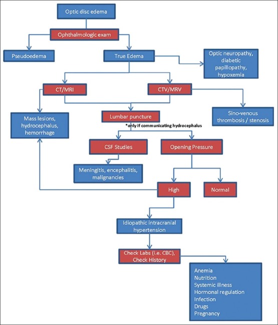
When a patient presents with optic disc swelling, it is important to obtain a thorough history, physical exam, and directed testing to determine if neurosurgical intervention is required. Optic disc swelling should distinguish between true disc edema and pseudo-edema via an ophthalmologic exam[23] [Figure 3]. The latter is most commonly caused by optic disc drusen.[23] Pseudo-edema resembles true disc edema, but lacks the distinguishing characteristics of true disc edema such as obscured disc vasculature, prominent nerve fiber layer elevation, venous congestion, exudates/cotton wool spots, and the absence of late cup, circumpapillary light reflex, and venous pulsations.[23] True disc edema could be caused by a local pathology such as optic neuropathy or by a systemic pathology such as increased ICP.[23] For the latter scenario, optic disc edema is specifically termed papilledema.[23]
Figure 3.

Approach to optic disc edema (adapted from Lee et al.[23])
Etiologies leading to papilledema include PTC/IIH, hydrocephalus, shunt failure, mass lesions, trauma, venous sinus obstruction, and infection.[23] Typically the patient with papilledema receives a CT or MRI. A CTV or MRV can help rule out sinovenous thrombosis or stenosis. Lumbar puncture may also be indicated to measure opening CSF pressure. Abnormal CSF studies could indicate infectious etiologies such as meningitis/encephalitis.
Once serious neurosurgical causes are excluded, the physician should consider more unusual causes of optic disc edema. If the patient has increased ICP with no clear etiology, PTC/IIH is a potential diagnosis. Classically, PTC/IIH is diagnosed in the setting of papilledema when the following conditions are met: (1) Normal imaging studies (usually MRI); (2) normal CSF studies; (3) elevated opening pressure; and (4) other signs and symptoms secondary to elevated ICP (such as uni-or bilateral disc edema or a sixth nerve palsy).[23]
Although PTC/IIH is considered idiopathic, it has been associated with many conditions, including nutrition (such as hypervitaminosis A and vitamin deficiencies), drugs (such as phenytoin and anabolic steroids), hormonal regulation (such as thyroid disease, Addison's disease, and hypoparathyroidism), systemic illness (such as lupus, malignancies, and renal failure), and infections (such as HIV and Lyme disease).[23] Consideration of these associations may prevent unnecessary medications and surgical procedures such as CSF shunting.
In Case 1, it is important to note that the patient had significant anemia, and to consider the role of medications in inducing anemia. This patient was taking 6-MP, which acts as a false metabolite of the purine hypoxanthine. It has been used as an immunosuppressive drug for inflammatory bowel disease (IBD)[35] and as an antineoplastic drug for acute lymphoblastic leukemia[15] and chronic myelomonocytic leukemia.[36] There have been documented cases of severe myelosuppression,[22,48] aplastic anemia,[45] and hemolytic anemia.[36] Typically, IBD patients undergo thiopurine S-methyltransferase (TPMT) tests before administration of azathioprine/6-MP since low TPMT-activity is associated with drug-induced side effects such as hematoxicity and hepatotoxicity.[46] TPMT is an enzyme that inactivates thiopurine drugs.[46] Even for patients with normal TPMT test results, cytopenia has been reported.[24] We believe that the patient in Case 1 likely suffered from bone marrow suppression secondary to 6-MP. Although tests involving TPMT were normal, the patient's counts increased after the medication was discontinued, which indicates that 6-MP bone marrow suppression was a likely reason for the pancytopenia.
In a review of 77 patients with clinical evidence of intracranial hypertension, Mollan et al.[31] found that 8 of the 77 had microcytic anemia. Seven of the eight patients with microcytic anemia had resolution of their symptoms after treatment for anemia; the other patient required placement of a ventriculoperitoneal shunt and exhibited improved symptoms. Similarly, Stiebel-Kalish et al.[43] reported anemia in 15 of 96 patients who presented with clinical intracranial hypertension. Many types of anemia have been associated with intracranial hypertension, including hemolytic anemia,[49] aplastic anemia,[19,25,30,33] sickle cell anemia,[16] iron-deficiency anemia,[8,21,41] anemia secondary to paroxysmal nocturnal hemoglobinuria,[1] and pernicious anemia.[38] In addition to optic disc edema, other ocular findings in patients with anemia include cotton wool spots, nerve fiber layer/preretinal hemorrhages, vitreous hemorrhages, and central retinal vein occlusion.[29]
The mechanism between anemia and intracranial hypertension has remained rather nebulous. Iron-deficiency anemia has been regarded as a hypercoagulable condition and linked to cerebral venous thrombosis.[3,4,20,34,44] Moreover, iron-deficiency contributes to thrombosis by inducing reactive thrombocytosis, possibly via erythropoietin production,[10,12] leading to a higher risk of thrombovascular occlusions. In addition, erythrocyte deformability is reduced in patients with iron deficiency,[50] increasing the viscosity of the blood. The hyperviscosity could intensify venous pressure without evidence of venous sinus thrombosis on neuroimaging.[6]
Hypercoagulability has also been reported in various conditions related to hemolytic anemia, including beta-thalassemia,[32] paroxysmal nocturnal hemoglobinuria,[1] and sickle cell disease.[2] It is thought that microparticles derived from the destruction/hemolysis of blood components (RBC, platelets, endothelial cells, and monocytes) may play a role in the hypercoagulable status present in hemolytic anemias.[2]
Regarding Case 2, papilledema is a relatively rare complication of leukemia. Several reports exist concerning the relationship between papilledema and acute myeloid leukemia,[5,26] acute lymphoblastic leukemia,[7,9,11,17,47,52] chronic myelogenous leukemia,[14] and chronic lymphocytic leukemia.[39] In a study of 82 children with acute leukemia, Reddy et al.[37] found that only 3 children complained of eye symptoms. However, ocular manifestations of the disease were discovered in 14 children on examination. In addition to papilledema, other ocular lesions observed included proptosis, intraretinal hemorrhages, white-centered hemorrhages, cotton wool spots, macular hemorrhage, subhyaloid hemorrhage, vitreous hemorrhage, cortical blindness, sixth nerve palsy, and exudative retinal detachment with choroidal infiltration.[37]
Headache is the most frequent side effect of ATRA therapy.[40] Although uncommon, intracranial hypertension is also a complication. Notably, the issue has been prevalent mainly in younger populations, especially patients who have taken doses ranging from 45 and 80 mg/m2 daily.[13] The mechanism to explain the connection between ATRA therapy and intracranial hypertension has remained elusive, but studies focusing on the mammalian nervous system have demonstrated the influence of retinoids and the RARα receptor on the development of the nervous system.[27,28] Consequently, there has been speculation that ATRA could affect the structures involved in the production (choroidal plexus) and/or drainage (arachnoid villi) of CSF, inducing intracranial hypertension.[51] To rationalize the age dependence with ATRA-related intracranial hypertension, researchers have postulated either a reduction in the RARα receptors with age or a reduction in the response of these receptors to retinoid stimulation.[13,51]
Moreover, the administration of intrathecal Ara-C has been associated with papilledema. Sommer et al.[42] reported three patients inflicted with acute myeloid leukemia without evidence of central nervous involvement who received four doses of 50 mg of intrathecal liposomal cytarabine for maintenance therapy. After the third or fourth dose of cytarabine, the patients developed bilateral papilledema. With protracted dexamethasone therapy, the adverse reaction resolved. Jabbour et al.[18] also reported neurotoxic reactions from using intrathecal liposomal cytarabine in combination with high-dose methotrexate in patients with acute lymphocytic leukemia. Of 31 patients with newly diagnosed acute lymphocytic leukemia, one patient suffered from papilledema.
CONCLUSION
Shunt placement and operative intervention are not always mandated in the treatment of papilledema. Nonsurgical causes for papilledema should be considered in the differential diagnosis.
Footnotes
Available FREE in open access from: http://www.surgicalneurologyint.com/text.asp?2013/4/1/60/110686
Contributor Information
Ha Son Nguyen, Email: hsnguyen@mcw.edu.
Kathryn M. Haider, Email: khaider@iupui.edu.
Laurie L. Ackerman, Email: lackerma@iupui.edu.
REFERENCES
- 1.Aktan S, Kansu T, Kansu E, Zileli T. Papilledema in paroxysmal nocturnal hemoglobinuria. J Clin Neuroophthalmol. 1984;4:47–8. doi: 10.3109/01658108409019496. [DOI] [PubMed] [Google Scholar]
- 2.Ataga KI. Hypercoagulability and thrombotic complications in hemolytic anemias. Haematologica. 2009;94:1481–4. doi: 10.3324/haematol.2009.013672. [DOI] [PMC free article] [PubMed] [Google Scholar]
- 3.Balci K, Utku U, Asil T, Buyukkoyuncu N. Deep cerebral vein thrombosis associated with iron deficiency anaemia in adults. J Clin Neurosci. 2007;14:181–4. doi: 10.1016/j.jocn.2005.09.020. [DOI] [PubMed] [Google Scholar]
- 4.Belman AL, Roque CT, Ancona R, Anand AK, Davis RP. Cerebral venous thrombosis in a child with iron deficiency anemia and thrombocytosis. Stroke. 1990;21:488–93. doi: 10.1161/01.str.21.3.488. [DOI] [PubMed] [Google Scholar]
- 5.Beslow LA, Abend NS, Smith SE. Cerebral sinus venous thrombosis complicated by cerebellar hemorrhage in a child with acute promyelocytic leukemia. J Child Neurol. 2009;24:110–4. doi: 10.1177/0883073808321057. [DOI] [PubMed] [Google Scholar]
- 6.Biousse V, Rucker JC, Vignal C, Crassard I, Katz BJ, Newman NJ. Anemia and papilledema. Am J Ophthalmol. 2003;135:437–46. doi: 10.1016/s0002-9394(02)02062-7. [DOI] [PubMed] [Google Scholar]
- 7.Caca I, Unlu K, Ari S, Dirier A. Unilateral optic disc edema in a patient with acute lymphocytic leukemia: A case and review of the literature. Ann Ophthalmol. 2005;37:303–5. [Google Scholar]
- 8.Capriles LF. Intracranial hypertension and iron-deficiency anemia; report of four cases. Arch Neurol. 1963;9:147–53. doi: 10.1001/archneur.1963.00460080057008. [DOI] [PubMed] [Google Scholar]
- 9.D’Angio GJ. Papilledema, presenting as reversible loss of vision, in a child with acute lymphoblastic leukemia–comment. Pediatr Blood Cancer. 2005;45:734. doi: 10.1002/pbc.20545. [DOI] [PubMed] [Google Scholar]
- 10.Dahl NV, Henry DH, Coyne DW. Thrombosis with erythropoietic stimulating agents-does iron-deficient erythropoiesis play a role? Semin Dial. 2008;21:210–1. doi: 10.1111/j.1525-139X.2008.00435.x. [DOI] [PubMed] [Google Scholar]
- 11.de Fatima Soares M, Braga FT, da Rocha AJ, Lederman HM. Optic nerve infiltration by acute lymphoblastic leukemia: MRI contribution. Pediatr Radiol. 2005;35:799–802. doi: 10.1007/s00247-005-1440-8. [DOI] [PubMed] [Google Scholar]
- 12.Franchini M, Targher G, Montagnana M, Lippi G. Iron and thrombosis. Ann Hematol. 2008;87:167–73. doi: 10.1007/s00277-007-0416-1. [DOI] [PMC free article] [PubMed] [Google Scholar]
- 13.Guirgis MF, Lueder GT. Intracranial hypertension secondary to all-trans retinoic acid treatment for leukemia: Diagnosis and management. J AAPOS. 2003;7:432–4. doi: 10.1016/j.jaapos.2003.08.005. [DOI] [PubMed] [Google Scholar]
- 14.Guymer RH, Cairns JD, O’Day J. Benign intracranial hypertension in chronic myeloid leukemia. Aust N Z J Ophthalmol. 1993;21:181–5. [PubMed] [Google Scholar]
- 15.Hawwa AF, Millership JS, Collier PS, McCarthy A, Dempsey S, Cairns C, et al. The development of an objective methodology to measure medication adherence to oral thiopurines in paediatric patients with acute lymphoblastic leukaemia–an exploratory study. Eur J Clin Pharmacol. 2009;65:1105–12. doi: 10.1007/s00228-009-0700-1. [DOI] [PubMed] [Google Scholar]
- 16.Henry M, Driscoll MC, Miller M, Chang T, Minniti CP. Pseudotumor cerebri in children with sickle cell disease: A case series. Pediatrics. 2004;113:e265–9. doi: 10.1542/peds.113.3.e265. [DOI] [PubMed] [Google Scholar]
- 17.Iqbal Y, Palkar V, Al-Sudairy R, Al-Omari A, Abdullah MF, Al-Debasi T. Papilledema, presenting as reversible loss of vision, in a child with acute lymphoblastic leukemia. Pediatr Blood Cancer. 2005;45:72–3. doi: 10.1002/pbc.20369. [DOI] [PubMed] [Google Scholar]
- 18.Jabbour E, O’Brien S, Kantarjian H, Garcia-Manero G, Ferrajoli A, Ravandi F, et al. Neurologic complications associated with intrathecal liposomal cytarabine given prophylactically in combination with high-dose methotrexate and cytarabine to patients with acute lymphocytic leukemia. Blood. 2007;109:3214–8. doi: 10.1182/blood-2006-08-043646. [DOI] [PubMed] [Google Scholar]
- 19.Jeng MR, Rieman M, Bhakta M, Helton K, Wang WC. Pseudotumor cerebri in two adolescents with acquired aplastic anemia. J Pediatr Hematol Oncol. 2002;24:765–8. doi: 10.1097/00043426-200212000-00018. [DOI] [PubMed] [Google Scholar]
- 20.Kinoshita Y, Taniura S, Shishido H, Nojima T, Kamitani H, Watanebe T. Cerebral venous sinus thrombosis associated with iron deficiency: Two case reports. Neurol Med Chir (Tokyo) 2006;46:589–93. doi: 10.2176/nmc.46.589. [DOI] [PubMed] [Google Scholar]
- 21.Knizley H, Jr, Noyes WD. Iron deficiency anemia, papilledema, thrombocytosis, and transient hemiparesis. Arch Intern Med. 1972;129:483–6. [PubMed] [Google Scholar]
- 22.Kubota D, Takishima C, Ishii K, Kawamura T, Matsumoto T, Itsui Y, et al. A case report: Severe bone marrow suppression caused by 6-mercaptopurin in Crohn's disease patient. Nihon Shokakibyo Gakkai Zasshi. 2006;103:1044–9. [PubMed] [Google Scholar]
- 23.Lee AG, Brazis P. Clinical pathways in neuro-ophthalmology: An Evidence-Based Approach. 2nd ed. New York, NY: Thieme Publishers; 2003. [Google Scholar]
- 24.Leung M, Piatkov I, Rochester C, Boyages SC, Leong RW. Normal thiopurine methyltransferase phenotype testing in a Crohn disease patient with azathioprine induced myelosuppression. Intern Med J. 2009;39:121–6. doi: 10.1111/j.1445-5994.2008.01855.x. [DOI] [PubMed] [Google Scholar]
- 25.Lilley ER, Bruggers CS, Pollock SC. Papilledema in a patient with aplastic anemia. Arch Ophthalmol. 1990;108:1674–5. doi: 10.1001/archopht.1990.01070140028014. [DOI] [PubMed] [Google Scholar]
- 26.Lin YC, Wang AG, Yen MY, Hsu WM. Leukaemic infiltration of the optic nerve as the initial manifestation leukaemic relapse. Eye. 2004;18:546–50. doi: 10.1038/sj.eye.6700701. [DOI] [PubMed] [Google Scholar]
- 27.Maden M, Holder N. The involvement of retinoic acid in the development of the vertebrate central nervous system. Development. 1991;(Suppl 2):87–94. [PubMed] [Google Scholar]
- 28.Maden M, Ong DE, Chytil F. Retinoid-binding protein distribution in the developing mammalian nervous system. Development. 1990;109:75–80. doi: 10.1242/dev.109.1.75. [DOI] [PubMed] [Google Scholar]
- 29.Mansour AM, Salti HI, Han DP, Khoury A, Friedman SM, Salem Z, et al. Ocular findings in aplastic anemia. Ophthalmologica. 2000;214:399–402. doi: 10.1159/000027532. [DOI] [PubMed] [Google Scholar]
- 30.Mohla A, Oworu O, Hutchinson C. Aplastic anaemia presenting with features of raised intracranial pressure. J R Soc Med. 2006;99:315–6. doi: 10.1258/jrsm.99.6.315. [DOI] [PMC free article] [PubMed] [Google Scholar]
- 31.Mollan SP, Ball AK, Sinclair AJ, Madill SA, Clarke CE, Jacks AS, et al. Idiopathic intracranial hypertension associated with iron deficiency anaemia: A lesson for management. Eur Neurol. 2009;62:105–8. doi: 10.1159/000222781. [DOI] [PubMed] [Google Scholar]
- 32.Mustafa NY, Marouf R, Al-Humood S, Al-Fadhli SM, Mojiminiyi O. Hypercoagulable state and methylenetetrahydrofolate reductase (MTHFR) C677T mutation in patients with beta-thalassemia major in Kuwait. Acta Haematol. 2010;123:37–42. doi: 10.1159/000260069. [DOI] [PubMed] [Google Scholar]
- 33.Nazir SA, Siatkowski RM. Pseudotumor cerebri in idiopathic aplastic anemia. J AAPOS. 2003;7:71–4. doi: 10.1067/mpa.2003.S1091853102420149. [DOI] [PubMed] [Google Scholar]
- 34.Ogata T, Kamouchi M, Kitazono T, Kuroda J, Ooboshi H, Shono T, et al. Cerebral venous thrombosis associated with iron deficiency anemia. J Stroke Cerebrovasc Dis. 2008;17:426–8. doi: 10.1016/j.jstrokecerebrovasdis.2008.04.008. [DOI] [PubMed] [Google Scholar]
- 35.Present DH, Korelitz BI, Wisch N, Glass JL, Sachar DB, Pasternack BS. Treatment of Crohn's disease with 6-mercaptopurine.A long-term, randomized, double-blind study. N Engl J Med. 1980;302:981–7. doi: 10.1056/NEJM198005013021801. [DOI] [PubMed] [Google Scholar]
- 36.Pujol M, Fernandez F, Sancho JM, Ribera JM, Milla F, Feliu E. Immune hemolytic anemia induced by 6-mercaptopurine. Transfusion. 2000;40:75–6. doi: 10.1046/j.1537-2995.2000.40010075.x. [DOI] [PubMed] [Google Scholar]
- 37.Reddy SC, Menon BS. A prospective study of ocular manifestations in childhood acute leukaemia. Acta Ophthalmol Scand. 1998;76:700–3. doi: 10.1034/j.1600-0420.1998.760614.x. [DOI] [PubMed] [Google Scholar]
- 38.Reid HA, Harris W. Reversible papilloedema in pernicious anaemia. Br Med J. 1951;1:20. doi: 10.1136/bmj.1.4696.20. [DOI] [PMC free article] [PubMed] [Google Scholar]
- 39.Saenz-Frances F, Calvo-Gonzalez C, Reche-Frutos J, Donate-Lopez J, Huelga-Zapico E, Garcia-Sanchez J, et al. Bilateral papilledema secondary to chronic lymphocytic leukaemia. Arch Soc Esp Oftalmol. 2007;82:303–6. doi: 10.4321/s0365-66912007000500010. [DOI] [PubMed] [Google Scholar]
- 40.Sano F, Tsuji K, Kunika N, Takeuchi T, Oyama K, Hasegawa S, et al. Pseudotumor cerebri in a patient with acute promyelocytic leukemia during treatment with all-trans retinoic acid. Intern Med. 1998;37:546–9. doi: 10.2169/internalmedicine.37.546. [DOI] [PubMed] [Google Scholar]
- 41.Schwaber JR, Blumberg AG. Papilledema associated with blood loss anemia. Ann Intern Med. 1961;55:1004–7. doi: 10.7326/0003-4819-55-6-1004. [DOI] [PubMed] [Google Scholar]
- 42.Sommer C, Lackner H, Benesch M, Sovinz P, Schwinger W, Moser A, et al. Neuroophthalmological side effects following intrathecal administration of liposomal cytarabine for central nervous system prophylaxis in three adolescents with acute myeloid leukaemia. Ann Hematol. 2008;87:887–90. doi: 10.1007/s00277-008-0521-9. [DOI] [PubMed] [Google Scholar]
- 43.Stiebel-Kalish H, Kalish Y, Lusky M, Gaton DD, Ehrlich R, Shuper A. Puberty as a risk factor for less favorable visual outcome in idiopathic intracranial hypertension. Am J Ophthalmol. 2006;142:279–83. doi: 10.1016/j.ajo.2006.03.043. [DOI] [PubMed] [Google Scholar]
- 44.Stolz E, Valdueza JM, Grebe M, Schlachetzki F, Schmitt E, Madlener K, et al. Anemia as a risk factor for cerebral venous thrombosis? An old hypothesis revisited. Results of a prospective study. J Neurol. 2007;254:729–34. doi: 10.1007/s00415-006-0411-9. [DOI] [PubMed] [Google Scholar]
- 45.Tajiri H, Tomomasa T, Yoden A, Konno M, Sasaki M, Maisawa S, et al. Efficacy and safety of azathioprine and 6-mercaptopurine in Japanese pediatric patients with ulcerative colitis: A survey of the Japanese Society for Pediatric Inflammatory Bowel Disease. Digestion. 2008;77:150–4. doi: 10.1159/000140974. [DOI] [PubMed] [Google Scholar]
- 46.Teml A, Schaeffeler E, Herrlinger KR, Klotz U, Schwab M. Thiopurine treatment in inflammatory bowel disease: Clinical pharmacology and implication of pharmacogenetically guided dosing. Clin Pharmacokinet. 2007;46:187–208. doi: 10.2165/00003088-200746030-00001. [DOI] [PubMed] [Google Scholar]
- 47.Tourje EJ, Gold LH. Leukemic infiltration of optic nerves-demonstration by computerized tomography. Comput Tomogr. 1977;1:225–8. doi: 10.1016/0363-8235(77)90005-9. [DOI] [PubMed] [Google Scholar]
- 48.Ugajin T, Miyatani H, Demitsu T, Iwaki T, Ushimaru S, Nakashima Y, et al. Severe myelosuppression following alopecia shortly after the initiation of 6-mercaptopurine in a patient with Crohn's disease. Intern Med. 2009;48:693–5. doi: 10.2169/internalmedicine.48.1736. [DOI] [PubMed] [Google Scholar]
- 49.Vargiami E, Zafeiriou DI, Gombakis NP, Kirkham FJ, Athanasiou-Metaxa M. Hemolytic anemia presenting with idiopathic intracranial hypertension. Pediatr Neurol. 2008;38:53–4. doi: 10.1016/j.pediatrneurol.2007.08.012. [DOI] [PubMed] [Google Scholar]
- 50.Vaya A, Simo M, Santaolaria M, Todoli J, Aznar J. Red blood cell deformability in iron deficiency anaemia. Clin Hemorheol Microcirc. 2005;33:75–80. [PubMed] [Google Scholar]
- 51.Visani G, Bontempo G, Manfroi S, Pazzaglia A, D’Alessandro R, Tura S. All-trans-retinoic acid and pseudotumor cerebri in a young adult with acute promyelocytic leukemia: A possible disease association. Haematologica. 1996;81:152–4. [PubMed] [Google Scholar]
- 52.Wolfe GI, Galetta SL, Mollman JE. Spontaneous remission of papilledema and sixth nerve palsy in acute lymphoblastic leukemia. J Neuroophthalmol. 1994;14:91–4. doi: 10.3109/01658109409024031. [DOI] [PubMed] [Google Scholar]


